Mapa mental
CELL
Conteúdo do mapa
CELL
Encompassed in biomembrane
- Up to 50% of the membrane can be proteins
- Fluid
- Dynamic at 37º
- biomembrane as to be able to move things around
- 2 dimensional fluids
- Lateral diffusion
- within the leaflet Phospholipids in one leaflet can move within that leaflet with ease common, easy, rapid
- Transverse Diffusion
- Between bilayers is very slow & unlikely you never get flip-flopping you rarely get phospholipid from one leaflet going into the other
- Lateral diffusion
- Temperature & composition dependent
- Fluidity regulation
- Sterols, cholesterol
- break apart van der waal interactions = more fluid
- Composition of phospholipids
- # of double bonds
- # of phospholipids
- Proteins
- transmembrane proteins break van der waal forces = more fluid
- Sterols, cholesterol
- Fluidity regulation
- Measuring fluidity
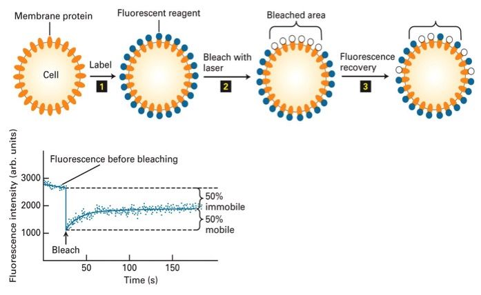
- FRAP
- using fluorescent proteins proteins in PM are potentially floating around & laterally diffusing in the PM bleached area cannot be seen on microscope recovery- does bleached area recover some of it's fluorescence?
- Goal: look for recovery
- only way to look for recovery is if proteins from periphery diffuse into this area & recover fluorescence protein diffusion related to amount of diffusion in the membrane more fluid = more recovery
- Graph
- Certain level of fluorescence, which is 100% (starting point) when you bleach, it drops to 0 (100-0 real quick) look for recovery, which eventually plateaus you end up with 50% recovery in this case, which means 50% are motile & 50 are non-motile 50% of the membrane has fluidity
- reason for non-motility
- protein linked to cytoskeleton, which is tethered to membrane & does not let proteins move
- FRAP
- Dynamic at 37º
- Closed compartments
- membrane-bound organelles
- Plasma membrane
- Cytosolic face
- Internal
- BUT FOR VESICLE MEMBRANE CYTOSOLIC = EXTERNAL FACE
- Internal
- Exoplasmic face
- proteins in lumen
- Carbs are found exclusively here
- Cytosolic face
- Asymmetric
- ASYMMETRY TILL YOU DIE
- Present with all proteins, not just lipid bilayer
- Proteins within membrane are same orientation
- Cadherins have binding domain in EC domain ALWAYS
- ETC
- Membrane proteins
- integral
- Proteins go through PM sizes of domain vary
- transmembrane domain
- most important domain inner part of lipid bilayer transmembrane made up of hydrophobic amino acids it has fatty acid carbon tails
- Spans lipid bilayer, hydrophobic region
- Different structures
- Alpha helix
- chain of amino acids making a simple little helix 20-25 amino acids
- Beta barrel
- complex structure hydrophobic amino acids make it to span the membrane
- Alpha helix
- cytosolic side
- Arg & lys
- Help anchor the protein
- if you try to pull protein through membrane, these charged proteins resist going into hydrophobic region
- Help anchor the protein
- Arg & lys
- Exoplasmic side
- glycosylated
- can add sugars to transmembrane proteins on extracellular domain
- glycosylated
- Cytosolic & exoplasmic domain
- peripheral
- Proteins somehow attached to membrane Does not tell you anything about its function
- Example: Dystrophin is bound to transmembrane protein You never find dystrophin outside of a cell
- attached non-covalently to something that is covalently linked to membrane
- Ionic interactions, hydrogen bonds protein-protein interactions van der waals forces
- Attached to transmembrane protein, integral membrane protein or lipid-linked
- integral protein can be linked to cytoskeleton via peripheral membrane protein
- ECM can be bound by peripheral membrane protein
- ECM can bind to integrins, which are transmembrane protein, thus making it a peripheral membrane protein
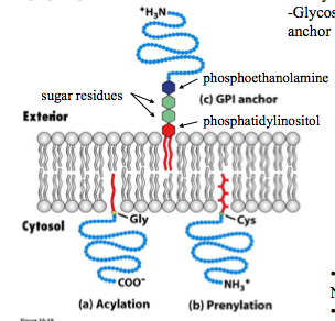
- Lipid-Linked
- Proteins physically linked to one of the phospholipids on the membrane
- Acylation
- N-term Glycine linked to phosphate
- Prenylation
- C terminal Cysteine or one that is close to C-terminal domain
- GPI linked
- Link to exterior surface
- PI (phosphlipd & phoshoglyceride)
- In order to link protein to membrane you have to link to PI LINK IS CALLED GPI ANCHOR
- have a signal on their C-term so they can be linked to GPI anchor
- Doesn't have to be transmembrane domain
- Some NCAM have GPI anchors
- NCAM = IG superfamily molecules have transmembrane domain, others have GPI anchors remember: both are cell adhesion molecules but they can do different things
- does not interact with cytoskeleton
- NCAM transmembrane molecules do interact with cytoskeleton
- Some NCAM have GPI anchors
- INSERTION OF PROTEINS INTO MEMBRANE
- Translation of any protein starts in cytosol
- ribosome is in cytosol
- Need signal to leave cytosol
- not necessarily sequence specific
- WHENEVER YOU HEAR SIGNAL IT IS BEING TAKEN TO ER
- Topogenic sequences
- found in proteins that can be translated
- Its not the exact amino acids that are signals, its the shapes they form, which are recognized by other proteins such as signal recognition particles
- N terminal Sequence
- found on N-term & is cleaved Recognized by signal recognition particle- taken to ER
- Signal Anchor Sequence (SA)
- anchored in ER N-term signal that is NOT cleaved
- Hydrophobic C-terminus
- acts as a signal to get protein to ER
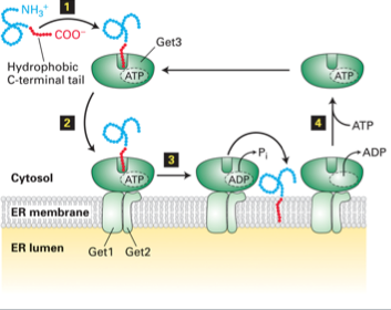
- Tail-anchored protein
- translation starts at N-terminal in the cytoplasm
- after translation finishes, a Hydrophobic C terminus made
- Occurs in cytosol, which has a hydrophilic environment
- Protein folds to get out of hydrophilic environment of cytosol
- Forms a structure recognized by GET3
- GET 3 in cytoplasm GET 1 & 2 - ER MEMBRANE
- GET3 recognizes C terminus
- Docks with Get 1 & 2 and uses ATP to shove hydropobic tail into ER membrane
- Result: protein that has hydrophobic C term tail in the membrane and N term domainin cytosol
- DOES NOT HAVE EXTRACELLULAR DOMAIN BUT HAS TRANSMEMBRANE DOMAIN
- Result: protein that has hydrophobic C term tail in the membrane and N term domainin cytosol
- Docks with Get 1 & 2 and uses ATP to shove hydropobic tail into ER membrane
- Forms a structure recognized by GET3
- after translation finishes, a Hydrophobic C terminus made
- translation starts at N-terminal in the cytoplasm
- Stop-transfer/Membrane anchor (STA)
- Where-ever its being transferred, we transfer & stop it anchor it there
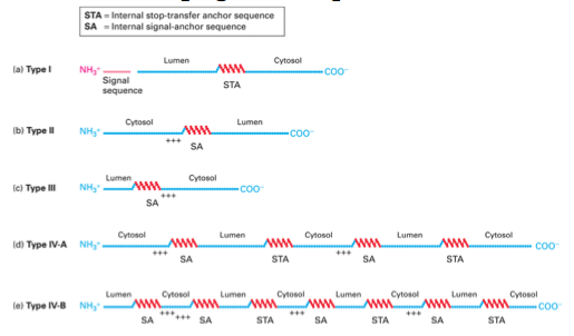
- Type 1, 3, 4B the N terminal are luminal
- Type 2 & 4A N terminal is cytosolic
- TYPES OF PROTEINS
- ALL TYPES OF PROTEINS ARE TRANSMEMBRANE PROTEINS
- Synthesis of Type I
- typical transmembrane proteins has hydrophobic domain
- N term sign go into lumen & is cleaved C term remains in the cytoplasm
- translation occurs in cytoplasm N terminal translated first
- N term signal is hydrophobic & begins to fold (stops translation)
- Signal recognized by signal recognition particles (goes to ER)
- N term goes via translocon into ER lumen
- N term sequence cleaved- mature protein does not have signal
- Translation continues into ER lumen
- N term sequence cleaved- mature protein does not have signal
- N term goes via translocon into ER lumen
- Signal recognized by signal recognition particles (goes to ER)
- N term signal is hydrophobic & begins to fold (stops translation)
- hydrophobic domain = STA,
- Synthesis of TYPE II & III
- No n terminal signal they have integral signal
- translation starts in cytosol with N term domain
- make N term domain, until you hit SA, which tells you to go to ER
- signal region = hydrophobic it becomes the transmembrane domain , but the thing is you already have this N terminal domain translated
- Does the N term domain stay in cytosol, or do you shove it into the lumen?
- depends on charges around the SA SA = hydrophobic, its going to be in the membrane
- cytosolic amino acids are often charged, which helps anchor the protein in the membrane & helps get protein to the right side of the matrix
- TYPE II
- Positive charged amino acids on N term side
- N term stays in cytosol
- translocon flips translation & lets C term domain go into lumem
- N term stays in cytosol
- Positive charged amino acids on N term side
- Type III
- No/very little charge on amino acids on N term domain
- N term goes into lumen
- Charged amino acids on Carboxyl term side of SA thus it stays in cytosol
- N term goes into lumen
- No/very little charge on amino acids on N term domain
- make N term domain, until you hit SA, which tells you to go to ER
- Synthesis of Type IV
- STA means protein stops transferring into ER
- Protein goes back & forth between signal, STA, signal, STA
- in & out of ER
- # of domains varies & position of N term domain can be cytosolic or luminal
- in & out of ER
- Protein goes back & forth between signal, STA, signal, STA
- STA means protein stops transferring into ER
- Translation of any protein starts in cytosol
- Transport across Membrane
- Passive Diffusion
- partition coefficient AND concentration gradient
- membranes are semi permeable & allow small uncharged molecules through
- partition - measures hydrophobicity of molecule
- How soluble a molecule is VS how soluble it is in an aqueous solution
- how fast a moelcule goes through biomembrane
- relatively high coefficient
- some solubility in membrane
- remember: do not want it too lipid soluble
- K = 0, not cross
- no energy
- driven by existing concentration different across PM or any membrane HIGH TO LOW
- partition coefficient AND concentration gradient
- Active Transport
- Needs energy
- primary active transport
- secondary active transport
- uses a situation that's already arisen using primary transport
- doesn't really require ATP, so not necessarily ATP powered
- Antiporters
- Symporters
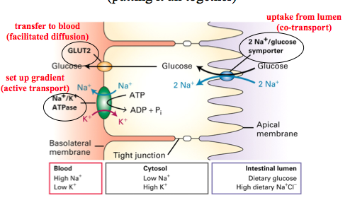
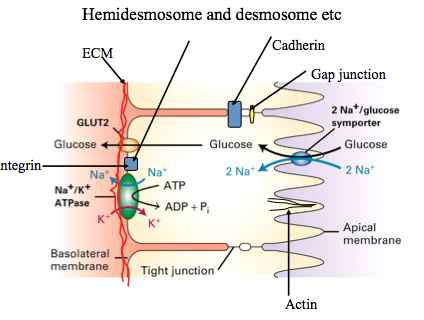
- Na/Glucose
- Na high outside cell- (+) charge Glucose = outside cell High glucose = inside cell, energy source for cells
- want more glucose inside cell, but you already have a high concentration of it Glucose moves against its gradient, which requires energy
- Energy is provided by symporter & uses the fact that Na wants to come in (down its gradient) Glucose uses the energy provided by Na
- 2 free energies
- 1) Concentration gradient of Na more sodium outside, it wants to get inside
- 2) Charge gradient of cell positive charge on outside of cell
- Both energies enough to move 2 Na down & 1 glucose against gradient
- Na/Glucose
- Use already established graident to tranport themselves
- Use one molecule that goes with concentration gradient & one against gradient
- pumps that use ATP to move molecules against concentration gradient
- ions are charged, cannot get them across the membrane, need to pump ions
- V & F
- responsible for pumping, they use ATP to pump H+ across membrane
- Important in mitochondria & chloroplasts, that is where a lot of H gets pumped around for ATP synthesis
- ABC type
- Move variety of small molecules
- Have ATP binding cassette
- specific domain that binds ATP in specific way
- Not restricted to ions
- Can flip from one leaf to other
- need ATP
- CFTR
- just pumps Cl-
- P
- move different types of ions two types first one is called muscle-calcium ATPase
- Muscle ATPase pumps 2 Ca out of cytoplasm per ATP
- in this case it pumps it into SR as soon as you trigger muscle contraction
- Calcium release is voltage gated channel, as soon as calcium comes out P-class pump kicks in & gets it back into SR
- END: cystosol has little or no Ca
- pump works against concentration gradient END: lots of Ca in SR, so when channel opens again Ca can flow out
- Na/K ATPase pumps 3 Na out per 2 K in per ATP
- working all the time in our cells
- Lots of Na outside of the cell & K inside of cell because pump is always working - pumps ions against concentration gradient
- Antiporter
- Facillitated transport
- pore/channel
- holes in membrane formed by some sort of protein
- Selective
- proteins line at pore, creating holes through membrane
- keep things out, let things in
- K+ resting channel
- K+ can go through the membrane leaving water molecules behind
- but its bound to water molecules, so it has to have the right driving force
- in this example: bound to 4 oxygen molecules, so it has to break bond with all 4 in order to pass through channel
- Needs concentration gradient
- channel's have oxygen that are same spacing as water shell
- K+ release itself from water & binds to Oxygen lining channel
- goes through channel & once its released to other side it picks up water molecules again
- Channel specific to K+, because if you look at Na even though it has hydration shell it is different size & spacing
- Results in creation of membrane potential
- more positive on one side
- Results in creation of membrane potential
- Channel specific to K+, because if you look at Na even though it has hydration shell it is different size & spacing
- goes through channel & once its released to other side it picks up water molecules again
- K+ release itself from water & binds to Oxygen lining channel
- channel's have oxygen that are same spacing as water shell
- K+ can go through the membrane leaving water molecules behind
- proteins line at pore, creating holes through membrane
- Uniporter
- movement of one molecule
- Glucose binds to one side of uniporter (GLUT1)
- conformational change, opens up, allows glucose to enter
- gate
- something that opens & closes
- opened/closed
- GLUT1 is basically a gate
- LIgand gated channel
- still with a concentration gradient
- Voltage gated channels
- opens with change in membrane potential
- EXAMPLE: channel that releases Ca+ from SR in response to nerve impulses
- nerve impulses travel down muscle cell & trigger voltage chennel on SR
- opens & calcium comes down concentration gradient
- nerve impulses travel down muscle cell & trigger voltage chennel on SR
- concentration gradient
- HIGH TO LOW moving through pore/channel such as protein-lined, so they don't come into contact with hydrophobic interior
- faster than passive
- Saturable
- only certain number of molecules can get through in any period of time
- limit to the speed
- pore/channel
- uniporter, symporter, antiporter
- do not tell you about type of transport, just the number of molecules & which way they are moving
- uni
- one mole moving
- sym
- two molecules moving same direction
- Anti
- two molecules moving different direction
- Co-transporter In Epithelial Cells
- Passive Diffusion
- integral
- Leaflets
- Semi-permeable
- Small & uncharged or hydrophobic
- YES YOU CAN ENTER
- Large &/or hydrophilic
- NO. DIPSHIT
- Ions & glucose
- Small & uncharged or hydrophobic
Amphipathic
- main component is phospholipid
- Micelles
- Liposomes
- CELL IS A LARGE LIPOSOME its a hallow sphere, where all of the tails form a bilayer structure
- lipid bilayer is not just a lipid
- Transmembrane proteins
- transmembrane receptors such as cadherins & integrins have transmembrane domains Dystrophin links transmembrane protein to cytoskeleton
- Transmembrane domain is the most imporatant domain
- Peripheral proteins
- EC domains
- integrins have domains outside of the cell to allow it to bind to ECM
- Composed of two leaflets
- row of phospholipids on each side hydrophobic tails facing inside
- lipid composition varies within each half of bilayer
- depending on composition, certain properties arise
- EXAMPL: PC or PE can be used in the membrane
- If PC is cyclindrical & PE has a comb-like structure, then you get bilayers of different shapes PC would form a straight bilayer, but as soon as you put in PE (tail wider than head), you introcude curves
- Transmembrane proteins
- Fatty Acid
- basic building block of phospholipid Cx:y x = carbon molecules y = double bonds
- Phosphoglyceride
- Base = glycerol
- 2 esterified fatty acids attached to glycerol
- different heads groups, ALL attached by a phosphate
- depending on head group you have PE, PC, PS, PI
- Plasmalogen
- One of its fatty is not esterified
- Base = glycerol
- Saturated vs. unsaturated
- Temperature
- Humans = 37ºC Palmitate (C16:0)- TM = 63ºC at 37 it forms a fat (solid) thus not a good building block
- longer chain = higher TM
- more carbons interact with each other, if you stack them together there are van der waal interactions TEMPERATURE CANNOT BE TOO HIGH BECAUSE IT BECOMES A SOLID AT 37ºC
- More double bonds = lower TM
- double bonds introduce kinks, you cannot stack them & as a result get less attraction between chains becomes more liquid
- Cis double bonds
- PM is semi-fluid because it uses combination of saturate & unsaturated
- Temperature
- Sphingolipids
- base = sphingosine
- 3 carbons that kind of look like glycerol sphingosine already has this carbon chain, so you are only adding ONE fatty acid
- Glycolipid
- sugar group as polar head
- can/cannot use phosphate group
- can be a phospholipid if there is no phosphate used to attached head group its just a sphingolipid
- base = sphingosine
- Sterols
- 4 ring hydrocarbons intercalate themselves into lipid bilayer with polar hydroxyl groups facing same direction as polar head groups
- Cholesterol
- No long carbon chain (tail)
- Thickness may change
- PC + cholesterol = thickens membrane
- Sphingomylein + cholesterol = does not change thickness
made up of lipids, sterols, proteins