
Notas
Mycobacteria
Conteúdo das notas (1)
Página 1
They are unicellular rod shaped obligate aerobes (only grow in aerobic conditions) 2-4 micrometers length x 0.2-0.5 micrometers in width they are non motile, non capulated and non sporulating. There are more than 100 species but only a few cause disease. They are SLOW GROWING ~20hrs to divide and the cell wall has a HIGH LIPID CONTENT ~60%
Phylum: actinobacteria
Family Mycobacteriaceae
Genus: Mycobacterium
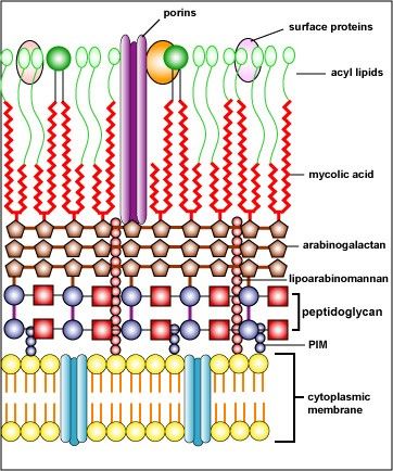
Cell Wall
Has high glycolipid content (mycolic acid) which is very resistant so it can't be stained by gram positive so special techniques have to be used. Other lipids include surface acyl lipids, lipoarabinomannan, phosphatidy inositol mannosides (PIM and cord factor (TDM). Cord factor allow the micro organism to survive in macrophages and divide. They are ACID FAST so can withstand decolourisation.
Ziehl Neelson stain
This is for ACID FAST bacteria
Stain with concentrated CARBOL FUCHSIN and heat to stain cells. STAIN PINK
Destain with 1% acid-alcohol (HCl/ethanol) or 20% H2SO4. mycobacteia remain PINK background is decourised.
Counterstain with METHYLENE BLUE to stain background
Infections caused by mycobacteria
M.bovis causes TB from unpasturised milk and M.tuberculosis causes TB. It is often asymptomatic and causes weight loss, a temperature, cough and blood in sputum
M.avium causes disseminated TB in immunocompromised patients e.g HIV. The symptoms include lung infection that spreads to bones, joints and blood. It frequently ends in death.
M.leprae cause leprosy which results in lesions on face and limbs, there is a loss of peripheral nerves in secondary infection.
Tuberculosis
1720: Benjamin Marten- consumption caused by very small living creatures
1882: Robert Koch discovered the cause of it was by an infectious agent
1825: Augustos Granville - diagnosed an egyption mummy with ovarian tumour
21st Century: UCL identified that granvilles 600BC mummy had TB not an ovarian tumour.
Currently 1/3 of the worlds population have TB most of which are in sub-Saharan africa with 8.4 million new cases each year and 1.5 million deaths. The number of cases in the UK are rising as although there was a vaccine introduced levels of HIV are increasing
Risk Factors
Close contact with large population
Poor nutrition
IV drug use
Alcoholism
HIV infection is the main factor!
TB infection is a droplet nuclei ~ 5 micrometers with 3 bacilli, it can remain suspended in the environment for hours. Its small size means it can bypass the mucocillary lining.
Nuclei droplet inhaled
(7-21 days) MTB mulitples within MACROPHAGES, these then secrete IL-12 and present MTB antigen. Eventually they burst releasing MTB.
IL-12 stimulates T-lymphocytes to infiltrate and recognise MTB antigen, they become sensitized and release inflammatory factors. This forms TUBERCLE the primary lesion
MTB multiples within unactivated macrophages expanding the tubercle
In healthy individuals the cell mediated response(CMI) heals the primary lesion forming a dormant lesion called GHON FOCUS that can reactivate.
Symptoms
90% of people are unaware they have it as a healthy immune system keeps the primary lesion at bay, the other 10% develop primary tuberculosis and experience symptoms.
Lower respiratory tract: Cough, weight loss, night sweats, fatique and a fever
Meningitis
Blood posioning
Kidney infection
joint infection
Patients are HYPERSENSITISED
Secondary Infection (Reactivation)
Any impairment of the CMI can cause DORMANT PRIMARY FOCUS to become NEOCROTIC and liquidify distributing M.tuberculosis into the lungs causing widespread infection
Diagnosis
M.tuberculosis is a catagory 3 mirco-organism which means it can cause serious human disease/hazard but there is treatment. It requires sputum to be analysed in a class 3 lab under class 1 safety cabinet. The sample is stained using ZIEHL NEELSON STAIN, alot of microbes are needed for it to be visulised. The sample can also be cultured in an agar called LJ slope contaning egg, glycerol, minerals, potato flour, antibiotics and malachite green which is incubated at 37c for 12weeks. The antibiotics kill any other bacteria. The colonies are yellow and irregular in shape, they form a distinctive sepentine cords caused by cord factor.
Treatment
Six months treatment consisting of ISONIAZID, RIFAMPICIN, PYRAZINAMIDE, ETHAMBUTOL. The side effects include hepatitis and gastrointestinal disturbances.
Prevention
BCG vaccination (1953) is a weak form of m.bovis that is 50%-80% effective and lasts 10-15 years. it was orignally given to everyone until 2005 since when only at risk individuals recieve it. tuberculin skin testing demonstrates effective vaccination or previous exposure/infection
0.1ml of tuberculin is injected, sensitizd T-cells react if previous exposure to antigen producing a pale elevation of skin (wheal) 6 -10mm diameter. It is examined 48-72 hours later, result is positive if wheal is bigger than 10mm.
Informações
- Descrição
- University Microbiology Notas sobre Mycobacteria, criado por Beckie Thorne em 22-04-2015.
- Criado por
- Beckie Thorne
- Visualizações
- 0
- Avaliação