
Microscopes
Slides do conjunto (9)
-
- Maximum resolution of 0.2 micrometers - cannot be used to view organelles smaller than this e.g. ribosomes, endoplasmic reticulum and lysosomes
- Maximum useful magnification is x 1500
-
- Maximum resolution of 0.0002 micrometers (about 1000 times higher than an optical microscope)
- Maximum useful magnification of x 1 500 000
- Produce black and white images but these are often colored by a computer
-
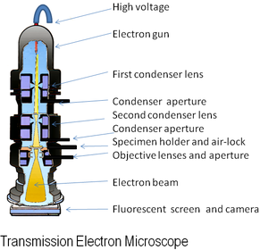
Transmission Electron Microscope
- Use electromagnets to focus a beam of electrons, which is then transmitted through the specimen.
- Denser parts absorb more electrons and so will appear darker on the final image.
- Give high resolution images allowing the internal structure of organelles to be seen.
- Can only be used on thin specimens in a vacuum i.e. not on living organisms
-
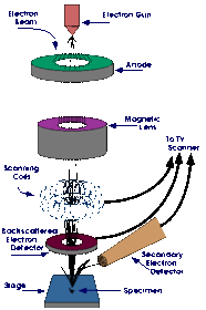
- SEMs scan a beam of electrons across the specimen knocking off electrons from the specimen. These are gathered in a cathode ray tube to form an image.
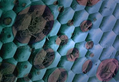
- Images show the surface of the specimen and they can be 3-D
- Can be used on thicker specimens that TEMs.
- Lower resolution than TEMs.
-
- Pipette a small drop of water onto the centre of the slide.
- Use tweezers to place a thin section of specimen on top of the water droplet (specimen must be thin enough to let light through.)
- Add a drop of stain.
- Add the cover slip.
-
- Artefacts are things that can be seen through a microscope that aren't part of the specimen.
- e.g. dust, air bubbles, fingerprints.
- Artefacts are usually made during the preparation of the specimen.
- Especially common in electron micrographs as specimens need a lot of preparation to be viewed under an electron microscope
-
- Homogenisation - can be done in several different ways e.g. vibrating the cells or grinding them in a blender, to break down the plasma membrane and release the organelles into solution. The solution must be kept ice-cold to reduce enzyme activity that could break down organelles, isotonic to prevent damage of the organelles through osmosis, and a buffer solution should be added to maintain pH.
-
- 2) Filtration - Homogenised cell solution is filtered through a gauze to remove any cell or tissue debris. As the organelles are much smaller than the debris they can easily pass through the gauze.
-
- 3) Ultracentrifugation - The cell fragments are poured into a tube which is put into a centrifuge and is spun at a low speed. The heaviest organelles get flung to the bottom of the tube and form a thick sediment at the bottom - the pellet. The rest of the organelles stay suspended in the fluid above - the supernatant. The supernatant is drained off, poured into another tube, and spun in the centrifuge again at a higher speed. Again, the heaviest organelles form a pellet, and the supernatant is drained off and spun again. This process is repeated at higher and higher speeds until all the organelles are separated out.
- The organelles are separated out in the order: Nucleus, chloroplasts, mitochondria, lysosomes, endoplasmic reticulum, ribosomes.
Informações
- Descrição
- As-Level AS-Level Biology (Topic 2A: Cell Structure and Division) Slides sobre Microscopes, criado por Heather Jones em 27-03-2016.
- Criado por
- Heather Jones
- Visualizações
- 0
- Avaliação