17626826
2. Cell Adhesion and Communication
Description
No tags specified
Slide Set by Chloe Cavarretta, updated more than 1 year ago
More
Less
|
|
Created by Chloe Cavarretta
over 6 years ago
|
|
Resource summary
Slide 1
Learning Outcomes
Describe with examples the structure and function of different classes of cell-cell and cell-matrix interactions
Give an overview of how cell adhesion molecules can interact with components of the cytoskeleton
Give an overview of the role integrins play in cell signalling events
Slide 2
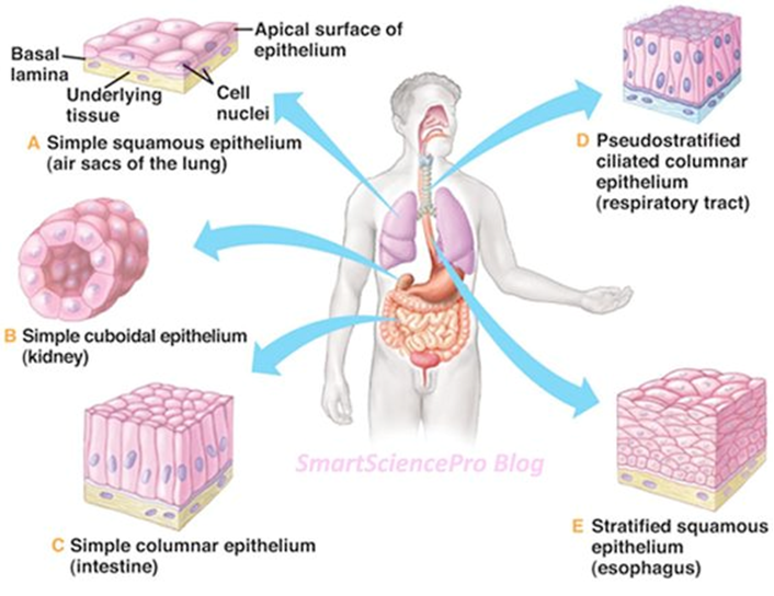
Epithelial Tissue
Covers the internal and external surface of organs
Contains a layer(s) of cells on top of basement membrane
Cells adhere to each other laterally as well as to basement membrane
Slide 3
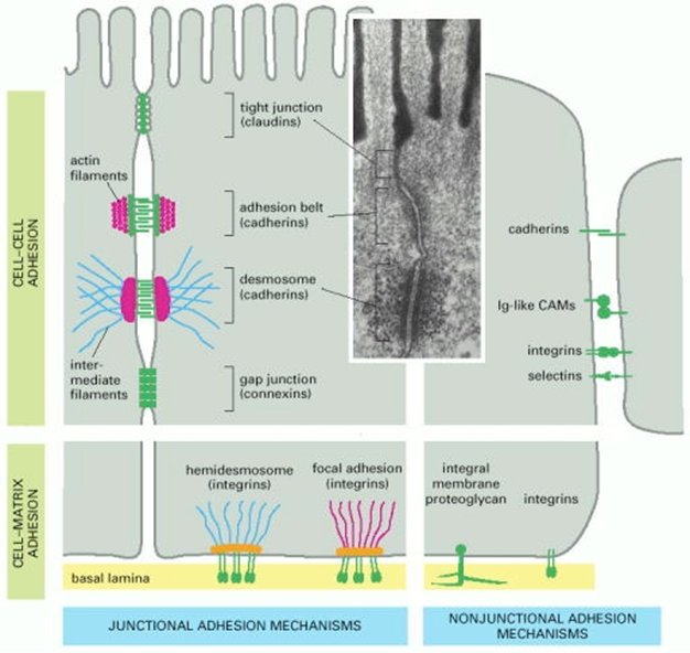
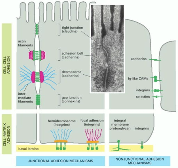
Cell adhesion overview
Lateral Cell-Cell Adhesion established via:
Tight junctions (junctions are specialised modifications of cell surface in neighbouring cells)
Adherens junctions
Desmosomes
Gap junctions
Non-junctional adhesions (proteins such as cadherins, selectins, integrins)
Cell-Matrix Adhesion established via:
Hemidesmosomes
Focal adhesions
Non-junctional adhesions (integrins)
3 distinct classes of cell adhesion junctions:
Occluding (tight) junctions= forms a barrier between epithelial cells
Attachment junctions= links cell cytoskeleton to that of its neighbouring cell or to matrix
Communication junctions= directly connect cytoplasm of adjacent cells
Slide 4
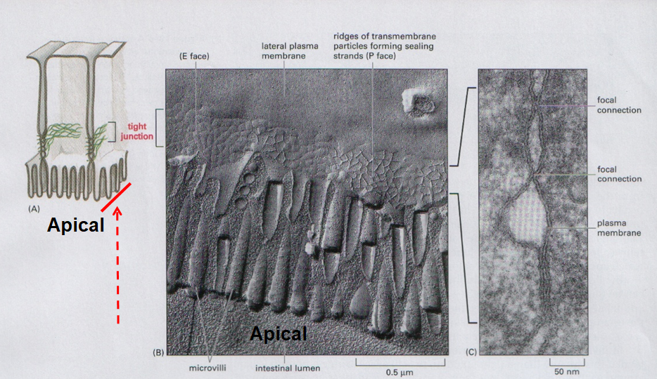
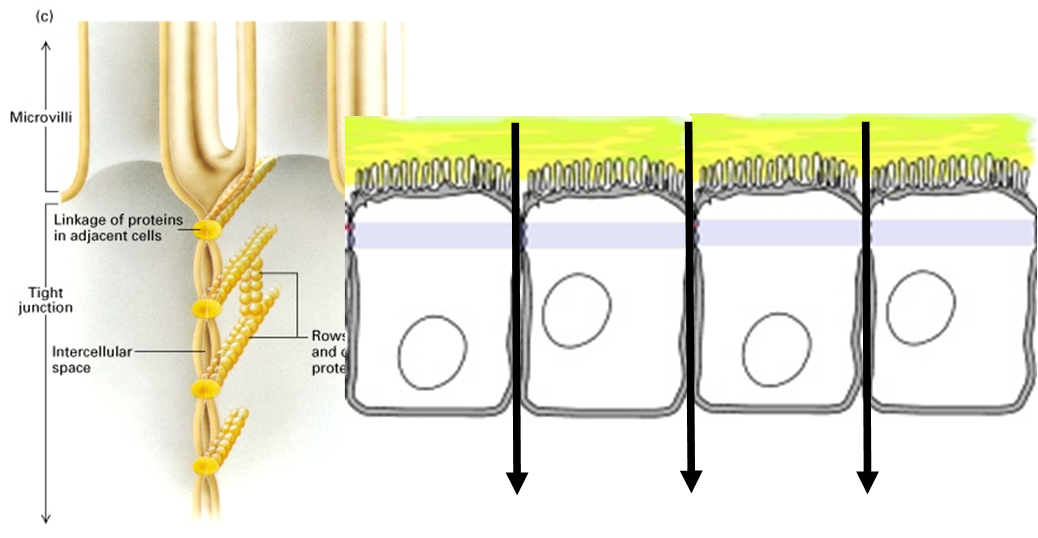
Tight junctions: Structure
Structure: Proteins
Seal adjacent epithelial cells in a narrow band beneath their apical surface
Bands made up of interconnected (sealing) strands of transmembrane proteins that wrap around the cell
3 types of transmembrane proteins:
Claudins = core proteins in tight junction fibrils
Occludins = position and stabilise claudins
Junctional adhesion molecules (JAMs) = position and stabilise claudins
Proteins in the sealing strands attach stably to structural proteins to interconnect the sealing strands, also transiently to signalling proteins
Caption: : Electron micrograph- network of thin fibrils where protein embedded (left) 2 plasma membranes of adjacent cells joined together by tight junctions (right)
Slide 5
Tight junctions: Selectivity
Tight junctions allow the passage of small ions, solutes, electric currents between epithelial layers
Differing junctions allow passage of differing solutes through
Dependent on size (4-40A) and charge, different tissues have different selectivities)
Selectivity determined from different claudins- humans have 24
Eg expression of claudin 16 restricted to ascending limb of loop of Henle, mutations associated with conditions caused by Mg and Ca ions unable to pass through tight junctions
Slide 6
TJ: Role- regulate paracellular transport
Transfer substances across epithelium by passing through intracellular space between cells
Act as molecular sieves rather than completely impermeable ("occluding")
Property of TJs which regulates this process= selective permeability
Slide 7
TJ: Role- regulate transcellular transport
Transfer substances across epithelium by passing through cells, crossing apical and basolateral membranes
Allows substances against concentration gradients eg Glu absorption
Property of TJs which regulates this process=separation of apical and basolateral membrane
Slide 8
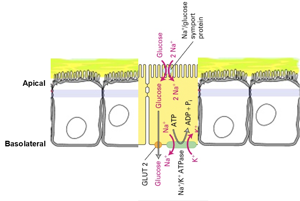
Transcellular transport: Glucose absorption
If absorption of glucose across intestinal epithelium relied on paracellular transport we would not gain all the glucose from our diet:
Sodium-potassium pump ATPase in basolateral membrane generates Na gradient by pumping Na out of the cell against conc gradient
Low conc of Na in cell compared to outside
Energy stored in gradient is used to bring glucose into cell from lumen of SI via Na/Glu symporter
High conc Glu in cell compared to bloodstream
Glu enters bloodstream via facilitated diffusion through Glu trasnporter (GLUT2 in baolateral membrane)
Tight junctions play a role in maintaining separate identity of 2 membranes and membrane proteins cant cross the barrier they create:
Essential that ATPase and GLUT2 are present on basolateral membrane and not apical
Essential that Na/Glu symporter is on apical and not basololateral
Slide 9
Tight Junctions: Summary
Formed by interconnected sealing strands of claudin, occludin and junctional adhesion molecules that encircle the cell
Act as molecular filters thus regulating paracellular transport across epithelial tissue
Maintain identity of apical and basolateral membranes by preventing diffusion of proteins and lipids between the two
Slide 10
Attachment Junctions: Adherens
Caption: : Electron micrograph- thick, dark bands close to PM of neighbouring cells, bridged by rod-like structures projecting into extracellular space
Serve as bridges: link actin filaments (through catenin) of adjacent cells
Zonula adherens: found immediately beneath tight junctions in epithelial tissues
Other types found joining cell types
Slide 11
All adherens junctions have 2 common properties:
Contain cadherins:
TM proteins that bind to identical cadherins on neighbouring cells
Role in determining tissue organisation- enable cells to find appropriate binding partner and determine junction strength
Link to actin cytoskeleton:
Regulate changes in cell shape and/or to sheer stress (important in cytoskeleton dynamics)
Through catenins (role in signalling : beta-catenin can act as TF)
Catenins link cadherins to MFs eg link cytoplasmic tails of cadherins to MF in zonula adherens, mutations in catenins lead to cancer
Slide 12
Attachment junctions: Desmosomes
Caption: : Electron micrograph- thick patches of electron-dense material on cytoplasmic side of PM, connected to int filaments in cytosol
Also add structural integrity to tissues by linking cytoskeleton of adjacent cells
Through intermediate filaments
Slide 13
2 common properties:
Contain cadherins (desmocollins and desmogleins) that link adj cells together:
Cadherins bridge intracellular space on one side of membrane and serve as docking sites for cytosolic proteins (desmoplakin) that bind int filaments on the other side
Link to int filaments through adaptor proteins( plagoglobulins/desmoplakin):
Attachment to filaments through adaptors regulated by cell signalling
Can be initiated by activated growth factor for receptors (coordination of cell growth and adhesion)
Attachment junctions: Desmosomes
Slide 14
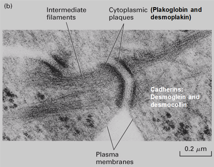
Attachment junctions: Desmosomes
Electron micrograph of desmosome between 2 keratinocytes:
Shows intermediate filaments linking to cytoplasmic plakoglobulin and desmoplakin that, in turn link to TM cadherins desmoglein and desmocolin
Formation of bridge between plasma membrane of neighbouring cells
Slide 15
Attachment junctions: Hemidesmosomes
Located on basal surface of epithelial cells
Providing structural stability to epithelial sheets
Anchor cells to underlying basement membrane through cytoplasmic 'plaques' that connect to intermediate filaments
Plaques contain integrins (TM protein) which connect to int filaments through adaptor protein plectin
Extracellular region of integrins binds to components of basement membrane
Slide 16
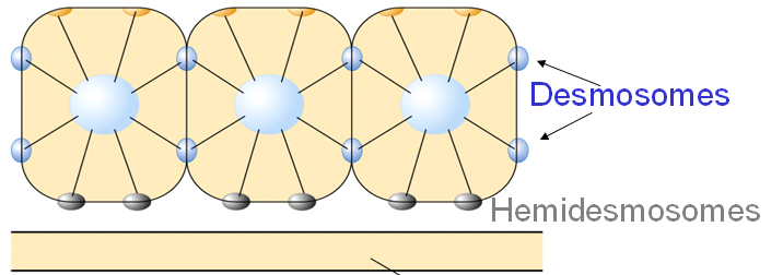
Arrangement of hemi- and desmosomes
Organises intermediate filaments into robust network
Mutations that disrupt desmosomes and/or hemidesmosomes -> severe blistering diseases
Hemi and desmosomes both link intermediate filaments but are structurally very different
Slide 17
Integrins: general
Heterodimers receptors composed of alpha and beta subunits: 18 different alpha, 8 beta, 24 known different heterodimers
Bind to ECM components:
Different heteromers bind different ECM components- different cell types express different integrins so binding of a cell to its ECM is determines by the integrins present on its basolateral membrane
Specificity of which ECM component an integrin binds is determined by integrin alpha subunits
Complexity: Most heterodimers can bind more than one ECM component and each component binds more than one integrin
All known binding sites on ECM proteins contain acidic amino acid (asp) and many contain sequence : Arg-Gly-Asp
Also bind to components of focal adhesions- linking actin cytoskeleton to ECM
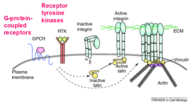
Slide 18
Changes within the cell affect affinity of the integrin for its ligands
Activation of adaptor proteins (eg talins) -> link integrins to actin cytoskelton focal adhesions:
Integrin doesnt bind to actin cytoskleton or ECM until talin binds
Signalling via GRCP or RTK activates talin and induces dimerisation
Talin binds to beta-intgrin subunit which causes conformational change in integrin extracellular binding domain, enabling ECM binding
On binding, talin also links actin filaments
Inside-out signalling: signalling pathways inside cell affect role of integrins outside
Integrins: Signalling
Slide 19
Integrins: Signalling
Outside- in signalling: transduce information outside of the cell to the inside
Binding of cell to underlying matrix can cluster integrins together
Makes their cytosolic domains form a docking site for signalling molecules
Integrin signalling pathways regulate many cell physiologies:
Cell differentiation
Inhibition of programmed cell death
Cytoskeletal rearrangements: importance in cell migration across the ECM and not sticking to it eg during wound-healing focal adhesions promote remodelling of actin cytoskeleton to drive leading edge forward
Slide 20
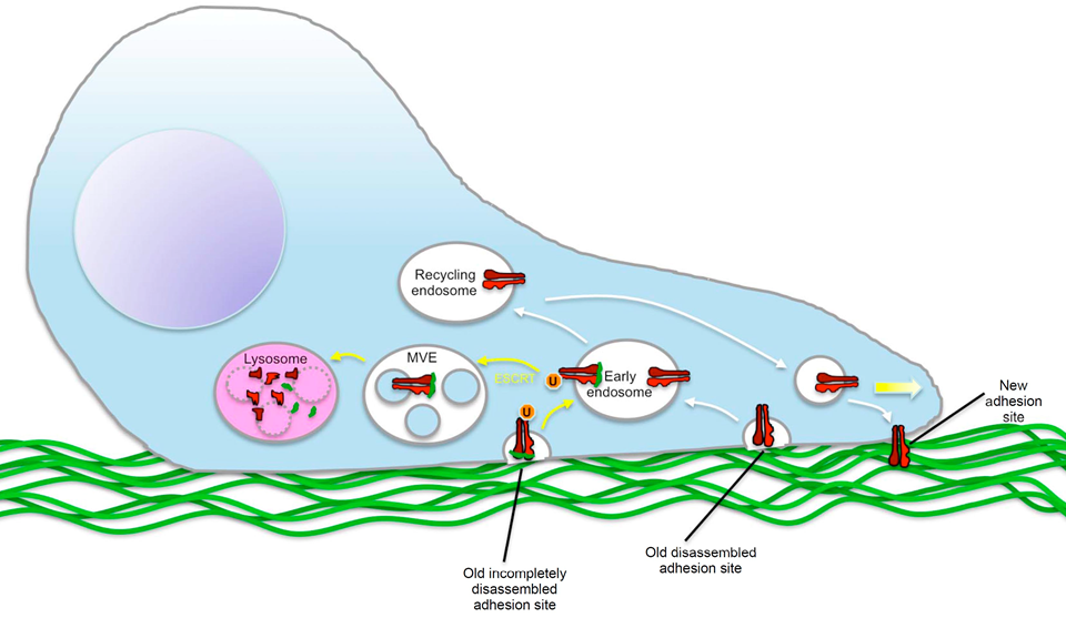
Trafficking of integrins
Trafficking of integrins used to regulate cell movement: formation/disassembly of focal adhesions
Endocytosis of integrins:
Can break attachment to substrate
Internalised integrin can be recycled and establish new attachments in direction of movement
Or integrin can be trafficked to the lysosome and degraded causing the cell to loose attachment = dysregulation in cancers
Slide 21
Communication junctions (Gap)
Gap junctions facilitate direct transfer of ions and small molecules between adjacent cells
Found in most vertebrate and invertebrate cells and contain upto thousands of channels- can form large or small junctions
Only known means of direct cell-to-cell transport in animals
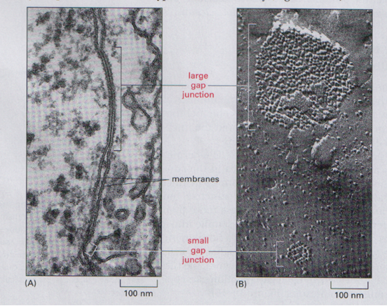
Caption: : Regions where PMs are closer together than surrounding areas with gaps bridges by channels projecting out of PM
Slide 22
Gap junctions
Gap junction channels consist of 2 halves (hemichannel or connexons):
2 connexons on opposing membranes of adjacent cells dock togther to form gap junction
In intracellular gap creates 2-4nm pore for ions/small molecules to pass through
Subunit of connexon= connexin
6 connexins form hetrameric connexon = channel cluster
Connexin subunit = tetraspan, 4 TM alpha helices
Gap junction channels have different permeability depending on connexins present: Homomeric/homotypic, heteromeric, heterotypic
Transport: inorganic ions, sugars, amino acids, nucleotides, vitamins, cAMP and IP3, NOT macromolecules (proteins, NA, polysaccharides)
Slide 23
Gap junctions: Function and Regulation
Function: Used for rapid communication
Reflexes reactions: in brain mediated by neurons linking gap junctions, allowing potential to be spread rapidly, avoiding delay at chemical synapses
Synchronisation of cardiomycetes signal to contract: communicated through gap junction, mutations in connexins lead to arrythmia
Regulation
Alternate between open and closed states
Closed- through phosphorylation of connexins when large increase in intracellular Ca ion conc and/or pH
Eg cell damage causes elevation of Ca in cell and releases damaging metabolites
Closing prevents further damage to neighbouring cells
Slide 24
Summary
Connections between cells can be established via:
Tight junctions
Adherens junctions
Desmosomes
Gap Junctions
Synapses
Connections between cell & their underlying matrix can be established via:
Hemidesmosomes
Focal adhesions
Non junctional adhesions
Want to create your own Slides for free with GoConqr? Learn more.