6. Protein Degradation, Autophagy

Description

Cell Biology L14
Chloe Cavarretta
Slide Set by Chloe Cavarretta, updated more than 1 year ago
Chloe Cavarretta
Created by Chloe Cavarretta over 7 years ago
3
0

Resource summary

Slide 1

    Learning Outcomes: L14
    Assess depending upon the location and type of protein how a protein might be degraded Identify and contrast common factors between protein degradation routes Identify the role of protein complexes in protein degradation

Slide 2

    Why Degrade Proteins?
    Incomplete or mis-sense proteins: Cellular errors including incorporation of inaccurate aa or analogues Proteins with disruptive mutations Products of premature termination Products of proteolytic cleavage Post-synthetic damage: Mis-folded proteins  Protein ageing eg O free radical damage Denaturation eg heat shock    
    Unwanted proteins: Inactive or used- activated and want to unactivate Free subunits of multimeric complexes- excess subunits Other proteins made in excess Any protein in wrong place at wrong time

Slide 3

    Intracellular Degradation Routes
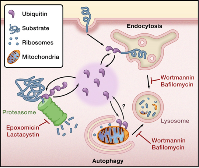
    Two sites for protein degradation: Cytosolic structure=proteosome : Degrades s​​​oluble cytosolic proteins Membrane bound organelle= lysosome (endocytic or autophagic delivery) Degrades membrane bound protein via receptor mediated endocytosis Degrades lumenal proteins that enter by endocytosis/phagocytosis Soluble proteins, organelles and bacteria (that escape into cytoplasm) degraded via autophagy in lysosome

Slide 4

    Ubiquitin Targets Proteins Degradation
    Ubiquitin targets proteins for degradation at proteosome and lysosome: Soluble protein= add ubiquitin, sends to proteosome  Receptor =taken into endosome and ubiqutinated, targeted to lysosome  Damaged mitochondria in cytoplasm= ubiquitinated which targets to autophagosome and delivery to lysosome Ubiquitination is a commonality of all pathways of degradation (proteosome, endocytic or autophagic delivery to lysosome)  

Slide 5

    Ubiquitin
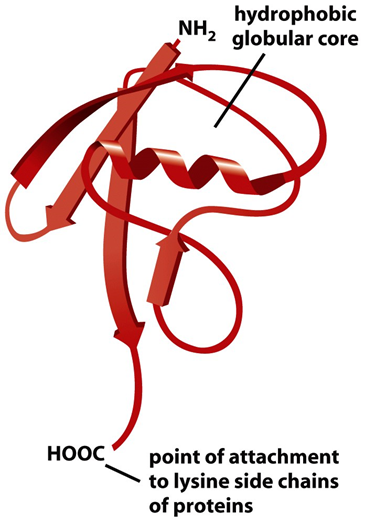
    76 aa pp that labels proteins  (~8kDa) Named ubiquitin because so common in every organism  Majority of ubiquitination occurs to amino group of lysine residues in proteins (attachment point is C- of ubiqutin)

Slide 6

    Ubiquitin Conjugating Enzymes
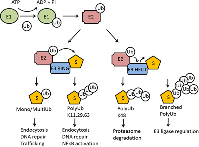
    Ubiquitin modification is ATP-dependent process carried out by 3 classes of enzymes: Ubiquitin activating enzyme (E1)= Puts ubiquitin into activated state (bound to E1) Ubiquitin conjugating enzyme (E2)= E2 recieved ubiquitin from E1 and uses E3 ligase to transfer ubiquitin  Ubiquitin ligase (E3)= transfer ubiquitin directly to substrate (E3 ring) or via E3 hect domain Reactions of the 3 enzymes are sequential

Slide 7

    Many Flavours of Ubiquitination
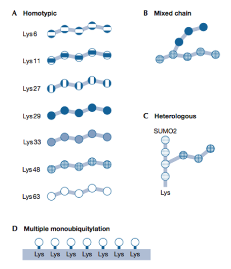
    Ubiquitin itself can be ubiquitinated Monoubiquitination= protein gets one ubiquitin added Polyubiquitination= each ubiquitin gets ubiquitinated Occurs on same lysine for every ubiquitin eg 48 poly-Uq chain (all on lys 48) Can get mixed chains eg Uq + Uq(K48) + Uq(K63)= causes branching  Differences between poly and mono ubiquitination, and differences within polyubiquitination can determine where protein will be degraded and rate of degradation  Often poly on lys-48 causes degradation at proteosome

Slide 8

    Many Flavours of Ubiquitination
    Polyubiquitination can have homotypic or mixed chains  Heterologous= sumoylation is anotehr modification, can get ubiquitination within sumoyl groups  Multiple monoubiquitination can look like polyubiquitination

Slide 9

    Ubiquitination Reversible
    Ubiquitin removed by de-ubiquitinases (DUBs) DUBs break down chains into single molecules Ubiquitin recycled, not degraded with target protein (removal before degradation) Can be removed for protein editing and to rescue protein from being degraded  

Slide 10

    Proteins that Recognise Ubiquitin Signal
    Different ubiquitin conformations will give a molecular tag that enables protein to be targeted for degradation Proteosome has 2 proteins that recognise ubiquitin signal  Intrinsic receptors: cap proteins (part of proteosome)- directly bind ubiquitinated protein to proteosome Shuttling receptors: Uq protein bind shuttle which target to proteosome by binding caps (intermediary protein) bringing uq protein to proteosome Autophagy: Uq proteins bind intermediary protein that targets to autophagosome (Intermediary protein has BD for Uq and autophagosome) Endocytic: intermediary protein binds Uq protein and targets to endosome   

Slide 11

    Degradation via Proteosome
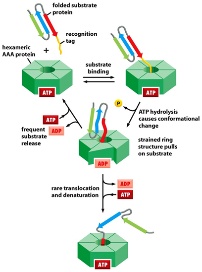
    Central cylinder contains proteases and cap proteins Cap proteins are hexameric ATPases (AAA-ATPase) Energy dependent process to unfold and degrade Recognition tag targets protein to cap of protein, uses ATP hydrolysis to unfold protein and allow to be thread through proteosome and degraded into constitutive aa 

Slide 12

    Example of Proteosome Degradation
    Tumour supressor p53 regulated by ubiquitination: p53 and Mdm2 form a well established autoregulatory loop  p53 gene causes mdm2 transcription, increase of mdm2 inactivates p53 mdm2 is a E3-uq ligase- ubiquitinates and causes its degradation Mono-uq causes p53 to export to cytoplasm, poly-uq targets to proteosome The ubiquitinating-enzymes and the proteosome are targets for cancer therapy drugs Human papilloma virus encoded E3 ligase that ubiquitinates p53 causing cellular division... tumour

Slide 13

    Degradation via lysosome
    Lysosome characteristics: Membrane bound organelle contains hydrolytic enzymes (optimally active at acid pH) ~0.5um in diameter, often with an electron dense core Only when lysosome receives material from autophagy/endocytosis- delivery of lumen water activates hydrolases  Dependent on fluid environment Disorder- in MLIV cells cant degrade, lysosome swollen, internal membrane no longer electron dense
    Lysosome function: Degradation  Autophagy Phaocytosis Disease

Slide 14

    EGF Receptor Degradation
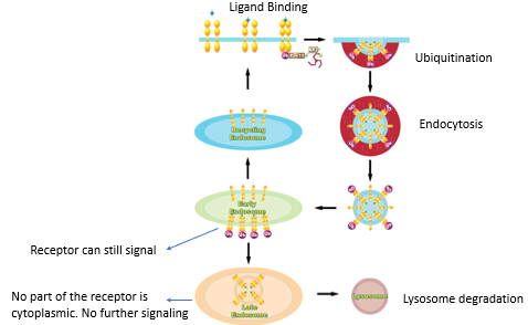
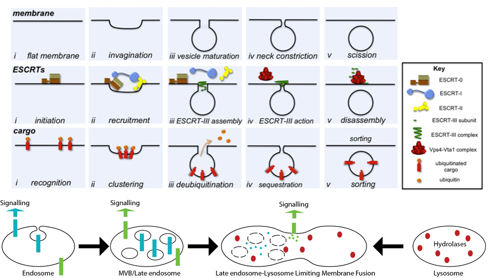
    To fully degrade receptor, need all parts of receptor available to lysosomal hydrolases: If extracellular part of receptor sticking in cytoplasm, can still signal into cell Transition of early endosome into late endosome (MVB) Invagination of endosome surface to give multi vesicular body (MVB) Requires ubiquitin and uses ESCRT proteins

Slide 15

    ESCRT
    Endosomal Sorting Complexes Required for Transport (ESCRT) Complex of proteins that recognise membrane bound ubiquitinated cargo and aids in their sorting into MVBs Ensure cargo is internalised in lumen of late endosome to allow efficient protein degradation Receptor in vesicle inside endosome

Slide 16

Slide 17

    Autophagy (self-eating)
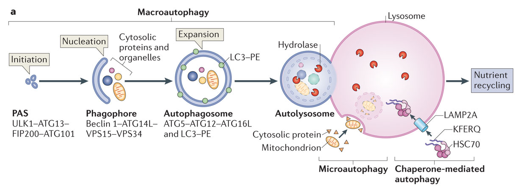
    Stimulated by stress, starvation(when low in nutrients), free radicals, damage, ammonia stress Cells capture their own cytoplasm and organelles ( in autophagosome) and deliver to lysosome for nutrient release Selective autophagy often captures ubiquitinated cargo Macroautophagy: Numerous proteins (complex) involved in generating phagophore Initiation of membrane, nucleation, expansion so it completely seals = autophagosome Selective autophagy- uq-protein brought to phagophore membrane by binding uq binding proteins on phagophore and incorporated into autophagosome Autophagy re-branded depending upon material sent to be degraded: Mitophagy (mitochondria), aggrephagy (protein aggregates), pexophagy (peroxisomes) 

Slide 18

Slide 19

    Autophagy
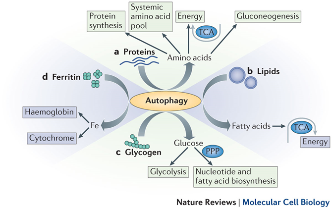
    Autophagy regulates energy homeostasis: Autophagy known as starvation response- material degraded to the building blocks to regulate energy Produce free aa, carbohydrates and lipids to be used to generate energy (before you develop ketosis)

Slide 20

    Autophagy and Disease
    Autophagy regulates cellular homeostasis Consequences of impaired autophagy: lack of nutrients, aggregate formation, immune activation, ROS accumulation, chronic infection Eg ROS- damaged mitochondria not cleared produce ROS Eg chronic infection- bacteria normally sent to autophagosome for clearance can replicate in cell  
Show full summary Hide full summary

Similar

Function and Structure of DNA
Elena Cade
Cells And Cell Techniques - Flashcards (AQA AS-Level Biology)
Henry Kitchen
Cells - Biology AQA B2.1.1
benadyl10
Biology AQA 3.1.3 Cells
evie.daines
GCSE AQA Biology - Unit 2
James Jolliffe
Cells and the Immune System
Eleanor H
Biology Unit 2 - DNA, meiosis, mitosis, cell cycle
DauntlessAlpha
Cell Transport
Elena Cade
Mitosis
Selam H
Unit 1 flashcards
C R
Cells, Tissues and Organs
yusanr98