
Notizen
CNS
Inhalt der Notizen (18)
Seite 1
CNS overview and gross anatomy
CNS vs PNS
Cluster/aggregate of neuronal cell bodies
CNS- nucleus
PNS- ganglion
Bundle of axons
CNS- tract
PNS- nerve
Revisit intro to NS
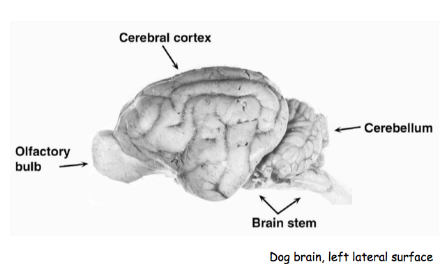
Brain
Surface anatomy + basic function
Cerebrum
Underlying deep nuclei
Basal ganglia control initiation of movement
Limbic system establish memory + emotional aspects of experience
Ofactory bulb
Involved in smell
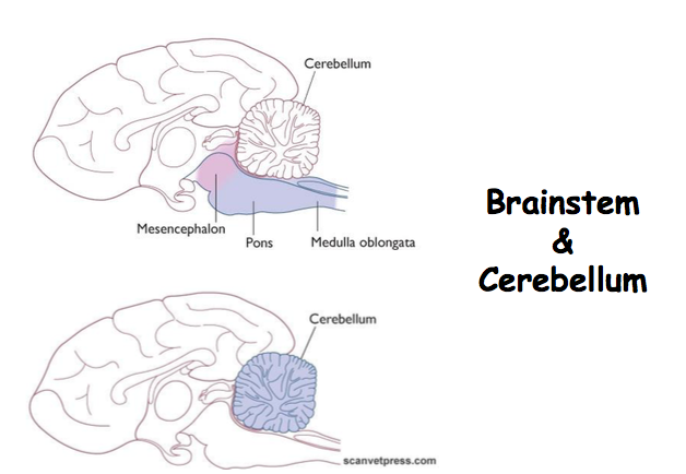
Cerebellum
Folded
Involved in balance, co-ordination and the refinement of movement
Brain stem ?
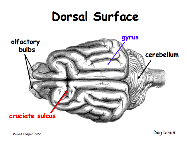
Cerebral cortex
Cerebral hemisphere
Symetrical mainly
In humans LHS diff due to speech
Gyrus(p)
Gyri (s)
Elevated ridges
Sulcus
Sulci (s)
Shallow grooves
Medial Longitudinal fissure
Deep sulcus between hemispheres
Cruciate sulcus
Bisects medial longitudinal fissure
Separates the primary motor cortex from primary sensory cortex
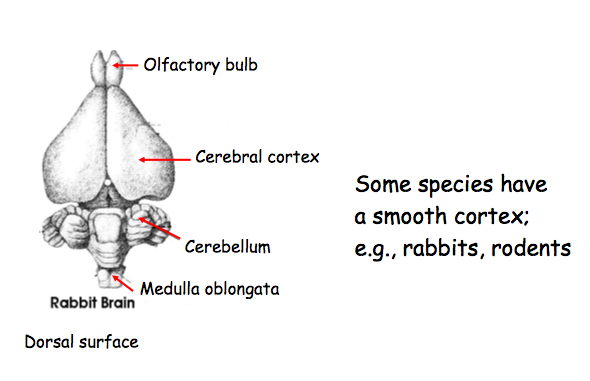
Rabbit brain
No folded cortex
Relationship between folding and intelligence ?
Not fully accepted
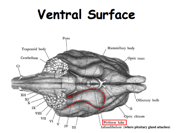
Olfactory bulb
Receive input from olfactory nerves
Olfactory peduncle
Optic tract?
Hypothalamus
2 mammillary bodies
Brain stem
Optic and Olfactory nerves DONT come from brain stem
Others DO
Midbrain
Visual function
Pons (Bridge)
Allows communication between spheres of cerebellum
Medulla oblongata
Where lots of cranial nerves come from
Trapezoid body
To do with hearing
Periform lobe
Processing smell
Species variation in relative sizes + Cortical folding
Size depends on relative importance of each structure to that species
Degree of cortical folding
Intelligence
Constraints of brain expression within constrained space during development
SA increased
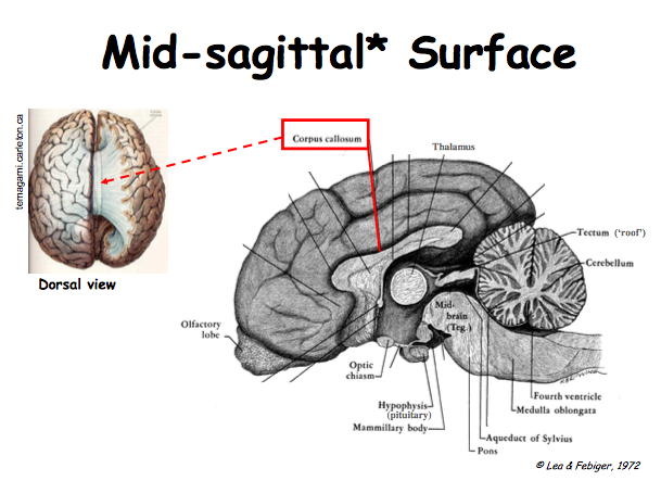
Corpus callosum
Giant bundle of axons that joins two hemispheres
Allows communication
Thalamus
Sensory processing
All sensory info eventually finds its way to thalamus
Relays info
Tectum
Part of midbrain
V developed in birds
Ventricle System
Shock absorber
Nutrient provision?
??
Fourth ventricle
Clinical importance
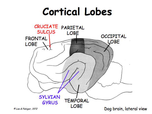
Frontal lobe
Rostral to cruciate sulcus
Sylvian gyrus is another useful
Parietal Lobe
Occipital lobe
Temporal lobe
Landmarks
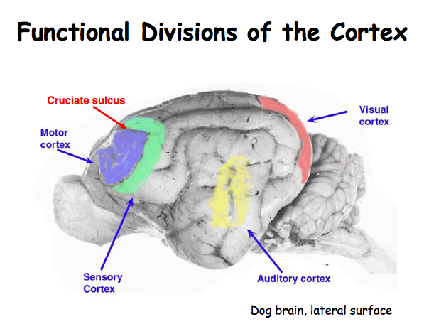
Lesions in any of these areas there will be corresponding defects
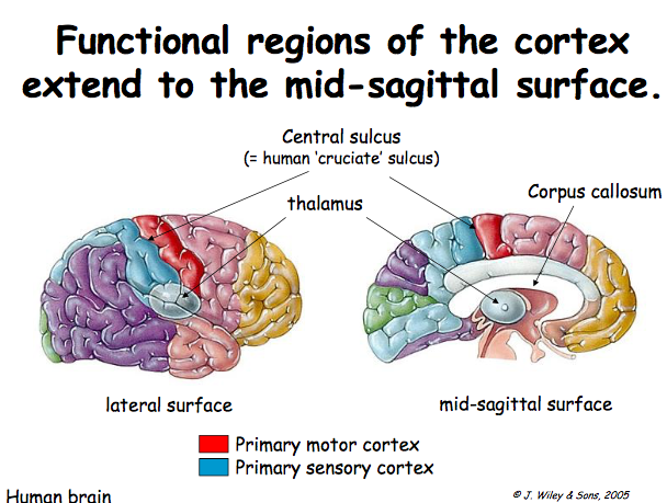
Species variation in relative size of cortical areas
Association cortex dominates in humans
Primary cortical areas represent relatively larger proportions in other mammalian brains
Rat - V large olfactory cortex, more reliance on smell
Man - V smal olfactory cortes , less reliance on smell. Larger association cortex
Stimulation of association cortex gives unpredictable response
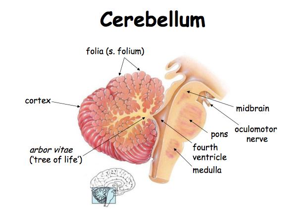
Highly folded
Folds narrower than cerebral cortex
Single fold = folium
Arbor vitae = white matter
Axons leaving cerebellum found here
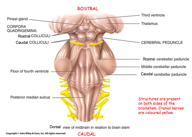
Peduncles
3 attach cerebellum to brain stem
Rostral
Caudal
Cerebral
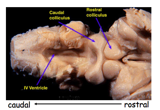
Colliculi
Rostral and caudal?
???
Deep Anatomy
Brain - subcortical structures
Integrative systems
?
Spinal cord
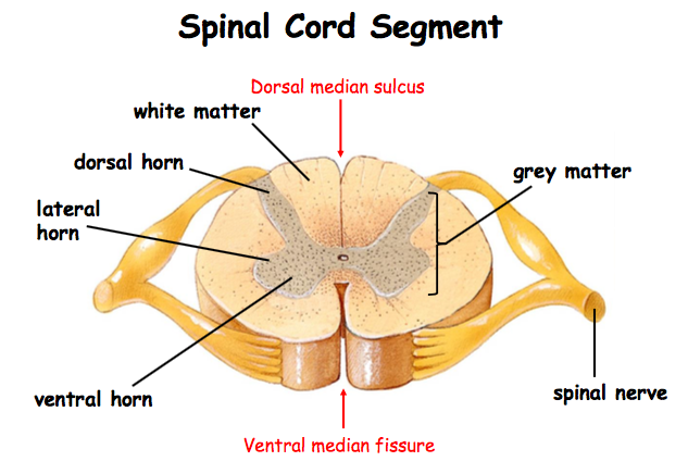
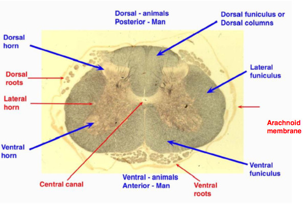
Grey matter
Neuronal cell bodies
Some unmyelinated axons
Split into horns
Dorsal horn receives somatic and visceral sensory neurones
Cell bodies reside in dorsal root ganglion
Lateral horn contains the cell bodies of preganglionic autonomic motor neurones
Ventral horn is the site of cell bodies of somatic motor neurones
White matter
Myelinated axons
Split into funiculi which are separated by motor + sensory roots
Funiculi - bundles of axonal fibres which project rostrally or caudally
Lateral = mixed
Dorsal = sensory
Ventral = motor
Spinal nerve
Dorsal median sulcus
Ventral median fissure
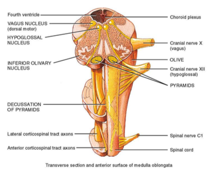
Sensory and motor sections as found in spinal cord
Cell bodies of neurons in the motor cranial nerves lie within brain stem in nuclei
Cell bodies of neurons within sensory cranial nerves within ganglia outside the brainstem
Cranial nerves innervate the head and neck
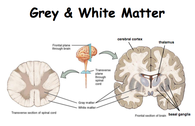
Grey and shite matter also found in brain
White matter on outside and inside
White matter on inside
Basal ganglia is grey matter
Movement
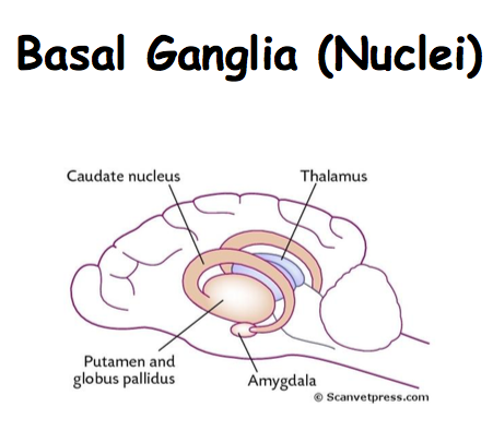
Caudate nucleus
?
Putamen and globules pallidus
Movement disorders in humans associated
Amygdala
To do with behaviour
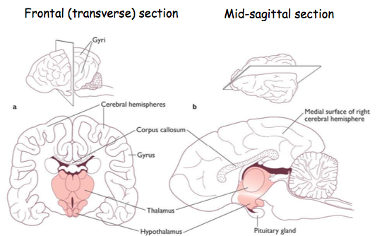
Deep structures can be observed from different aspects
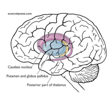
Deep cerebral nuclei
Produce movement disorders when damaged
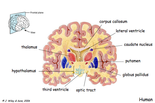
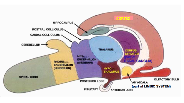
Seite 2
CNS embryology
3 layers of the embryo
Ectoderm
NS develops from here (Neuro-ectoderm)
Mesoderm
Endoderm
Neural tube formation
Neural plate forms from the ectoderm
Neural groove forms
Groove deepens and dorsal lips fuse forming neural tube
Proliferation of superficial ectoderm gives rise to the neural CREST
Neural tube pinched off from surface
Neural tube = CNS
Superficial ectoderm = PNS
Notochord = cylinder of mesodermal cells
Responsible for inducing formation of neural plate through secretion of chemical signals
Stimulate there mesodermal cells to develop into vertebral bodies + intervertebral disc
Spina Bifida
Occurs when caudal neuro-pore fails to fuse
Impossible for vertebral arches to form + fuse normally above spinal cord
Not always serious
Manx cats
Bulldogs
Damage to underlying neural tissue can cause serious problems
Incontinence
Paralysation of hind legs
Maturation of spinal cord
Initially 3 layers of neural tube
Ventricular zone
Mantle layer
Marginal layer
Neurones and support cells made in ventricular zone
Then they migrate into mantle layer
Mantle layer forms grey matter
Myelinated axons lie in marginal layer
Sulcus limitans divides spinal cord into alar + basal plates
Alar plate forms dorsal horns of grey matter
Basal plate forms ventral horns of grey matter
Control of neural cell differentiation
Cells within neural tube secrete signalling factors that affect neighbouring cells
Somites responsible for development of skin , muscle and connective tissue?
Roof plate secretes bone morphogenetic proteins BMPs
Floor plate and notochord release Sonic hedgehog SHH
Floor and roof plates involved in partition of spinal cord into sensory and motor regions
Development of brain regions and ventricles
Subdivision occurs
Prosencephalon (forebrain)
Telencephalon
Forms the cerebral hemispheres
Diencephalon
Optic cups grow outwards
Forms thalamus and hypothalamus
Mesencephalon (midbrain) no division
Rhombencephalon (hindbrain)
Metencephalon
Forms pons
Forms cerebellum
Myelencephalon
Mature medulla oblongata
Mature brainstem formed from rhombencephalon and mesencephalon
Cylopia
Embryonic prosencephalon fails to subdivide properly
Division of the orbits of the eyes into separate cavities also fails
Malformed face
Nose either missing or malformed and non functioning
Due to faulty genes or intake of plant toxins
Corn lily
Failure of developing cells to respond to SHH
1/16000 born animals
1/250 embryos
Most spontaneously aborted
Development of the brain
slides?
7 wks
Folding occurs
Subdivision also occurs
11wks
Cerebral hemisphere enlarged
Subdivision ?
At birth
Cerebral hemispheres and cerebellum v folded
Dicenp
Thalamus
Hypothalamus
Brainstem
??
Embryonic brain flexures
Expansion greater than cranial capacity
Initially 2 bends
Cephalic (Between mid brain and hind brain )
Cervical (Occurs at top of spinal cord)
3rd later in development = pontine
Cervical flexure less pronounced in quadrupeds than bipedals
image?
Spinal cord vs brainstem development
Lumen of neural tube expands to form cerebral ventricular system
Ventricles of brain are continuous with the central canal of the spinal cord
I wasn't listening - slide
Not a clue
Seite 3
Blood supply meninges + CSF
Meninges
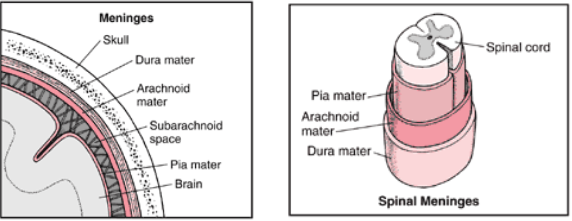
Membranes = meninges
3 layers
Physical protection of brain + spinal cord
Facilitate flow of CSF
Provide a framework for blood vessels to supply CNS tissues
Outside layer - Duramater?
Two indistinguishable layers within the skull
Outer=endosteal
Inner=meningeal
Separated to form two folds which course into fissures between brain tissue
Falx cerebri (between hemispheres)
Tentorium cerebelli (between cerebral and cerebellum)
Contain venous sinuses
Pain sensitive
Own blood supply
Arachnoid
Potential space between this layer and duramater
Avascular
Space underneath - subarachnoid space
Blood vessels here
?
Piamater ?
V thin
Tightly adherent to tissue
Highly vascular
Bring blood vessels to Brain tissue
Dura separates from endosperm within vertebral canal creating the epidural space
Denticulate ligaments ?
Cuff zones
??
Meninges follow nerves up to a point then blend into perinuerium
This is the cuff zone
Clinical relevance
Cerebrospinal fluid
Communicates with ISF of neural tissue freely
Cell-free, protein-free, low amino-acid content, low and stable K+ content and relatively low glucose content
Must be kept stable
Provides nutrition
Shock absorber
Acts as a volume buffer
Accommodates some swelling
Facilitates flow of neurotransmitters
Circulates through subarachnoid space, ventricles of brain and central spinal canal
Ventricles and central canal lined by ependymal cells which possess cilia
Communication through inter ventricular foramen
Image? Stream?
CSF production and drainage
Choroid plexi ?? stream? all this slide?
Choroid plexi main source of CSF
Small amount from ultra filtrate of pill blood
CSF
Blood:brain barrier maintains CSF in a steady state
BBB prevents changes in blood affecting CSF
Tight junctions between endothelial cells
Thick BM
Astrocytes
Transporting ependymal cells covering plexi pump solutes into the CSF to draw water in by osmosis
Lipid soluble substances readily across into CSF
Drugs must be lipid soluble
Abnormal BBB means some drugs could enter - CARE
Water soluble materials must be actively transported into CSF
Drugs vary in their ability to pass into the CSF and the permeability of the BB varies in different disease processes
CSF sampling
Sub arachnoid space
Cerebellomedullary system
Lumbar system
V sterile
CARE - negative pressure cause problems
Death
Arterial supply
Cerebral arterial circle
Located at ventral part of brain
5 arteries come off this?
Image
Dog input from Basilar and internal carotid arteries
Source to circle differs between species but derived from combination
Internal carotid
Basilar artery
Maxillary artery
Vertebral artery
Source matters in terms of slaughter techniques- Halal, Shechitah
Position of animal during slaughter has effect
Either directly supplies circle or does so via rate mirabile
Slows blood down , loses some of pulse pressure
Cools blood down
Dog and horse - blood from internal carotid and and basilar arteries
Sheep and cat - blood mainly from maxillary artery via rete mirabile
Ox- blood from maxillary artery + vertebral artery via rete mirabile
Functional end arteries
There are a few inter-arterial anastomoses + little collateral circulation in the CS?
Spinal cord blood supply
Branches of vertebral artery and aorta??
Venous drainage of the brain
Blood filled spaces that receive venous blood of vein
Flow very slow
Vulnerable to bugs
2 dorsal sinuses
Straight
Dorsal sagittal sinus
Ventral sinuses
Cavernous sinus
Most rostral
V vunerable for infection
Drains nasal cavity and orbit
Petrosal sinuses
Dorsal drains to transverse
Ventral drains to basilar sinus
Basilar sinus
Transverse sinus
Confluence of sinuses
Drains whats left?
Systemic circulation in vertebral canal?
Venous drainage of the spinal cord
Drain out through signal vertebral veins
Azygous ein
Vertebral vein
Caudal +Cranial vena cava
Seite 4
Eye anatomy and vision
Structure of Mammalian Eye
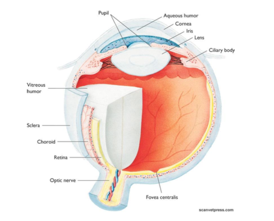
Something to do with nerves? stream
Layers of the Eyeball
3 tunics
Retina = inner
Vascular (urea) = middle
Fibrous = outer
Development of brain regions
Nervous layer of retina
Light-sensitive layer (innermost layer)
Photoreceptor cells (Neuroepithelial cells)
Outer segments made up of disks which form ...
Rods
Cones
Bipolar neurons
Ganglion cells
Development of Eye
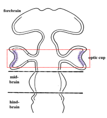
Represents a section through the developing brain as seen from above
Neural tube region containing the retinas starts to grow back towards the surface of the head , forming optic cups
Optic cups contain photoreceptor cells
Note how retinas end up back to front , cilia of neuroepithelial cells are at the back of the cup
Optic cup
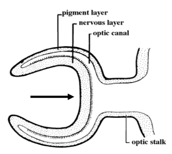
Represents a section through the optic cup as seen from above
Narrowing forms optic stalk
Layers? Stream
Lateral indentation has now deepened
Cup also indents ventrally to form the choroid fissure
Axons of retinal ganglion cells form the optic nerve which . along with blood vessels supplying retina- becomes entirely enclosed when the lips of the choroid fissure
??YES stream
Failure of choroid fissure to fuse can lead to haemorrhage and blindness
Pigment layer of retina
Outerlayer
Epithelial cells
Inner layer
3 layers including light sensitive cells
Fovea? stream
Optic disc -
Where optic nerves leave eye
No photoreceptors
Blind spot
Rods ???
Low light condition
Night
Black and white
Cones
Bright light
Day
Colour
Fact file
Cats + Dogs 95% rods +5% cones
Light and shade but not much colour
Optic nerve may be as many as a million axons
More in both optic nerves than ALL dorsal roots of spinal cord
Stream?
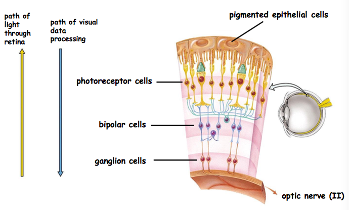
Histological layers of the retina
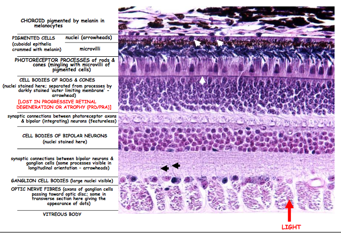
Number of cells in each layer increases the further back you go?
Progressive retinal degeneration or atrophy
Image?
Greater visual sharpness cones? stream
Retina viewed through the pupil
Opthalamoscope
Fovea
Straight behind lens
High density of cones
Optimal visual axis (no blood vessels)
Blind spot
Where ganglion cell axons leave retina
No sensory cells
?stream
Progressive retinal atrophy
Caused by loss of Rods and Cones
Loss of rods - Night blindness
Incl
Irish (red) setter
Abyssinian + Siamese cats
Blood vessels progressively atrophy leading to blindness
Pupils dilate to compensate
Appear able to see in day but bump Inyo things at night
Vascular layer
Choroid
Heavily supplied by blood vessels
Made up of collagen
Heavily pigmented - absorbs stray light
Iris
Continuous with ciliary body
Ciliary body ? stream?
Smooth muscle
Once optic nerve leaves eye it becomes myelinated
More nerves in optic nerves than all of PNS
Image?stream?
Optic Neuritis
Optic disc appears fuzzy
Inflammation of optic nerve
Oedema
Gives animal blurred vision
Tapetum Lucidum
Cells containing crystals lie between pigment layer of retina and choroid
Cells have no pigment - crystals instead
??stream
Triangular area of greenish-yellow, light reflecting , iridescent cells usuallydorsal to point where optic nerve leaves eye
Vitreous chamber
Contains vitreous humor
Maintains shape of eye
Holds retina back against the eye
Detached retina
Nervous portion of retina detaches from pigmented layer
Optic disc ALWAYS remains attached
Deprived of O2 and nutrients supplied by choroid
Without interventions = death
Causes
Blow to head
Bleeding in eye wall
Damage to vitreous humor
Sclera
Supporting
Continuous with cornea
Made of collagen
Thick layers running in many different directions
Ora serrata
Where photoreceptors finish
Lens
Sits behind pupil and iris
Image formation
Three processes involved
Refraction of light
Accommodation of lens
Constriction of pupil
Most refraction occurs at cornea and lens
Images focused in retina are inverted
Brain learns early in life to co-ordinate images with orientations of objects + corrects this
Accommodation
Distance vision
Relaxed ciliary body relaxed
Lens fibres taut
Lens flat
Close vision
Ciliary body contracted
Fibres slack
Spherical lens
Posterior chamber
Where?
Anterior chamber
Where?
Cataract
Lens becomes cloudy due to age, genetics or diabetes
Can be accompanied by lens luxation
Treatment = irreversible unless cataract removed but animal then has distance vision only. This can be restored by replacement with prosthetic lens
Diabetic cataracts
?
Drainage of aqueous humor
Humor formed by filtration of blood plasma by the ciliary processes
Fluid leaks through pupil from posterior chamber into anterior chamber
Fluid returns to circulatory system via venous plexus at transition between cornea + sclera
Stream this?
Proper notes needed from here on
Glaucoma
Raised intraocular pressure
Aqueous humour drainage blocked at limbus but production continues
Extreme pain
Blindness
Indirect causes
Pressure impedes circulation in ye , killing retinal cells and may compress and damage optic nerve
Treatment
Via eye drops
Drugs diffuse into eye, red humor secretion or improve drainage
In cats??
Cell types in cornea
Layers of collagen
Stratified simple epithelium ? on top
Healing v slow if wound deep into badly vascularised cornea
However cornea v innervated -- why things are v painful if they get in eye
stream?
Vascularisation + Keratinisation of the cornea
Keratoconjunctivitis sicca (dry eye)
Cornea starts to dry and becomes opaque
Autoimmune disease causing failure of tear production
Treatment: Artificial tears
?
Corneal ulceration
Deep wound into cornea
Treatment -
Bringing in blood supply
3rd eyelid over
Cornea transplant
Doesn't bring about immune reaction as no blood supply
Conjunctivitis
Inflammation of conjunctival membranes: sometimes discharge
Many causes:
Vrial and bacterial infection
Disorders of tear production
Exposure to foreign material
Allergy
Treatment
Tear drops
Ointment
Visual pathways + reflexes
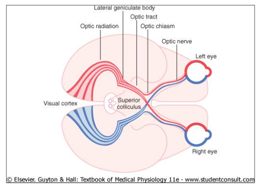
Stream?
Cross at optic chasm
Stream?
Left visual field processed by right visual cortex
Right visual fields processed by left visual cortex
what
More fibres that cross over - better depth vision they have
Some axons?
Visual field
Horizontal field of vision
Carnivore eyes = frontal position = restricted monocular lateral field and large central binocular area
Herbivore eyes = lateral position = wide monocular lateral field and narrow binocular area
Pupillary light reflex
2 sets of Smooth muscle
Circular
Inner layer
Constricts pupil
Radial
Outer layer
Dilates pupil
Nerves supplying these muscles
Rostral
Some sympathetic
Healthy animal
BOTH pupils should constrict when light shone in eyes
If not something along visual pathway is wrong
Eyeball Positioning + strabismus
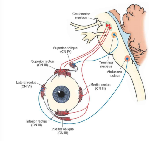
Work out from eye positioning where problem is in brain stem?
stream?
Which cranial nerves are problem?
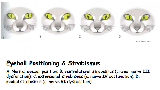
Seite 5
Spinal cord and segments
Protective structures
Vertebral column
Meninges
Connective tissue (covers spinal nerves)
Foramen Magnum
Large hole at base of skull through which spinal cord passes
Spinal nerves emerge from vertebral foramina
Stream?
Autonomic nerves branch of into sympathetic chain
Transverse section through spinal cord
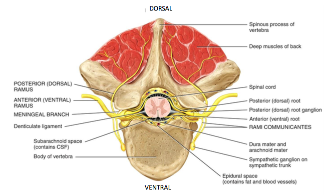
stream?
External Anatomy
Intumescences
Enlarged areas
More nuerones supplying area
Conus medullary
Caudal tapering
Cauda equina
Lumbar cistern
Spinal nerves
?
Number of pairs of nerves in each region varies with species
Anatomical + Functional region of canine spinal cord
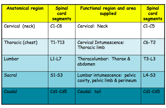
Stream?
Spinal cord + Initial sections of spinal nerves in a mammal
Spinal column is longer than spinal cord itself
Spinal nerves angled downwards
This forms the cauda equina
Thoracic level = Epidural space filled with fat
Clinical relevance = site for injection of anaesthetic
Lumbar level = Lumbar cistern
Good place for sampling CSF
??STREAM?
Internal anatomy
?
Spinal cord segment
Each segment gives rise to one spinal nerve on each side, innervating muscles in that body segment
The number of segments = number of spinal nerves
Stream?
Motor axons exit via ventral root?
Transverse Sections of spinal cordite different levels
Images
Intumesenses?
Larger diameter at lumbar and cervical region
Stream?
Spinal Nerves
Branches = rami
Networks = plexuses
Brachial plexus supplies forelimbs
Lumbosacral plexus supplies hindlimb
Spinal nerve plexus
? stream
Dorsal and ventral branches??
Anatomical + Functional spinal cord regions + Main nerve plexi
Plexuses are generally to obvious or important except for ventral ones which form enlargements opposite the origins of the limbs = brachial + lumbosacral plexuses. These give rise to nerves distributed to forelimb and hindlimb structures respectively
Innervation of thoracic + pelvic limbs
Occurs from intumescences via plexi
Cranial part of the intumescence + plexus supplies the cranial and proximal muscles of the limb
Caudal part of the intumescence supplies the caudal and distal muscles of the limb
Clinical relevance
Injection sites for anaesthetic
Loss of feeling - where problem is
SEE adendum
Distribution of dermatomes in the dog
Regions of skin supplied by sensory parts of the skin
Clinical importance?
Spinal cord physiology
Sensory + motor pathways
Definition / concept
Nomenclature (origin and destination)
Ascending = sensory
Descending = motor
Locations pf major tracts
Sensory + Motor nerve tracts
Sensory nerve tracts from skin and musculoskeletal system to cerebral cortex
3 sets of neurones
Dorsal column
Spinothalamic tract
Motor nerve tracts from cerebral cortex to skeletal muscles
Stream??
Spinal cord segment: Regions
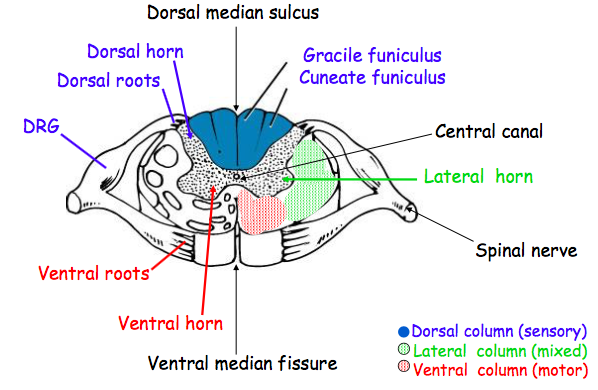
Stream ?
Locations of major sensory + motor tracts
?
Seite 6
Development of the head
Intro
12 pairs of cranial nerves
Numbered as well having a name according to either function, anatomical characteristics and distribution
Clinically important to know functional anatomy
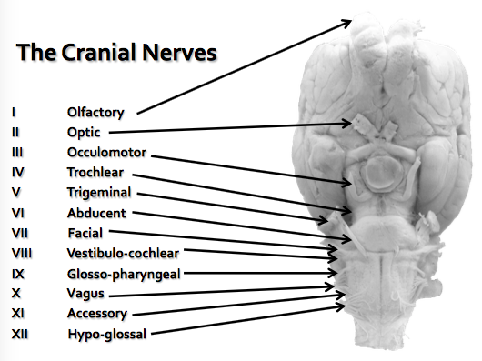
Development
In trunk cavitation of mesoderm results in splanchnic and somatic lateral plate mesoderm enclosing a coelomic cavity
In head cavitation doesn't occur
Lateral plate mesoderm remains as solid belt of tissue surrounding gut tube
Intermediate mesoderm fails to develop
Segmental Head
Adult trunk is clearly segmental
Head appears not to be but embryonically it was once
Seven segments
Ectoderm
Somites
Endoderm
Lateral plate mesoderm
Segmental arteries
Neuraxis
Nerves
Ectoderm
Form outer epithelial covering
Function of somatic sensation
Innervated by somatic afferents
Segments marked externally by lateral indentations (pharyngeal cleft)
1st segment exception
Equivalent to gill slits in fish
Deeply invaginated into segments 1 + 2
Forming stomodeum
Consequently 1+2 don't have ectoderm
Oral plate is thin plate tissue where stomodeal ectoderm meets pharyngeal endoderm
Lies at boundary between 2+3
Endoderm
Forms inner epithelial covering
Sensation supplied via autonomic afferents (visceral afferents)
Motor (sympathetic/parasympathetic) supplied via autonomic efferents
Cephalic part of fore-gut becomes pharynx
Flattened dorso-ventrally
Marked off into 6 divisions by 6 internal gill slits
In mammals theses are pharyngeal pouches
In fish these perforate into external gills slits
In mammals no perforation occurs
Lateral plate mesoderm
Belt of striated skeletal muscle which surrounds pharynx of all segments except 1
Each belt forms a donut
Separated from each other by pharyngeal cleft and pharyngeal pouch
Each belt known as pharyngeal arch
Forms special visceral muscle of the gut tube
Innervated by special visceral efferents
Also forms skeletal elements
Somites
Each segment has a pair of somites
Form striated muscle of eyeball + tongue
Innervated by somatic efferents
Segmental arteries
All but 1st segment have a pair of segmental arteries
Forms 6 pairs of aortic arches
Embedded in lateral plate mesoderm
Innervated by autonomic afferents /efferents
Neuraxis
Differentiated into brain
Each segment of neurosis referred to as a neuromere
Obliteration and migration
Obliteration
Somites 4 + 5 +associated nerves
Pharyngeal arch 5
Arterial arches 1+2
Pharyngeal clefts/ pouches 5+6
Ectoderm of segments 6+7
Fails to develop further
Endoderm of arch 2
Ectoderm of segments 3-5
Migration
Somites 6+7
Oral + nasal cavity
Vertical dotted line is boundary between ectoderm and endoderm
Paired nasal pits form
Right and left separated by septum
Nasal cavity breaks through into primitive oral cavity
Palatine process grows
Soft palate endodermal
If lateral palatine folds fail to develop= cleft palate
Tongue
Epithelium
Front of tongue = ectoderm of arch 1
Back of tongue = endoderm of arch 3
Muscle
Migration of paired somites 6+7
Pituitary + Thyroid glands
Pituitary gland
Anterior + intermediate lobes arise as diverticulum of ectoderm from arch 1
Posterior develops from brain stem
Thyroid gland
Arises as diverticulum of endoderm from arch 2
Thyroglossal duct is remnant of this
Larynx, Trachea + Lungs
Depression in floor of arches 4+6 forms laryngo-tracheal groove
Trachea + lungs develop from this
Epithelial linings = endodermal
Pharyngeal pouches + Clefts
Cleft 1 = external ear
Boundary = ear drum
Pouch 1= middle ear cavity / Eustachian tube
Clefts 2-4 disappear
Pouch 2 = palatine tonsil
Pouch 3 = parathyroid + thymus
Pouch 4 = parathyroid, thymus ultimobranchial body
Ectoderm
1 = skin of top of head
2 = the rest of skin of head
3,4 +5 = small area of skin in external ear
6+7 fail to develop
Somites
1-3 = extrinsic muscles of eye
1 = all rectus muscles (except lateral rectus) + ventral oblique
2= dorsal oblique
3 = lateral rectus + retractor oculi
4-5 disappear
6-7 = muscles of tongue
Intrinsic = tongue itself
Extrinsic attach tongue to other structures
Lateral plate mesoderm
Arch 1 = chewing muscles / malleus+ incus
Arch 2 = facial muscles / stapes + hyoid
Arch 3 = stylopharyngess muscle/ hyoid
Arch 4 = pharyngeal muscles + cricothyroid muscle/ laryngeal cartilages
Arch 5 disappears
Arch 6 = laryngeal muscles/ laryngeal cartilages
Aortic arches
1+2 disappear
3 = internal carotid, carotid sinus + body
4
Right = right subclavian
Left = aortic arch, aortic baroreceptor site + aortic bodies
5 disappears
6 = pulmonary trunk + ductus arterioles on left side
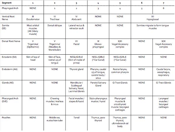
Each segment has a pair of cranial nerves
Each structure of the head requires a nerve supply
Seite 7
Cranial nerves
Segmental trunk nerves
Somatic nervous system under voluntary control
1 = afferent sensory
2-3 = inter neuron
4 = efferent motor
Primary afferent cell bodies in dorsal root ganglion
Dorsal and ventral roots fuse to form single spinal nerve carrying all 4 nerve types
Autonomic nervous system under involuntary control
1= afferent sensory
2 = inter-neuron
3 = pre-ganglionic efferent
4 = post-ganglionic efferent
Has additional ganglion where pre- and post-ganglionics synapse
Segmental head nerves
Similar of runs EXCEPT
Autonomic efferent neurons leave via dorsal root
Dorsal and ventral roots do NOT unite
Head as 5 th neuronal component
Striated muscle arising from somites supplied by somatic efferents as in trunk
Striated muscle arising from lateral plate mesoderm supplied by SPECIAL VISCERAL EFFERENTS leaving via dorsal root
Summary
Segment 1 has no pharyngeal arch therefore it borrows its dorsal root nerve from segment 2 = ophthalmic branch of V
Due to development of mouth dorsal root nerve of segment 2 splits into 2 major branches
Segments 4 + 5 lose their somites so don't have a ventral root nerve
Segment 6 loses its pharyngeal arch so doesn't have a dorsal root nerve
Segments 6+7 somites unite to form tongue muscles, so does their ventral root nerve
Segments 5+7 share dorsal root nerve
Cranial nerves
Special sense nerves
CN I - Olfactory nerve
Olfaction/smell
CN II - Optic nerve
Vision /sight
CN VII - Vestibulo-cochlear nerve
Balance + hearing
The other cranial nerves
Each segment should have a dorsal and a ventral root
Dorsal roots innervate all structures
Contain SA, AA, AE + SVE neurons
Ventral roots innervated somites
Contain SE neurons only
Except CN III has small AE component to ciliary muscles + SVE to leave palpebrae superioris
Remember that during development pharyngeal arch 5 and somites 4+5 fail to develop as do their nerves
Segment 1
Ventral root nerve = III occulomotor
SE- Somite forming most orbital muscles
E to ciliary muscle
SVE to levator palpebrae superioris
Dorsal root nerve = ophthalmic branch of V
No pharyngeal arch so no dorsal root , borrows from segment 2
SA- ectoderm only, forms skin on top of head bone eyes
SVE, AA, AE - None
Segment 2
Ventral root nerve - IV trochlear
SE- somite forms dorsal oblique orbital muscle
Dorsal root nerve = V
Mouth splits trigrminal into 2 further branches - maxillary SA and mandibular SA + SVE
SA- ectoderm forms most of skin of face , rostral 2/3 of tongue
SVE - Pharyngeal arch 1 forms muscles of chewing
Segment 3
Ventral root nerve = VI abducent
SE- somite lateral rectus + retractor oculi
Dorsal root nerve = VII facial
SA - ectoderm forms small area of skin internal ear cavity
SVE- pharyngeal arch 2 forms muscles of facial expression
AA - taste from 2/3rds of tongue , palate
AE - to mandibular / subigual salivary glands, nasal cavity glands, lacrimal gland
Segment 4
Ventral root = NONE
Dorsal root nerve = IX glosso-pharyngeal
SA - ectoderm forms very small area in ear canal
SVE - pharyngeal arch 3 forms stylopharyngess muscle
AA - endoderm forms lining of pharynx / caudal 1/3 of tongue, aortic arch 3 forms internal carotid
AE - motor to parotid salivary gland
Segment 5
Ventral root nerve = NONE
Dorsal root nerve = X/XI vagal-accessory complex
SA- ? ectoderm forms very small area of ear canal
SVE - pharyngeal arch 4 forms rest of pharyngeal muscles + cricothyroid muscle
AA - endoderm forms lining of common pharynx and larynx , aortic arch 4 forms root of aorta/ right subclavian
AE- Endoderm of gastro-intestinal tract
Segment 6
Ventral root nerve = XII hypoglossal
SE- somite 6 forms muscles of tongue
Dorsal root nerve = NONE
Segment 7
Ventral root nerve = XII hypoglossal
SE- somite 7 forms muscles of tongue
Dorsal root nerve = X/XI vagal-accessory complex
SVE - pharyngeal arch 6 forms rest of laryngeal muscles
AA - endoderm forms lining of larynx caudal to vocal folds/ trachea/ lungs /gastro-intestinal tract , aortic arch 6 forms pulmonary artery/ ductus arteriosus
AE- endoderm of gastro-intestinal tract
Seite 8
Motor systems
Structures involved
Motor cortex
Basal nuclei
Thalamus + internal capsule
Red nucleus and substantial nigra
Nuclei in pons and medulla oblongata
Cerebellum
Cranial nerves, Spinal cord, spinal nerves
NMJ
Skeletal muscle
Somatic motor system
Upper motor neurone UMN
Lower motor neurone LMN
Neuromuscular junction NMJ
Skeletal muscle fibres
UMN + LMN
UMN exist wholly within the CNS
They synapse on LMN which have a cell body in CNS and axon in PNS
LMN run via spinal or cranial nerves to innervate skeletal muscle fibres
LMN for forelimb from cervical intumescence
LMN for hindlimb from lumbar intumescence
UMNs
UMNs initiate, regulate, modify nd terminate LMN activity
May synapse directly on LMN or via interneurons
LMNs can be influenced y >1 UMN
UMNs to LMNs supplying flexor muscles travel in the lateral funiculi of the spinal cord
UMNs to LMNs supplying extensor muscles travelling the ventral funiculi of the spinal cord
?
UMNs and LMNs
Extensor muscles are dominant in body to maintain posture against gravity
Tempered by effect of inhibitory UMNs on the LMNs which supply these muscles
Without this inhibition extensors would take over leading to spasticity
LMNs
Have cell bodies in the CNS, run from CNS to muscle fibres
AP in the LMN causes muscle contraction
Regulated UMNs - initiate , regulate and terminate LMN function
LMN can fire without UMN input - reflex action
In conscious animal LMNs have a constant sub-threshold depolarisation causes ACh release which causes muscle tone and trophic support
Motor unit
Motor unit = LMN + NMJ + skeletal muscle fibres
Small motor unit allows fine movement
Large motor unit = more force, less finesse
??
As long as it has an intact LMN , a muscle can be made to contract
The motor system is not an island
Motor function occurs in response to sensory input and/or through voluntary action governed by the cerebral cortex
Normal function of all components is required for the animal to mobilise normal
Somatic reflexes
A reflex is the functional unit of the nervous system
It is an innate reaction to a stimulus and is present from birth
Sensory input leads to motor output without input from higher centres - doesn't require UMN input
This is different to a response which is a learned behaviour
May be monosynaptic or polysynaptic
May send info to higher centres
May be ipsilateral or contralateral
May be intra segmental or intersegmental
Monosynaptic reflex
?
Patellar reflex
?
Muscle spindles and GTO
?
Voluntary movement
?
Polysynaptic reflex
?
Withdrawal reflex
?
Reflexes don't require the input of an UMN
Removal of UMN input often causes a reflex to be exaggerated ??
In an injury to the LMN , the muscle will lodestone, bulk and reflexes
In an UMN injury, reflexes will be normal or increases will tone, and any atrophy will be relatively ild and due to disuse. Co-ordination will be reduced , but strength will be normal
UMN tracts
Corticospinal tract fine motor skills
Coticonuclear tract from cortex to cranial nerve nuclei
Rubrospinal tract skilled movements
Cortical input means this can serve similar function to corticospinal traction non -primates
Tectospinal tract orientates head + eyes in response to sights + sounds
Vestibulospinal for maintenance of posture/balance
Reticulospinal stabilises body against gravity
Pyramidal + extrapyramidal
Corticospinal tract runs caudally in the ventral medulla. Tracts form triangular shape on the transverse section - called the pyramidal tract
All other UMN tracts are extrapyramidal
Pyramidal tract enables fine, voluntary movements
Extrapyramidal tracts mainly control posture + subconscious, rhythmic movements
Although rubrospinal tract does generate fine skilled movements in non-primate mammals
Feedback between different nuclei and tracts ensures balance between excitation and inhibition
Cerebellum
Coordinates and integrates sensory input and input from the cerebral cortex
Output to UMN nuclei
Cant initiate movement
Cerebellar cortical nuclei inhibitory to deep cerebellar nuclei
Deep nuclei excitatory UMN nuclei
Cerebellar dysfunction leads to loss of coordination
??
Synapses become more efficient with practise
Particularly important for rapid /complex movements
Seite 9
Somatosensory systems
Sensory cells
Adaptation
Sensory receptors
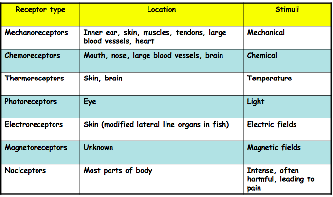
Sensory neurons
Rapidly adapting
Slowly adapting
Somatic sensations
inner ear= had in space
muscles tendons joints = body in space
Touch
Rapidly adapting
Meissners corpuscle
Tactile hair
Slowly adapting
Expanded tip receptor
Ruffini's end organ
Pressure
Itch
??
Thermal
Sensory perception
Acute pain
Chronic pain
Nociceptive pain
Neurogenic pain
Control of pain transmission
Gate cell scan inhibit pain sensations
By releasing enkephalins
Pain sensitisation
Treatment of pain = Analgesia
Somatosensory pathways
Decussation
Dorsal column-medial lemniscal
Spinothalamic pathway
Seite 10
Locomotion and proprioception
Gait
Span = distance you move whilst foot is on ground
Span is limited
Stride = distance moved whilst leg isn't in contact with ground
Stride can be increased by a suspension phase
walk = 4 beat
trot = 2 beat diagonals
pace = 2 beat different sides
canter = 3 beat
gallop = 4 beat with greater suspension phase
Proprioception = sense that enabled good motor function
Conscious proprioception
Intension tremor - try to do something + then tremor
3 neurone system
Question
Subconscious proprioception
2 neurone system
Question
Spinal tracts
Stream
Kinaesthesia
the sense of the position and movement of the body
??
Horse hasn't realised that leg won't support its body
Problem with fine movement, know where its leg is
Cerebellum again
Confounding factor
?
Question
subconscious hind legs spread, front legs narrow
dragging feet - conscious?
swaying
front legs moving at faster pace than back legs
info
Question
Not brain - neurotically no other abnormalities
Not lumbar - forelimbs are abnormal
Cervical - instability, squishes spinal cord
Woblers syndrome
Seite 11
Ear + Hearing
Outer ear
??
Becomes progressively more bony
Tympanic membrane = eardrum
Ear wax
Must be flushed if overproduction
Otherwise can lead to ulceration
Sedation necessary
Otitis externa
Treatment - ear drops
Also include pain killer
Middle ear
Auditory tube leads to back of throat (nasopharynx)
Equalises pressure on back of ear drum
stirrup connects to inner ear via oval window
Otitis media
Horner's syndrome
Conduction deafness
stream ?
Inner ear
transduction = conversion of one form of energy to another
Bony + membranous labyrinths
Membranous labyrinth
stream
Bony labyrinth
Bipolar neurones
Stream
Oval window is where stirrup foot plate makes contact with the inner ear
number of turns varies with species
Round window
Stream
Prevents perilymph to leak out
Allows movement of perilymph?
Organ of corti
?
??
Mechanics of hearing
?
?
Frequency discrimination in the cochlea
NB frequency measured in Hz
Narrow and stiff near round window , flexible at end of cochlea
Nerve Deafness
Noise trauma can kill off all cells
Auditory pathway
Difference in timing between noises
Crossing over of nerves
Time taken for sound to reach other ear
Allows animals to know where a sound is from
LGM - vision
MGN - auditory
Trapezoid body
Sits just above the pyramids
Seite 12
Vestibular system
Vestibular cell
Vestibular Organs
Crista
Senses changes in rotation of head
Macula
Senses changes in linear direction
Has gelatinous mass containing otoliths
Stream
Otoliths
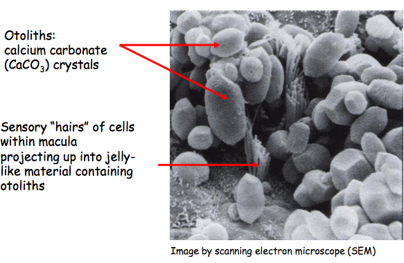
Simulation of otoliths
How they work?
Static balance
Head upright?
Head tilted/upside down
Force of gravity causes hairs to stretch , triggering nerve impulses
Location of inner ears
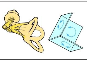
?
How ampullae work
Moves in opposite direction of rotation
Enables dynamic balance
Dynamic balance
Vestibular pathways
Connect with a number of diff nuclei and so give communicate with many diff pathways
All depusate
Communicate with many cranial nerves involved with eye
Relays in thalamus before finally vestibular cortex
Control of eye movements and posture
Righting reflex - allows animals to land on their feet when they're falling
Vestibular reflex
Tonic neck reflex
Righting reflex
Vestibulo-ocular reflex
Motion sickness
Vestibularsyndrome
Seite 13
Taste + Smell
Chemoreception
Pheromones - small smell particles within species
Papillae
species differences not exam
Gustatory
Mechanical
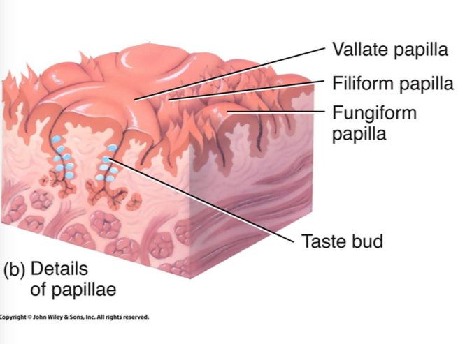
strem
Features of the tongue
Taste bud
Not shown in images e- stem cells
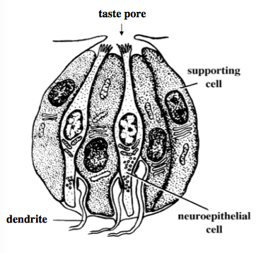
Tastants
Taste molecules
Bitter
Salty
Sweet
Umami
Sour
Gustatory pathway
Stream
Olfactory communication
Olfactory epithelium
Neuroepithelial cells are able to regrow their axons
Only part of CNS which can do this
Olfactory bulb neuron = mitral cells
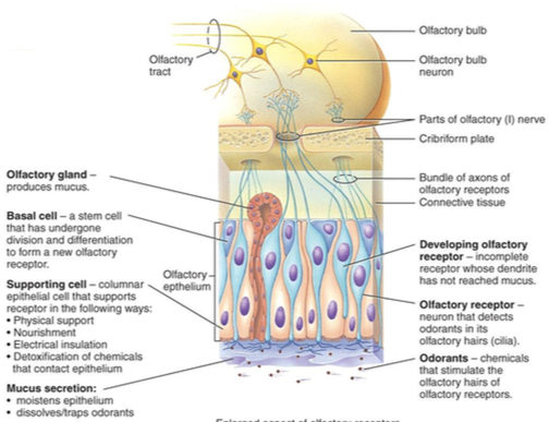
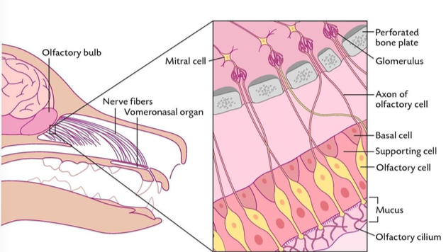
Vomeronasal organ - accessory organ
Epithelia lining lined with ?
One end blind?
Olfactory + vomeronasal structure + function
Flehmen reaction
Cuts of nasal pathway
Not all species
Olfactory pathways - Canine
Conscious and subconscious pathways
anosmia - loss of sense of smell
Loss of appetite
Hypothalamus v involved in controlling appetite
Nursing sick animals
Seite 14
Learning, Memory + Behaviour
Limbic system
Highly interconnected brain areas coordinating
Homeostasis
Motivation
Emotion
Includes olfactory cortex
Route for olfactory info re
Feeding
Sexual behaviour
The hippocampus forms part of the limbic system and is important for memory formation
Amygdala = seat of aggression
Abnormal behaviour - limbic/forebrain lesions
The limbic system includes regions of the forebrain associated with emotional responses
Forebrain includes the frontal lobe , the seat of planning , decision-making and personality
Lesions cause changes in an animals normal habits, personality, attitude, reaction to the environment
Clinical signs
Depression
Lethargy
Listlessness
Stupor
Destructive behaviours
Pacing
Circling
Rabies
Contagious disease of virtually all mammals
Dogs = main vectors for virus (saliva)
Virus passes along nerves from bite or scratch and reaches CNS, directly affecting the hippocampus and other structures
Lesions in limbic system alter level of aggression such that docile and wild animals become the opposite
Resulting in 'dumb form' and then 'furious form' = two stages the disease
Animals goes from being morose and sulky to violent, refusing food, snapping at anything passing , easily excited , vacant stare, great salivation and finally paralysis
Conformation = histology
Negri bodies are cellular inclusions often found in the hippocampus and purkinje cells of the cerebellum as well as the medulla oblongata and various ganglia .
Frontal lobotomy
Lesions the frontal lobe
Performed on humans and animals to alleviate violent and aggressive behaviours that are destructive in nature
Humans show extensive changes in personality and loss of intellect as well, so procedure is infrequently used
Results in dogs are variable
Performed as a last resort
Can be performed by inserting a sharp tool through eye socket
GA not needed - brain has no pain fibres
Amygdala
Experimental destruction of amygdala leads to variable alteration in animals' responses
In cats - unfriendliness, fear, rage , tendency to attack animate and inanimate objects
In other animals- tame them
Lesions in temporal lobe can cause psychic seizures bringing about bizarre and hallucinatory behaviour - aggressive response to things that aren't there
Seizure disorders - epilepsy
Spontaneous discharge of activity by hippocampal neurons
Characterised by fits
Sudden , complete lossof consciousness
Muscular convulsions
Eyeballs may roll
Lots of salivation
Particularly a disease of dogs
Causes
Genetic
Trauma
Infections
Treatment
Sedatives
Tranquillizers
Ascending reticular activating system (ARAS)
Reticular formation core throughout brainstem of neurons regulating vegetive function , such that lesions here are fatal .
A vast array to external and internal environmental information is passed via the RF to wide-ranging areas of cortex, to determine the arousal level of the animal and control the sleep-wake cycle
Consciousness + sleep
Transition sleep to consciousness due to activity in the RF
RF neurons recieven info from sensory organs and neurons commanding muscles also extend to cortex
Continuous sensory info will cause RF to bombard cortex + keep animal awake
When ready to sleep, animal lies quietly to reduce sensory stimulation
The RF is the main target of drugs for GA
ARAS functions by ranking incoming info in terms of importance
Enables animal to focus
Filters garage of sensory info
Clincally - decreased consciousness can be due to focal lesions of RF or diffuse lesions of cerebrum
Levels of consciousness
Normal - bright, alert + responsive
Obtunded - dull, tends to fall asleep
Stuporous - sleepy
Comatose - unconscious
Brain dead - no cortical electrical activity or brainstem function
Hypothalamic lesions
Abnormal
Water consumption
Appetite
Pica = eating non foods
Temperature regulation
Hemi-neglect = animal ignores one side of body
Causes not all fully understood
Memory and learning
Memory is the storage of info in the NS
To benefit, retrieval must also be possible
Learning is changes in behaviour based on previous experience
Forms of learning depend on consequences of different stimuli
Habituation - natural reactions to stimuli are suppressed
Occurs gradually over time
Sensitisation - natural reactions to stimuli are enhanced
Classical conditionings
Animal is trained to associate 2 different stimuli with one another
One is an unconditioned stimulus which causes a reflexive reaction
One is a conditioned stimulus that evokes the same reaction as the UCS
Operant conditioning - learning by trial and error
Obedience training based on this
Aid of repeated rewards at strategic timing
Forms of memory
Short term
Stores info from a few seconds to hours
Limited capacity - events registered here are either lost or transferred to long term memory depending on factors like concentration + motivation
Based on electrical activity in neuronal networks
Long term
Lasts for days and up to many years
Almost unlimited capacity
Result of physical or chemical changes in cells
Declarative
Information about what something is and what has happened
Primarily connected to cerebral cortex
Memory store in cortex aren't limit
Factual knowledge organised into networks making it simpler to remember new facts or events if they can be associated with others that are already remembered
As memory grows it actually becomes increasingly easy to remember new info
Procedural
Provides info about how something is done
WRT movements
Working
Includes info recently required to make decisions or solve current problems
Contains elements of both short and long term memory
Contents continuously being uploaded as individual faces new problems and has to make new decisions
Hippocampus
Where transition from short to long-term memory occurs,
Unsure how this occurs
Unsure how memories are received and transferred to working memory
Consolidation is the reinforcement of memory resulting from frequent retrieval of a piece of information
Plasticity is capability of brain to change functionally and structurally as a result of experience .
Deteriorates with age
Senility
Cognitive dysfunction
Collectively , cognitive disorders refer to dementia , failure to recognise owners, familiar environments and objects , inability to learn.
Cognition = act of knowing
Companion animals living long enough for these disorders to occur
Seite 15
Histology
Neuronal stains
Commonly - Nissl
Stains RER in cell body
Special stains needed to reveal ramify processes of neurons
Fuzzy appearance is due to dendritic spine
If nervous tissue gets lesion - retrograde degeneration occurs
Nerve degenerates from point of lesion back to cell body
Nissl disappears
Spinal Cord
Meninges
Pia mater - invests the tissue
Blood vessels sit in subarachnoid space
Sensory neurons in the DRG
Large + small cell bodies
Rounded
Unipolar neurons
Crowded at periphery of ganglion
Uninterrupted axon bundle
Motor Neurons in ventral horn
Huge cell bodies
Nucleus not always visible
In another plane of section
Multipolar
Polygonal
Many initial dendritic or axonal branches
Don't know which from routine stains
Ependyma
Type of epithelial cells
Cilia on apical surfaces that face into lumen of ventricles
Cells are cuboidal/columnar
Protective
Nutrient sensing
Keep concentrations of?? stable
Brain
H+E staining of grey matter in the cerebral cortex reveals the nuclei of glia and neurones but not neuronal processes
Nuclei of ?? appear granular due to DNA transcription
Dark staining nucleic from support cells
Astrocytes
Oligodendrocytes
Blood vessels present in subarachnoid space
Cerebellum
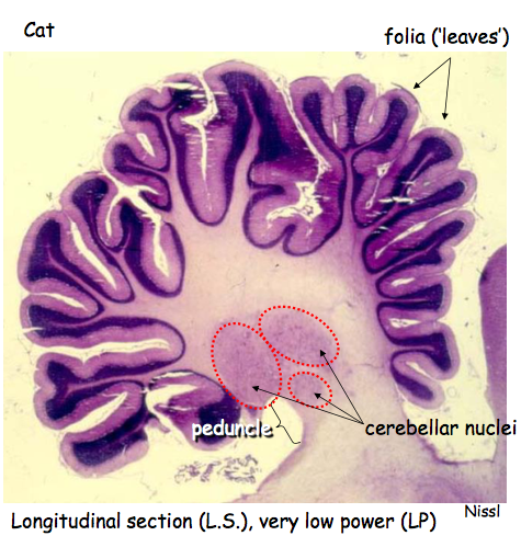
Nissl staining of a section through the cerebellum reveals
Layered + highly folded cortex
Cerebellar nuclei deep to cortex
Cerebellar peduncle
Cerebellar nuclei give rise to axons extending to other brain centres + spinal cord
Peduncle = stalk of cerebellum
Folds = folia (leaves)
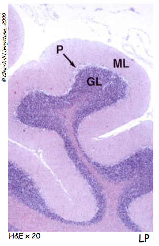
Arrangement of a cerebellar folium
Purkinje cell dendrites + granule cell axons lie in the molecular layers
Purkinje cell axons synapse on the cerebellar deep nuclei located within the white matter
Layers of cerebellar cortex
Molecular layer
Composed of granule cell axons + Purkinje cell dendrites
Purkinje neurons
Send their axons into underlying white matter providing sole output of cerebellum
Granular layer
Vast numbers of densely packed small neurons
Eye
Lacrimal Gland
Produces tears
Serous gland produces many lysozyme
Lumens hold tear
Protects surface of eye
Histologically lacrimal gland has similar appearance to serous salivary gland
Tears are secreted into the lumen of each secretory unit and drain onto the eye from each gland via many small ducts
Whole Eye
Choroid is distinguishable even at low magnifications due to its high pigment content
The suspensory ligament by which the lens attaches to the ciliary body isn't preserved
Cornea
Anterior epithelium
Moderately thin layer of stratified, non-keratinised squamous epithelium
Microvilli on top layer of cells?
Bowmans membrane
Anterior limiting lamina
Substantia propria (stroma)
Thick layer of highly regular, dense collagen in thin layers
Bright pink in H+E
Splitting of collagen plates to reveal small spaces is artifactual
Descemet's membrane
Posterior limiting lamina
Thick collagen/elastic basement membrane
Posterior epithelium
Squamous cells
Anterior chamber
Just right amount of hydration to maintain transparency
Ciliary body + surrounding structures
Cornea
Canal of Schlemm
Lined by squamous epithelium
Anterior chamber
Iris
Loose CT tissue
Trabeculae
Meshwork of fine collagenous material with slit-like spaces
Lined by simple squamous epithelium
Ciliary body
Main component is ciliary muscle
Bundled of smooth muscle fibres arranged in different directions
Ciliary processes
Lined with a double layer of cuboidal epithelium
Deep layer holy pigmented
Surface layer is not pigmented
Lens
Lens centre
Examination at high power shows cells devoid of organelles
Cells are living
Unknown how
Epithelium
Cuboidal
Single layer lining mature lens under the capsule
Secrete collagen
Capsule
Thick layer of collagen secreted by underlying epithelial cells
Layers in the wall of eyeball
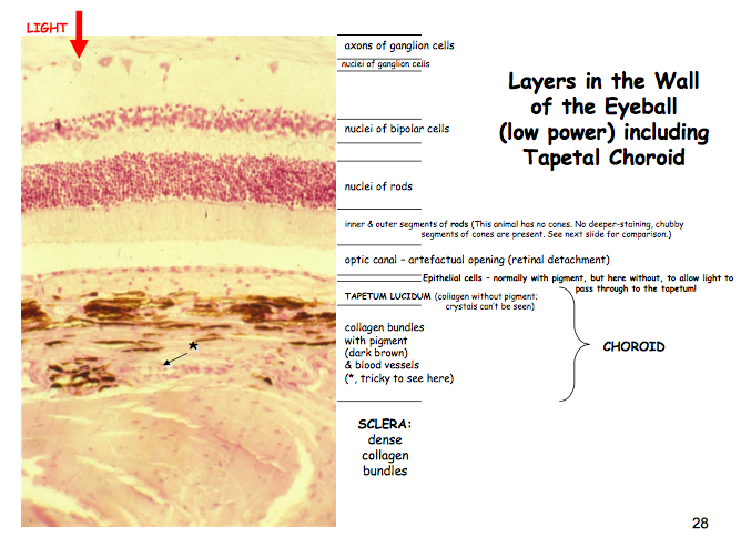
Choriod isn't highly pigmented in order to reflect light
Cones often not found in animals
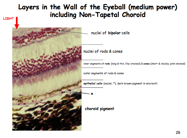
No tapetum is this region of choroid - heavily pigmented
Retina and optic nerve
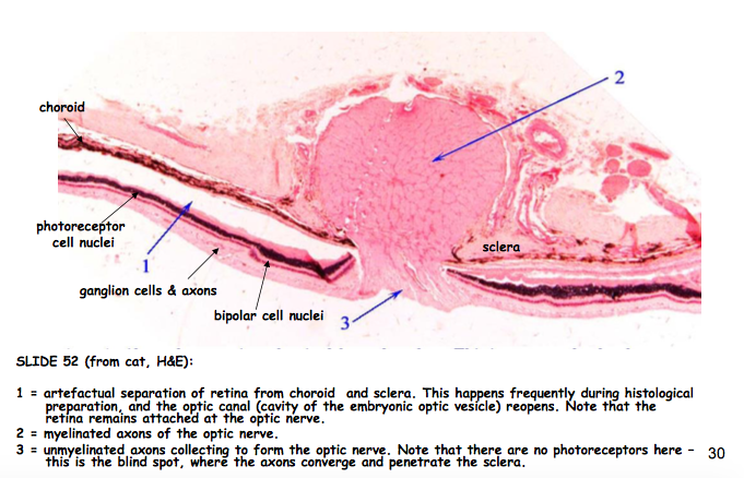
Myelination occurs outside eye
Seite 16
Neurology exam
Starts before they've entered the consult room
Gait
Posture
Appearance
Don't forget the full basic clinical examination
Top to toe examination
Tailor exam to patient
All body systems
Neuro signs can often be a manifestation of systemic disease
I.e liver disease
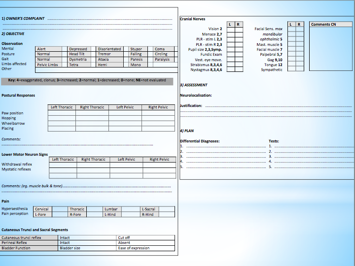
Mentation, Posture, Symmetry
Tilted to one side?
Turned to one side?
Let animal wander in consult room
Moving normally?
Gait
Space where owner can walk them up and down?
Dragging toes
Tracking up?
Turning corners?
Conscious proprioceptions
Information about fine positioning movements
Feet right way up?
Enough force?
Paw righting reflex
Normal = immediately correct
Don't use whole leg (subconscious proprioception)
Test motor neuron function
Subconscious proprioception
Information that allows you to maintain the right posture
Step reflex
Q
Dorsal
Spinocerebellar
Q
Subconscious
Q
Subconscious?
Q
Sight
UMN
LMN
Symmetrical movement
Muscle tone
Proprioception
Q
Can tell if she can feel it without looking at face to see if she's showing a pain response
Myotactic reflex
Pressure on tendon causes limb to extends
Q- Muscle spindle
Q- proprioception
Q- Golgi tendon organ
Q
No effect / increased
Q
Reflexes absent / reduced
Cutaneous trunci
Pinching skin should cause symmetrical contraction
Allows localisation
Perineal reflex
Testing muscles of perineum
Testing sacral segments of spine
Bowel + Bladder function
History v important
Episodic
Might not occur during exam
Squat properly
Emptying bladder properly
Vision + eyelid/facial movements
Menace response
Don't create a breeze or ear around skin
Tests facial response
A response
Tests optic response
Vestibular function + eye movements
Vestibular reflex
Abscense normal
Occurs at rest
Abnormal
Asses for strabismus
Check both eyes
Q
Cat
Round dark optic discs
Dog
Triangular
Pain?
Pressing on edges of page vertebra to see if it hurts
Animals often stoic
If sore
Stay rigid
If not
Dip down a bit
Remember
Not able to perform all tests on all patients
DO NO HARM
Asses for pain
There may be other things that inhibit / affect examination
Now what
Localise the lesion
Next steps are taken to endeavour to identify the disease process
Blood tests
Haematology/biochemestry
Endocrine testing
Serology
Toxicology
Urinalysis
Enables interpretation of blood findings
Assits identification of concurrent disease
Diagnostic imaging
Ultrasound
Of abdomen if non-CNS disease is suspected
Of eye to examine retina, rostral optic nerve and retrobulbar area
Radiography
Plain
Contrast study
E.g. myelogram
Not is raised intracranial pressure
After CSF tap
Don't show whats inside bones v well
Fluoroscopy
A moving radiograph
Alimentary tract mobility
Advanced imaging
Computed tomography (CT)
Magnetic resonance imaging (MRI)
Better for CNS
Magenets make protons in body line up , blast them with radio waves , then the protons give of radio signals that are used to make and image
CSF tap
?
Pharmacological testing
Stream
Electrical testing
Cataracts
Cant see back of eye
Test retina function
Electromyogrophy
Test muscle itself
Deafness? stream
Lesion = area of damage
Seite 17
Clinical cases
4 yr 5 m female entire JRT
Acute onset tetraparesis
2 days duration
Started suddenly no apparent cause
Vaccines up to date
Not been out of UK
3 months from last season
No known access to toxic substances
Feeding constant
No trauma
DUDE normal
No seizures / history of neurological issues
No paw righting reflex on right fore
Unable to initiate movement well
Left side more effective
UMN?
Patella reflex present
my-tactic reflex present
Feels pain in legs
Back leg + other side LMN normal
LMN deficits in right fore limb
Cutaneous trunci reflex reduced on RHS
Everything else normal
UMN signs all 4 limbs
LMN in right fore
Intact deep pain
Right sided loss of spinal segment reflexes caudal to forelimb
Normal cranial nerves and mentation
Cervical intumescence affected
UMN deficits in all legs
LMN deficits right fore limb
Everything else normal
Bloods and urine normal
Nothing detected on radiography
MRI
Oedema in spinal cord - C6
Haemorrhage on RHS
Diagnoses
Fibrocartilage??
?
Treatment
hydro + physiothearpy
5y 2m male neutered
Cut onset neck pain + head turn to left
Duration 4 days
No known cause
Only turns left
Vacant / depressed
Head carried low
Vaccinated and parasite control up to dat e
feeding nchanged
No known toxins
No known trauma
Onset overnight
DUDE
Quieter than normal
No righting reflex on right hind and right fore
testing conscious proprioception -- deficits
Uneven hops - more on right , irregularly spaced
No signs of weakness
Segmental spinal reflexes fine
Cutaneous truni normal
Cranial nerve examination showed reduced menace on RS
Rest was unremarkable
Reduced mentation
Neck pain
Left sided head turn
circle to left -
RHS conscious proprioceptive deficits
Reduced menace on right
Pathways affected
Proprioceptive
Optic
Linked only by brain
Forebrain
All cross
Left forebrain
All pass through thalamus
Left sided lesion of thalamus
Left Circling = hemineglect as left side of brain not working properly , can't see that right is there
Neck pain = swelling
Blood and urine wnl
MRI - oedema in brain around a mass I left thalamus
Neoplastic? Inflammatory?
NO CSF - Raised pressure
Treatment yon presumed inflammatory disease
Steriods
Gastro protectants
Gabapentin = pain
2 weeks later better
4 weeks corticosteroids reduced
Relapse
Treatment restarted
Doing well!
Seite 18
Informationen
- Beschreibung
- Vet Med 1 (NSF) Notiz am CNS, erstellt von Alice Butterworth am 11/02/2018.
- Erstellt von
- Alice Butterworth
- Aufrufe
- 0
- Bewertung