
Flashcards
Integumentary
Cards in this deck (145)
- 1What is the function of skin?Protect body from environment by acting as a barrier against:
Microorganisms, UV, radiation, loss of body fluids, stress of mechanical forces. regulating body temperature, immune surveillance, activation of Vit D, protection & pleasure via touch & pressure receptors, Fosters communal organisms, protect against pathological bacteria - 2What are the 2 major layers of skinEpidermis: superficial/outer layer
Dermis: Deeper layer/"true skin" - 3What is the subcutaneous layer of skin called and what does it contain?Hypodermis:
Contains macrophages, fibroblasts, fat cells, nerves, fine muscles, blood cells, lymphatics, hair follicle roots - 4What are keritinocytes and where are they located?Located in the epidermis. Same thing as scleroprotein. Protects from mechanical stress. Form the skin surface that you can see.
- 5What is the purpose of cornification/keratinization in the epidermis of the skin?Prevents dehydration of deeper skin layers. Prevents loss of body water
- 6What are the functional cells within the epidermis called?Langerhans cells
- 7What is the purpose of melanocytesLocated near base of epidermis. Synthesize & secrete melanin which is a hormone is response to UV light. Determines skin color.
- 8What causes vitilgo?Autoimmune disease. Loss of pigmentation results in patchy areas.
*Usually sharply demarcated - 9What is a Langerhans cell?KNOW THIS:
Type of dendritic (immune cell) in epidermis
Migrate to epidermis from bone marrow - 10What is the purpose/function of a Langerhans cell?INITIATE IMMUNE RESPONSE BY PRESENTING PROCESSED ANTIGEN TO T CELL
Provides defense against environmental antigens - 11What are Merkel cellsAssociated with touch receptors
Stimulated by damage to epidermis - 12The dermis is a haphazard arrangement of what type of tissue?Connective tissue
Collagen - skeletal part of the skin
Elastin - stretchy part of the skin
Reticulin - gel-like ground substance - 13What does the dermis contain?hair follicles
sebaceous glands
sweat glands
blood vessels
lymphatic vessels
nerves - 14The fibroblasts, mast cells, and macrophages are functional cells in what layer of skin?Dermis:
Fibroblasts - secrete connecive tissue matrix & collagen
Mast cells - release histamine & play role in skin hypersensitivity reactions
Macrophages - phagocytic, immune response, histiocytes - 15What is the Subcutaneous Layer of skin and what does it contain?Third layer of skin (hypodermis)
Contains fat cells (adipocytes)
Large blood vessels
SQ collagen continuous with dermal collagen - 16The nail is composed of which 4 structural unitsHyponychium (nail bed)
Nail plate
Proximal nail fold
Matrix (nail grows from it) - 17What is integrated unit with a hair follicle?Sebaceous gland
hair follicles ) sebaceous = integrated units - 18How does growth in nails differ from growth of hair follicles?Nails grow continuously
Hair grows in cycles - 19Where on the body are most sebaceous glands located?Face, chest, back
modified gland: eyelids lips, nipple, glans penis, prepuce - 20What do sebaceous glands secrete and what is the purpose of the secretion?Secrete sebum (composed mostly of lipids)
Holds in moisture, prevents drying - 21What stimulates the growth of sebaceous glands?Androgens
Early sign of puperty - 22WHAT IS THE PURPOSE OF ECCRINE GLANDS AND WHERE ARE THEY LOCATED?KNOW THIS:
Purpose - secretion important for thermoregulation
Cool body via evaporation
Where - all over body but concentrated on palms, soles, foreheads - 23WHAT IS THE PURPOSE AND DISTRIBUTION OF APOCRINE SWEAT GLANDS? WHERE ARE THEY LOCATED?Fewer in number than eccrine.
Purpose - very limited proven function
Located - folds of axillae, scalp, face, abdomen, genital area - 24What facilitates body temp regulation?Arteriovenous anastomoses in dermis
Sympathetic regulation, opening and closing arteriovenous anastomoses - 25What contributes to aging skin and loss of integrityGenetics
Cumulative environmental effects (UV radiation, inflammatory responses, tobacco smoke, gravity)
Structural changes ( thinner, less eleastic, direr, wrinkles, pigmentation changes) - 26WHAT CELLULAR ALTERATIONS CONTRIBUTE TO AGE ASSOCIATED CHANGES IN SKINDermoepidermal junction flattening = Shorter capillary loops
Decreased Melanocytes = Decreased Protection from UV radiation
Decreased Langerhans cells = Decreased Immune response - 27WHAT CELLULAR ALTERATIONS TO AGE ASSOCIATED CHANGES IN SKIN? (con'd)Decrased dermal thickness = Translucent, paper-thin quality
Loss of rete pegs = Smooth, shiny appearance
Decreased Elastin fibers = wrinkling
Decreased sensory production & perception - 28What is the essential 3 step diagnostic process?1) gather history
2) Perform a PE
3) Observe appearance & distribution of lesion - 29What is the Wood Lamp Examination?UV light skin fluorescence
- 30What is the KOH used for?For scabies & fungal exam
- 31What is the difference between primary & secondary lesions?Wiki:
Secondary lesions are a modification of primary lesions. - 32Telangiectasa, Macule, Patch Plaque, Wheal, Papule, Pustule, Vesicle & Bulla are what type of lesions?Primary lesions
- 33Scale, lichenification, Excoriation, Erosion, Ulcer, fissure, atrophy, scar, keloid are what type of lesions?Secondary lesions
- 34What must you make sure you are doing when assessing a lesion?Make sure you are palpating.
Remember there is no normal lesion
Need to see if it's superficial or deeper - 35WHAT IS LICHENIFICATION?KNOW THIS:
rough thickened epidermis due to rubbing/scratching
Often involves flexor surfaces of the extremities
MADE BY SCRATCHING
ex. chronic dermatitis - 36Describe telangiectasiaPrimary lesion:
Fine, irregular lines of capillary dilation
ex. rosacea, actinic damage - 37MaculePrimary lesion:
FLAT, circumscribed area of
color change < 1 cm
(freckle)
ex. freckles, flat moles, petechiae - 38PatchPrimary lesion:
FLAT, nonpalpable, irregularly-shaped macule > 1 cm
ex. vitilgo, cafe-au-lai spots - 39PlaquePrimary lesion:
ELEVATED, firm, rough lesion w/ flat top surface > 1cm
ex. psoriasis, seborrheic keratoeses - 40Wheal?Primary lesion:
ELEVATED, irregular area of cutaneous edema
Solid, transient, diameter varies
ex. insect bites, urticaria - 41PapulePrimary lesion:
ELEVATED, firm, circumscribed area,
<1 cm
ex. warts, raised moles - 42PustulePrimary lesions:
Elevated, superficial lesions filled w/ purulent fluid
ex. impetigo, acne - 43VesiclePrimary lesion:
ELEVATED, circumscribed, superficial, filled w/ serous fluid,
<1 cm
ex. chickenpox, HSV - 44BullaPrimary lesion:
vesicle > 1cm
ex. blisters, pemphigus - 45ScaleSecondary lesion:
Heaped up, keratinized cells
flaky, dry or oily
Irregularly-shaped, thick or thin, vary in size
ex. seborrheic dermatitis, dry skin - 46ExcoriationSecondary lesion:
Loss of epidermis
Linear, hollowed-out, crusted area
ex. abrasion, scabies - 47ErosionSecondary lesion:
loss of part of epidermis
Depressed, moist, glistening area after vesicle/bulla rupture
or chemical injury
ex. chemical burn - 48UlcerConcave loss of the epidermis and dermis, varies in size
ex. pressure ulcer, stasis ulcer - 49FissureSecondary Lesion
Moist or dry linear crack/break from epidermis to dermis
ex. athletes foot, keratoderma - 50AtrophySecondary lesion:
thinning of skin surface w/loss of skin markings
ex. aging skin, striae - 51ScarSecondary Lesion:
Thin to thick fibrous tissue, occurs after injury
ex. healed wound, healed surgery site - 52KeloidSecondary Lesion:
ELEVATED, irregularly shaped, enlarging scar
Grows beyond wound boundaries
Cause: excessive collagen formation during healing
ex. ear piercings, surgery sites, tattoos - 53Difference between purpura or petechiae?Petechiae does not blanch
Purpura - non-tender
Petechiae - circumscribed are of blood <0.5 cm
Purpura circumcised are of blood > 0.5 cm - 54BurrowNarrow, raised, irregular parasitic channel in skin
- 55ComedonePlug of sebaceous/keratinaceous material lodged in hair follicle opening
Open: black head
Closed: white head - 56What causes pressure ulcers?Ischemic ulcers:
Shearing force
friction
Moisture
UNRELIEVED PRESSURE IS MOST SIGNIFICANT CAUSE - 57What is a decubitus ulcer?ulcer/pressure sore from when individual lies/sits in one position for extended period of time
- 58WHAT IS THE TWO KEY FEATURES OF A A STAGE I PRESSURE ULCER?KNOW THIS:
Non-blanchable erythema
of intact skin - 59Name 4 stages of pressure ulcer grading/staging?I. NON-BLANCABLE ERYTHEMA OF INTACT SKIN
II. Partial-thickness skin loss
III. Full-thickness skin loss
IV. Full-thickness tissue loss - 60Characteristics of Stage I pressure ulcer: Where is it usually located? What does it look like?NON-BLANCHABLE ERYTHEMA WITH INTACT SKIN
Usually over bony prominence
Darkly pigmented skin may not have ANY VISIBLE BLANCHING - 61What are the characteristics of Stage II pressure ulcer? What layer(s) of skin are involved?Partial-thickness skin loss:
erosion or blistering
involving epidermis or dermis
Presents as shallow open ulcer w/
pink-red wound bed
No slough - 62What are the characteristics of Stage III pressure ulcer? What layer(s) of skin are involved?Damage or necrosis to SQ tissue
May extend to (but not through) underlying fascia - 63What are characteristics of Stage IV ulcer? What layer(s) of skin are involved?Exposure of muscle, bone, support structures (tendons, joint capsules)
Can include undermining, tunneling - 64Describe unstageable ulcer?Full thickness tissue loss w/ wound bed base obscured by slough and/or eschar
- 65What is a suspected deep tissue injury?localized area of purple/maroon discolored intact skin or blood-filled blister
Cause- underlying soft tissue damage d/t pressure or shear
Can be blister, erosion, non-blanchable red/dark skin
Reddish blue with deeper tissue damage - 66What are the risk areas and at risk people for pressure ulcers?Bony prominences: sacrum, heels, ischia, greater trochanters, behind ears, nasal bridge, forehead)
Recumbent, debilitated, sedated - 67What is pruritis and why is important?Acute or chronic itching
As aspect of eczema, psoriasis, infestations
DO NOT DISMISS CHRONIC ITCHING
can be manifestation of systemic disease: renal, liver, thyroid, neuropathy, malignancy - 68What is a another term for skin thi thickening?LICHENIFICATION
can lead to scarring
Increase risk of infection - 69What pathophysiology causes itching?Likely carried by unmyelinated C nerve fibers
Note: a neuropathic itch is an itch without stimuli - 70What did she equate inflammatory disorders to?THINK FIRE
- 71WHAT IS ECZEMA OR "DERMATITIS"?General term to describe inflammatory response in skin
- 72WHAT IS ALLERGIC CONTACT DERMATITIS?Common form of T-cell-mediated hypersensitivity
Delayed Type IV reaction
Genetic predisposition - 73WHAT ARE SENSITIZING AGENTS FOR ALLERGIC CONTACT DERMATITIS?KNOW THIS:
Formed by allergens (chemicals, drugs, latex)
Allergin binds to carrier protein forms Hapten-specific sensitizing antigen
Langerhans cell process antigen & present to Tcell - 74In allergic contact dermatitis what happens when the Langerhans cells process the antigen?LC present it to T cells
T cells become sensitized to antigen
Release cytokines & chemokines
Induction of leukocyte infiltration & inflammation - 75WHAT IS THE CLINICAL PRESENTATION OF ECZEMA OR "DERMATITIS"?Acute, subacute, chronic phases
Lesion with INDISTINCT BORDERS
Erythema, papules or scales
Pruritis
Epidermal changes from scratching
Location related to underlying cause - 76WHAT IS THE CLINICAL PRESENTATION OF ALLERGIC CONTACT DERMATITIS?KNOW THIS:
Several hours before immunologic response
ERYTHEMA & SWELLING w/ PRURITIC VESICULAR LESIONS
Pattern of distribution provides clue to allergen source - 77In irritant contact dermatitis, what is the inflammation related to?Concentration of irritant
Exposure time
Disruption of barrier
Age - 78What is atopic dermatitis?Allergic dermatitis (A = allergy)
Usually childhood but can be lifelong - 79What causes stasis dermatitis?Pooling of venous blood (venous stasis)
- 80What are clinical manifestations of stasis dermatitis?Worse on leg than the other
Worse on front than back
Staining from RBC in skin
Swelling evolves to redness, itching
Elevation & compression
Chronic condition
Crackly, dry, shiny, likes pretibial area - 81What is seborrheic dermatitis?AKA cradle cap
chronic skin inflammation
From yeast already on body
Nasal labial folds, brow, scalp
More common as you age
NO CURE - 82WHAT ARE THE CLINICAL PRESENTATION OF SEBORRHEIC DERMATITIS?KNOW THIS:
greasy, scaly, white-yellowish inflammatory PLAQUES
Scalp, eyebrow, eyelids, ear canals, nasolabial folds, axillae, chest, back
Mid+ pruritis
Periods of exacerbation/remission - 83WHAT IS PSORIASISChronic, relapsing, proliferative inflammatory d/o
Exacerbations/remission
May involve skin, scalp, nails, JOINTS
Any age - 84WHAT ARE THE CELLS INVOLVED IN THE PATHOPHYSIOLOGY OF PSORIASIS?KNOW THIS:
Th cell-mediated autoimmune dz
Derived from activated T cells, B cells, dendritic cells, macrophages, CYTOKINES
HLA-Cw6 gene - 85WHAT IS THE PATHOPHYSIOLOGY OF PSORIASIS?KNOW THIS:
KERATINOCYTE HYPERPROLIFERATION
"Making too much skin"
Impaired ability of skin, skin thinks there is a wound there, so it makes a scab & then puts extra healthy skin on top of scab - 86WHAT HAPPENS TO EPIDERMAL SHEDDING TURNOVER TIME WITH PSORIASIS?KNOW THIS:
DECREASES
TO 3 TO 4 DAYS
(Normal is 26 to 30 days)
rapid cellular proliferation means no time for cell maturation & keratinization
Keratin forced through makes skin peel off - 87Why is psoriasis particularly concerning?It's a risk for co-morbidities!
Cardiovascular
artherosclerosis
IBS
Metabolic syndrome
(vessels can become inflamed too) - 88WHAT IS THE CLINICAL MANIFESTATION OF PLAQUE PSORIASIS?KNOW THIS:
Well-demarcated, thick, silvery, erythematous plaque(s) surrounded by normal skin
(scalp, elbows, knees, sites of trauma) - 89What is inverse psoriasis?it's at the folds
Still SHARPLY DEMARCATED
Need to distinguish it between yeast - 90Guttate psoriasis?Usually kids
Think guttate = drops
Usually after strep infection
Usually self resovles - 91What is erythrodermic (exfoliative) psoriasis?Most serious
Widespread, red, scaling lesions
fever chills
Pain itching
Hard to maintain body temp
Can be caused by steroids - 92What is psoriatic arthritis?Involves joins in hands, feet, knees, ankles
JOINT STIFFNESS IN MORNING IS NOT NORMAL - RED FLAG - 93What is pityriasis roseaBenign self-limiting inflammatory d/o
Most often in young adults
Seasonal peaks in spring & fall
May be associated with herpes cluster
Bigger deal if PREGNANT - 94WHAT IS THE CLINICAL PRESENTATION OF PITYRIASIS ROSEA?KNOW THIS:
Begins with solitary lesion or HERALD PATCH (circular, demarcated, salmon-pink, 3-4 cm, usually on trunk)
2nd lesions develop in 14-21 days (extend over trunk, upper extremities)
Oval shaped
"Christmas tree pattern" - 95WHAT IS HERALD PATCH?KNOW THIS:
Pityriasis rosea
First solitary lesion
Circular, demarcated
salmon-pink
3-4 cm
usually on trunk - 96WHAT IS ACNE ROSACEA?KNOW THIS:
Chronic readily-exacerbated INFLAMMATORY skin dz - 97What is thought to be the cause of acne rosacea?KNOW:
Immune-mediated inflammation may be a factor (cause really unknown)
(may be associated with mite) - 98What is the clinical presentation of Acne Rosacea?KNOW:
Chronic inappropriate VASODILATION W/ FLUSHING & skin sensitivity
Develops in middle age adults - 99Where do lesions usually occur in acne rosacea?LESIONS OCCUR IN THE MIDDLE THIRD OF FACE: FOREHEAD, NOSE, CHECKS, CHIN +
INFLAMMATION OF BLOOD VESSELS & PILOSEBACEOUS GLANDS
(usually pustules) - 100What should you educate a lupus patient on?Not on study guide:
religious sun protection d/d UV initiates exacerbation of lesions - 101What is paraneoplastic autoimmune multiorgan syndrome?Most severe form of pemphigus (vesicubullous disorder)
Involvement of internal organs - 102What are the three types of skin infections?Bacterial
Viral
Fungal - 103What is folliculitis?Bacterial, viral or fungal infection of hair follicles
Clinical presentation:
pustules w/ surrounding erythema, scalp, extremities - 104What is a furuncle?AKA "boil"
inflammation of hair follicles, through follicular wall into surrounding dermis
Clinical presentation:
any skin area w/ hair lesion, deep red, painful - 105What is carbuncle?A collection of infected hair follicles (like a really bad foruncle)
Involves SQ tissue and lower dermis
Abscesses may develop - 106What is cellulitis?Infection of dermis and SQ tissue
Usually staph, MRSA
Risk factors: DM, edema, PVD
Can extend into lymph & blood - 107What is herpes simples virus (HSV)?viral skin infection of HSV
HSV-1 usually oral & eye infections
HSV-2 usually genital infections
Rash/clusters of inflamed painful vesicles - 108WHAT IS HERPES ZOSTER & HERPES VARICELLA?KNOW:
viral infections of skin caused by varicella-zoster virus (VZV)
VZV is primary infection (chickenpox)
Years later, virus reactivation causes herpes zoster (shingles) - 109WHAT ARE THE CLINICAL PRESENTATIONS OF VZV (HERPES ZOSTER)?KNOW:
immunosuppressed state
Initially pain/paresthesia localized to infected dermatome
NEXT, UNILATERAL VESICULAR ERUPTION ALONG DERMATOME
Lesions may scatter o/s dermatome, rarely cross midline - 110WHAT ARE VERRUCAE AND WHAT CAUSES THEM?KNOW:
Warts
Benign lesions caused by HPV
HPV infects stratum epithelium of skin & mm's (?)
Common in fingers of children - 111WHAT IS TINEA?KNOW:
FUNGAL superficial infection of skin caused by dermatophytes
Funi eat keratin present in stratum corneum, hair & nails - 112What is the clinical presentation of warts?May occur anywhere in body
Common on fingers of children
Lesions are round, elevated rough/grayish surface
Vary in size, shape, location - 113WHAT IS CLINICAL PRESENTATION OF TINEA?KNOW:
Classified according to body location
Tinea corporis (ringworm) = body, more in children
(also in nails - tinea ungium & feet - tinea pedis) - 114HOW DO YOU DIAGNOSE TINEA?KNOW:
KOH scrapings w/microscopy
Visualization of fungal hyphae (filaments & spores)
(can also use Wood lamp exam) - 115WHAT IS CANDIDIASIS?KNOW:
Fungal infection cause be yeastlike fungus Candida albicans
Normally found mm's, skin, GI & vagina - 116WHAT IS THE CLINICAL PRESENTATION OF CANDIDIASIS?KNOW:
AFFECTS ONLY OUTER LAYER OF MM'S & SKIN
MOUTH, vagina, uncircumcised penis, nail folds, large skinfolds
Initial lesion is pustule, whitish-yellow curdlike substance accumulateds over area - 117WHAT IS URTICARIA?AKA: hives, most commonly assoc w/ Type ! hypersensitivity reaction
- 118WHAT MEDIATES URTICARIA?KNOW:
Usually mediated by IgE (IgG too)
IgE stimulated release histamine, bradykinin, kallikrein, acetylcholine from MAST CELLS
(causes endothelial cells of skin blood vessels to contract) - 119What is the clinical presentation of urticaria?Pruiritic, circumscribed area of raised erythema w/central pallor
Blanches with pressure
Hive - 120WHAT IS SEBORRHEIC KERATOSIS?KNOW:
BENIGN proliferation of cutaneous BASAL CELLS - 121WHAT IS THE CLINICAL PRESENTATION OF SEBORRHEIC KERATOSIS?KNOW:
Smooth or warty elevated lesions
Color varies from tan to waxy yellow, flesh colored, brown, black, brown
Multiple lesions may be present on chest, back & face (all over) - 122WHAT IS ACTINIC KERATOSIS?KNOW:
A PREMALIGNANT LESION
CONSIDERED AN EARLY IN SITU SCC
Continue monitoring: HIGH RISK FOR SKIN CANCER, ESPECIALLY SCC
(felt more than seen) - 123What is basal cell carcinoma?surface epithelial tumor
- 124WHAT IS THE CLINICAL PRESENTATION OF BASAL CELL CARCINOMA?KNOW:
LESIONS USUALLY HAVE DEPRESSED CENTERS, ROLLED BORDERS
USUALLY FACE/NECK
metastasis extremely rare (no blood or lymph node invasion) - 125The incidence of melanoma on the rise? (T/F)True
- 126WHAT IS ONYCHOMYCOSIS?KNOW:
Fungal or dermatophyte infection of NAIL PLATE
(may require culture & microscopy) - 127WHAT IS THE CLINICAL MANIFESTATION OF ONYCHOMYCOSIS?Nail plate turns yellow or white, becomes elevated
Infection develops from dorsal surface
Hyperkeratotic debris develops & accumulate w/ nail plate - 128WHAT IS DIAPER DERMATITIS?KNOW:
FORM OF CONTACT IRRITANT DERMATITIS
PROLONGED EXPOSURE TO IRRITATION BY URINE/FECES
maceration by wet diaper
can also be candida albicans - 129What is clinical manifestation of dermatitis (diaper?mild erythematous papules
When secondary Candida present:
VERY erythematous patches w/sharp margins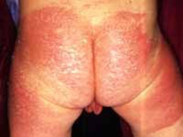
- 130What is impetigo contagiosum?Common BACTERIAL infection in infants, children
Increase incidence hot/humid climates
poor sanitary conditions, crowded living conditions, daycare facilitiesIP - 131WHAT IS THE CLINICAL MANIFESTATION OF IMPETIGO CONTAGIOSUM?KNOW:
YELLOW CRUST IS HALLMARK
usually on face, near nose & mouth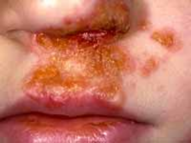
- 132What is ringwormAKA: mycoses
Caused by DERMATOPHYTES
Fungi that eat keratin
EPIDERMOPHYTON most common in kids
invade stratum corneum - 133WHAT IS THE CLINICAL MANIFESTATION OF TINEA CAPITIS?Varies by organism
Circular lesion, broken hairs, alopecia
Slight erythema with scaly inflammed borders - 134HOW DO YOU DIAGNOSE TINEA CAPITUS?KNOW:
POTASSIUM HYDROXIDE (KOH) EXAMINATIon
fungal culture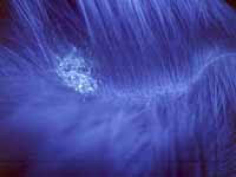
- 135WHAT IS TINEA CORPORIS?KNOW:
fungal infection of body, AKA ringwork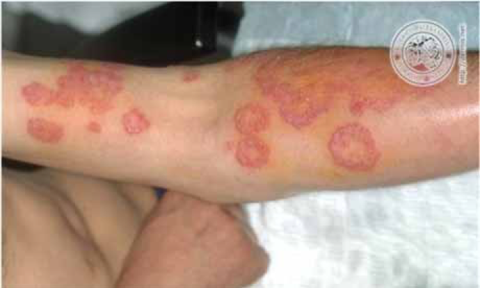
- 136WHAT ORGANISM CAUSES THRUSH?KNOW:
CANDIDA ALBICANS IN ORAL AND MUCUSOL MEMBRANES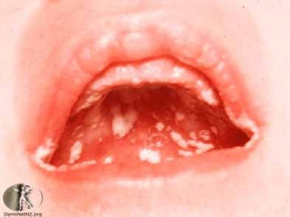
- 137What is the clinical presentation of of thrush?White plaques in mouth resulting in shallow ulcers
White dense covering of tongue - 138WHAT IS MOLLUSCUM CONTAGIOSUM?KNOW:
COMMON, HIGHLY CONTAGIOUS POX VIRUS INFECTION
school-aged kids, sexually active young adults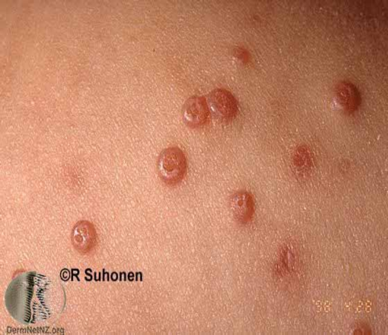
- 139What is the clinical presentation of Molluscum Contagiosum?Discrete, umbilicated, dome shaped papules anywhere on skin
- 140WHO CONTRACTS HERPES ZOSTER & chickenpox?KNOW:
HERPES ZOSTER (SHINGLES) OCCURS IN PARTIALLY IMMUNE PATIENTS WITH PREVIOUS EXPOSURE TO VZV (including exposure by vaccine)
Varicella (chickenpox) in VZV naive patients - 141What is clinical presentation of chickenpox/ varicella?1st itching/vesicles on trunk/scalp/face
Macules, papules, vesicles
Superficial vesicles easily rupture
Fever - 142WHAT CAUSES SCABIES?KNOW:
COMMON, CONTAGIOUS DZ CAUSED BY ITCH MITE SARCOPTES SCABIEI
colonize in human epidermis
transmission by direct contact & formites - 143What are the clinical manifestations of scabies?3-5 weeks post infestation
Female burrow tunnels into stratum corneum, deposit eggs,
Burrows, papules, vesicle lesions,
excoriations from scratching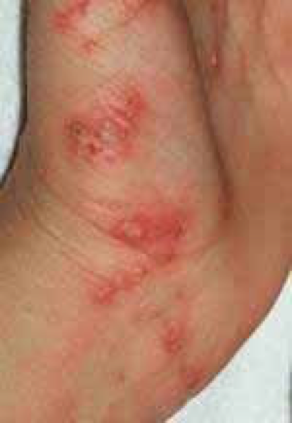
- 144Stevens-Johnson SyndromeNot in study guide or green
Rare, severe, immune drug reaction
Widespread epidermal apoptosis & detachment
Prodrome of malaise
Erythema/tenderness trunk, face, palms
Blister/bullae formation
Shedding of necrotic epidermis - 145—