
Mind map
Knee
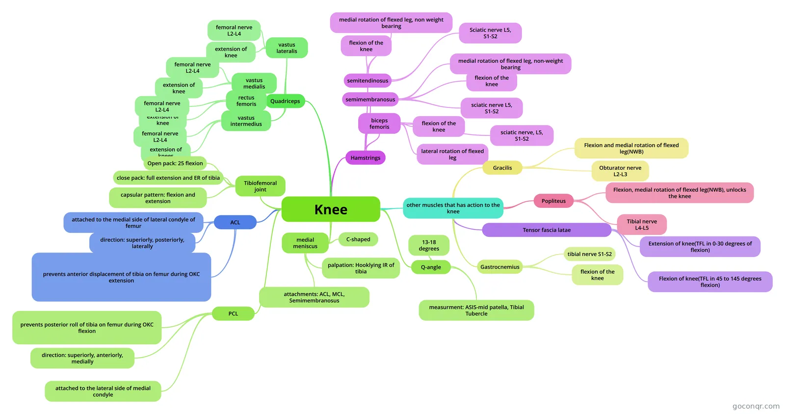
Mind map: knee anatomy
Map content
Knee
Quadriceps
- vastus lateralis
- extension of knee
- femoral nerve L2-L4
- vastus medialis
- extension of knee
- femoral nerve L2-L4
- rectus femoris
- extension of knee
- femoral nerve L2-L4
- vastus intermedius
- extension of knees
- femoral nerve L2-L4
- vastus lateralis
Hamstrings
- biceps femoris
- flexion of the knee
- lateral rotation of flexed leg
- sciatic nerve, L5, S1-S2
- semimembranosus
- flexion of the knee
- sciatic nerve L5, S1-S2
- medial rotation of flexed leg, non-weight bearing
- semitendinosus
- flexion of the knee
- Sciatic nerve L5, S1-S2
- medial rotation of flexed leg, non weight bearing
- biceps femoris
other muscles that has action to the knee
- Gracilis
- Flexion and medial rotation of flexed leg(NWB)
- Obturator nerve L2-L3
- Gastrocnemius
- flexion of the knee
- tibial nerve S1-S2
- Tensor fascia latae
- Flexion of knee(TFL in 45 to 145 degrees flexion)
- Extension of knee(TFL in 0-30 degrees of flexion)
- Popliteus
- Flexion, medial rotation of flexed leg(NWB), unlocks the knee
- Tibial nerve L4-L5
- Gracilis
Tibiofemoral joint
- Open pack: 25 flexion
- close pack: full extension and ER of tibia
- capsular pattern: flexion and extension
ACL
- attached to the medial side of lateral condyle of femur
- direction: superiorly, posteriorly, laterally
- prevents anterior displacement of tibia on femur during OKC extension
PCL
- attached to the lateral side of medial condyle
- direction: superiorly, anteriorly, medially
- prevents posterior roll of tibia on femur during OKC flexion
medial meniscus
- attachments: ACL, MCL, Semimembranosus
- C-shaped
- palpation: Hooklying IR of tibia
Q-angle
- 13-18 degrees
- measurment: ASIS-mid patella, Tibial Tubercle
Information
- Description
- knee Mind Map on Knee, created by otl_716 on 04/07/2014.
- Created by
- otl_716
- Views
- 24
- Rating