
Mind map
Shoulder
Map content
Shoulder
Bones
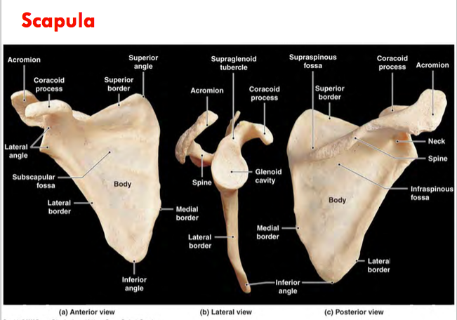
- Scapula
- Orientating based on Planes
- Sagittal
- Lateral
- Acromion
- Coracoid Process
- Lateral Border
- Glenoid Cavity/Fossa
- Glenoid Labrum
- Faces Anteriorly, Laterally and Superiorly
- Supraglenoid Tubercle
- Infraspinous Tubercle
- Medial
- Medial/Veterbrae Border
- Superior angle
- Lateral
- Coronal (Frontal)
- Anterior
- Subscapular Fossa
- Coracoid Process
- Posterior
- Infraspinous Fossa
- Supraspinous Fossa
- Scapular Spine
- Anterior
- Transverse (Horizontal)
- Superior
- Superior angle (medial)
- Supraspinous Fossa
- Inferior
- Inferior angle
- Infraspinous Fossa
- Superior
- Movement
- Retraction
- Protraction
- External (Lateral) Rotation
- Internal (medial) Rotation
- Elevation
- Depression
- Sagittal
- Classification
- Flat Bone
- Orientating based on Planes
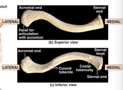
- Clavicle
- Movements
- Retraction
- Protraction
- Rotation
- Lateral Elevation
- Lateral Depression
- Curvatures
- The unusual shape of clavicle restricts the amount of medial (sternoclavicular) movement, to protect the axial skeleton and thus protect the organs
- Orientation based on Planes
- Sagittal
- Coronal (Frontal)
- Transverse (Horizonntal)
- Classification
- Long Bone
- Movements
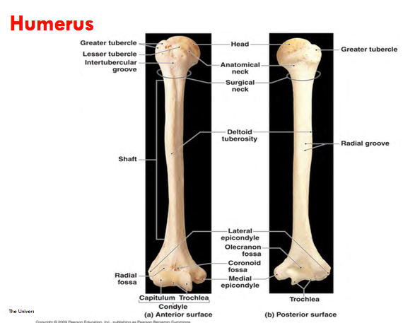
- Humerus
- Orientating based on Planes
- Coronal (Frontal)
- Anterior
- Lesser tubercle (Proximal)
- Radial Fossa (Distal)
- Coronoid fossa (Distal)
- Capitulum (Distal Projection)
- Trochlea
- Posterior
- Olecranon Fossa (Distal) - (connects to Olecranon part of Ulna)
- Anterior
- Transverse (Horizontal)
- Superior/Proximal
- Greater Tubercle
- Humerus Head
- Surgical Neck
- Anatomical Neck
- Inferior/Distal
- Medial Epicondyle
- Lateral Epicondyle
- Radial Fossa
- Mid-shaft
- Deltoid tuberosity
- Superior/Proximal
- Sagittal
- Medial
- Medial Epicondyle (Distal)
- Medial Supracondylar ridge
- Humerus Head (Proximal)
- Lateral
- Lateral Epicondyle (Distal)
- Greater Tubercle (Proximal)
- Lateral supracondylar ridge
- Medial
- Coronal (Frontal)
- Movements
- Abduction
- Adduction
- Flexion
- Extension
- Classification
- Long Bone
- Orientating based on Planes
- Scapula
Muscles
- Rotator Cuff
- Subscapularis
- Classifications
- Anterior/Coronal - Convergent
- Classifications
- Teres Minor
- Infraspinous
- Supraspinous
- Subscapularis
- Axio/Scapulo/Claviculo -Humeral
- Pectoralis Major
- Type
- Convergent
- Origin
- Insertion
- Actions
- Innervation
- Type
- Pectoralis Major
- Axiohumeral & Axio
- Rotator Cuff
Joints
- Glenohumeral Joint
- Joint Capsule
- = Greater Mobility
- Fibrous & loose capsule
- very thin & lax capsule
- Attaches to glenoid labrum
- Attaches around anatomical neck of humeru
- Reflects inferiorly onto medial shaft
- = Greater Mobility
- Ligaments
- Coracohumeral Ligament
- Attaches from Coracoid Process to Greater Tubercle
- Capsular (at connection to humerus) & Extracapsular
- Prevents Lateral movement of humerus and thus inferior dislocation
- Coracromional Ligament
- Prevents Superior dislocation of Humerus
- Extracapsular
- Transverse Humeral Ligament
- Goes in-between Greater and Lesser Tubercle of Humerus
- Capsular
- Keeps long head of bicep brachii in bicipital groove (intertubercular groove)
- Glenohumeral Ligament
- Capsular & Longitudinal
- Superior
- Prevents Lateral movement and thus stops inferior dislocation
- Limits external rotation
- Middle
- Limits external rotation
- 30% of people don't have
- Inferior
- Prevents anterior dislocation of Humerus when fully flexed or abducted
- Coracohumeral Ligament
- Classification
- Synovial, multiaxial, Ball and socket
- Articular Surfaces
- Covered in Hyaline Cartilage
- Humerus Head
- = Greater Mobility
- 1/2 Sphere area
- 3x size of glenoid fossa - large ball on small surface - allows joint to be multiaxial
- Only 25-30% contact between articular surfaces
- = Greater Mobility
- Glenoid Fossa
- Faces Anteriorly, Superiorly & Laterally
- Glenoid Lbarum
- Facilitates mobility without damaging any strutures
- Increases glenoid concavity
- Porvides attachment site for oint capsule, ligament & muscles
- Humerus Head
- Covered in Hyaline Cartilage
- Synovial Membrane
- Lines oint capsule and bony surfaces inside joint capsule which are not covered by articular cartilage
- Has no structure in-between articular surfaces
- Joint Capsule
- Acromioclavicular Joint
- Articular Surfaces
- Covered in fibro-cartilage
- Posteriolateral Acromial end of clavicle
- Anteromedial border of Acromion
- Covered in fibro-cartilage
- Joint Capsule
- Loose, Fibrous capsule attached to articular margins
- Reinforced by capsular ligaments (Superior & inferior Acromioclaviclular ligaments & trapzius)
- Partial Fibrocartilagenous intra-articular disc
- Partially divides the joint cavity - functions as a shock abdsorber & compensates for small degree of incongruity between the 2 joint surfaces
- Ligaments
- Acromioclavicular Ligament
- Capsular
- Prevents Dislocation of Clavicle at Acromial end
- Coracoclavicular Ligament
- Conoid part
- xtracapsular, Vertical & Posteromedial
- Produces posterior axial rotation of Clavicle
- Limits protraction of scapula
- Prevents Superior dislocation of Clavicle
- Trapezoidal part
- Extracapsular, oblique & anterolateral
- Limits retraction of scapula
- Prevents superior and lateral dislocation of clavicle
- Conoid part
- Acromioclavicular Ligament
- Articular Surfaces
- Sternoclavicular Joint
- Synovial membrane
- 2 synovial membranes (cavities) seperated by an intra-artiular fibrocartilagenous disc
- Intra-articular fibrocartilagenous disc
- When perforated it can allow communication between the synovial cavities
- Improves congruency of joint (fit together)
- Shock Absorber
- Prevents superior dislocation of Clavicle
- Acts as a pivot about which elevation and depression occur
- Attaches superiorly and posteriorly to upper border of sternal end of clavicle and inferiorly to 1st costal cartilage (1st rib)
- Articular Surfaces
- Sternal /Medial end of Clavicle
- Clavicular Notch of Superolateral angle of the sternum
- Adjacent upper medial surface of 1st Costal Cartilage (1st Rib)
- Joint Capsule
- Reinforced Anteriorly (ASC), Posteriorly (PSC) and Superiorly (Intraclavicular (IC)) by capsular ligaments
- Complete fibrous
- Strong
- needs to be in order to transfer weight and prevent dislocation
- Inferior part weak
- Ligaments
- Intraclavicular Ligament
- Capsular
- Prevents Lateral dislocation
- Connects Clavicles
- Anterior Sternoclavicular Ligament
- Capsular
- Prevents Anterior dislocation of Clavicle
- Reinforced by origin of sternomastoid (muscle)
- Posterior Sternoclavicular Ligament
- Capsular
- Prevents posterior dislocation of Clavicle
- Reinforced by Sternohyoid
- Costoclavicular Ligament
- Attaches from 1st Costal Catilage (1st Rib) to clavicle)
- Extracapsular & Bilaminar (2 layers - seperated by Bursae)
- Prevents superior dislocation of Calvicle
- Function
- Limits Retraction, Protraction & Elevation
- Provides stability at joint
- Intraclavicular Ligament
- Synovial membrane
- Scapulothoracic
- Articular Surfaces
- sUBSCAPULAR fOSSA
- tHORACIC cAGE
- FAKE JOINT - NO RELA CONNECTION BETWEEN ARTICULAR SURFACES
- Articular Surfaces
- Glenohumeral Joint
Images and files in the map
Information
- Description
- Anatomy Mind Map on Shoulder, created by Jack Henderson on 05/04/2018.
- Created by
- Jack Henderson
- Views
- 1
- History
-
- Created by Jack Henderson · Apr 5, 2018
- Copied by Jack Henderson · Apr 5, 2018
- Copied by Jack Henderson · Apr 5, 2018
- Rating