
Mind map
Retrovirus
Map content
Retrovirus
Replication
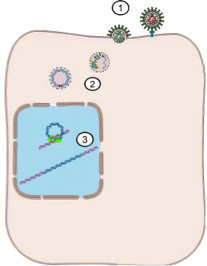
- 1. Virus attachment to plasma membrane
- Adhesion to cell surface by specific (protein) receptor
- Membrane fusion and release of nucleocapsid
- 2. Reverse transcriptase synthesises cDNA within the capsid
- → Initial reverse transcriptase – envelope removed, so ions and nucleases can get in
- Retrovirus nucleocapsid contains almost everything it needs to reverse transcribe its RNA
- Reverse transcription can take place in vitro, if virions are permeableised by mild detergents and the media contains a source of metal cations (Mg, Mn, Zn) and neucleotides
- DNA synthesis takes place inside the virus capsid, the virus does not induce interferon and is refractory to its effects.
- Retrovirus integrates into the host genome without de novo expression of antigen making it invisible to the immune system
- 3. Several copies of virus cDNA integrated into host genome at “random” loci
- → Can only insert where genome is unwound
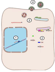
- 4. Synthesis of mRNA by host mRNA polymerase under control of strong virus promotor
- → Viral RNA – viral proteins made by ribosomes
- 5. Synthesis of new virus proteins
- 6. Synthesis of new genome by host RNA polymerase
- → +ve ss RNA – capped and poly-A tail (as made in mammalian cell), therefore looks different to all other viral and +ve ss RNA
- 7. Assembly and budding of new virus particles
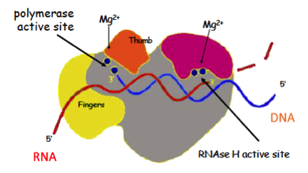
- Schematic of reverse transcriptase
- 50-100 copies per virus
- copies RNA into DNA (RNA used as a template)
- Can use RNA or DNA as a template, always producing DNA
- RNA/DNA runs through the centre of the template
- RNA is degraded at the RNAse H active site
- Produces ss DNA and bits of RNA attached
- Reverse transcriptase has both RNA dependent DNA polymerase and DNA dependent DNA polymerase activity.
- By a complex series of steps in makes a complete double stranded DNA copy of the virus genome
- Copies and degrades +ve sense RNA
- Restart at the other end and run through it again – end up with a complete copy
- Template switching copies -ve sense DNA to make duplex
- ds DNA produced, containing all genetic information in original RNA
- Classes of retroviruses
- Replication-competent retroviral genome
- Forms a Provirus ds DNA incorporated into the host genome
- Doesn’t produce new virus, just sits in host genome
- Strong viral Enhancer promoters
- (may promote a nearby c-onc)
- Cis-activating
- Forms a Provirus ds DNA incorporated into the host genome
- Replication-defective rapidly transforming
- (insertion of v-onc interfering with virus replication require helper virus)
- V-onc is therefore interfering with replication
- Double genome (means two pro-virus per cell)
- (insertion of v-onc interfering with virus replication require helper virus)
- Endogenous Retro Virus:
- A complete copy of a retroviral genome (pro-virus) without promotors transmitted in the germ line from parent to progeny present in every cell. (and can exist in every member of some species).
- Triangles indicate repeating sequences at either end
- These proviruses are under the control of cellular regulatory genes and are normally silent
- They usually do not contain v-onc (no ‘on’ gene).
- Not replication defective and can be induced by a variety of external stimuli (irradiation, mutagen, immune stimulus, retrovirus)
- Virus in genome that is silent can be activated by another virus particle with activation sequence
- Replication-competent rapidly transforming
- (Rous Sarcoma virus)
- V-onc has been inserted and will not affect replication
- Gene with v-onc causes tumour, therefore v-onc was the first oncogene to be found to cause tumours
- Combination of defective and competent
- Will replicate very well in a single host
- Will not pass on well to a new host
- (Rous Sarcoma virus)
- Replication-competent retroviral genome
- 1. Virus attachment to plasma membrane
Characteristics
- Genome
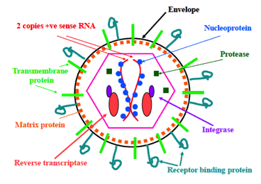
- 2 copies of +ve sense single stranded RNA
- Structure of retrovirus Genome (cf.mRNA)
- Only diploid virus
- (2 identical strands +ve sense RNA poly A tail and capped)
- Only +ve RNA not to act directly as an mRNA immediately after infection
- U3, R, U5 – strong promoters for translation process
- Only viral RNA to be synthesised and processed by host ENZYMES
- Only diploid virus
- Only viral RNA genome to be synthesised and processed by host ENZYMES
- GAG = group associated antigen
- Core protein in hexagonal area
- U3, U5 – untranslated protein
- Pol – reverse transcriptase, etc.
- Env – peplomers etc.
- Oncgenes
- Originally defined in retroviruses
- Viral genes that promote tumour development
- → ‘cancer causing genes’
- e.g. v-myc, v-mos, v-fos, v-src (>60)
- Written in italics when associated with a virus
- Myc = myelocytoma
- Fos = feline osteosytoma
- Many were found to have cellular equivalents
- e.g. c-myc, c-mos, c-fos
- Non-Italics - belong to host
- C = cellular
- V-Oncogens encode a range of proteins that affect growth promoting cell signalling pathways. (cell growth = virus growth)
- E.g. growth factor, translation factor
- Viral promotion of replication – if near cell growth genes, get a tumour
- v-onc differ from host genes
- They do not have introns
- Can be pushed out very quickly – made by reverse transcriptase of host RNA
- Are expressed out of their normal cellular context.
- Under the control of strong viral promotors.
- Have a high frequency of mutations. (generated during reverse transcription)
- The strong promotors of a retrovirus inserted close to a cellular oncogene may induce overexpression of the cellular oncogene (Cys-acting).
- They do not have introns
- Promoter acts on same strand of DNA, at lots of different points
- Structure
- Family: Retroviridae
- Genus Alpha retrovirus:
- Avian leukosis Viruses
- Rous sarcoma virus
- Genus Beta retrovirus:
- Pulmonary adenomatosis of sheep (Jaagsiekte)
- Several primate viruses
- Genus Gamma retrovirus:
- Feline leukemia virus
- Feline sarcoma virus
- Genus Delta retrovirus:
- Bovine Leukemia virus
- HTLV 1 & 2
- Simian T lymphotrophic virus
- Genus Lentivirus:
- HIV
- FIV
- BIV
- Maedi/Visna
- Caprine arthritis-encephalitis virus
- Equine infectious anaemia
- Genus Spumavirus:
- Bovine
- Feline
- Human Foamy virus (problem in cell culture)
- Genus Alpha retrovirus:
- Genome
Alpharetrovirus (avian)
- There are a large number of avian retroviruses which affect many species of birds including poultry.
- All chickens contain Endogenous Retro Viruses rarely expressed not pathogenic.
- Passed on in egg – present in fertilised ovum
- Avian leukosis virus : exogenous viruses, replication competent (no v-onc, cis acting slow transforming) they cause disease sporadically in birds >13 weeks
- Infect one to another
- One egg with retrovirus – will shed viral particle throughout life. Other chickens infected and virus gets oncogene – get tumour development
Feline leukaemia virus FeLV
- Infection occurs in high density cat populations via respiratory, grooming or bites
- Infection of adult cat
- Immune response controls virus infection.
- ± Latent provirus infection of lymphocytes
- Will ‘turn off’, go and sit in lymph node. Virus is in genome and it is in a ‘shut down’ cell
- Can’t get rid of virus totally by immune response, but can shut the disease down
- Infection of pregnant queen or new-born kittens
- 50% Immune response - control virus infection, latent infection of lymphocytes
- 50% No immune response – persistent virus infection, immuno-tolerant, productive infection of lymphocytes
- Will likely develop tumours
- Likely to trigger an oncogenic effect
- Source of infection for other cats
- Will likely develop tumours
- Infection at a late stage of pregnancy – still shedding at partuition
- More common in feral cat populations
- Cells infected with FeLV express Feline Oncovirus Cell Membrane Antigen (env gene product of ERV FeLVC)
- Immune competent cats
- Self-limiting infection associated with antibodies to viral antigens and FOCMA, remain non viraemic.
- Immune response to exogenous and endogenous infection
- Immune tolerant cats
- Persistent infection with viraemia, no antibodies to virus antigens or FOCMA.
- Infectious to other cats and develop disease.
- Immune competent cats
- FeLVA is a slow transforming (cis-acting) virus
- Wide range of diseases can be caused by this virus
- Depending on cell type/ oncogene involved
- Lymphosarcomas 30% of feline tumours; thymic, muticentric (T-cell), alimentary (B-cell)
- Scattered around – spleen, lymph nodes
- Myeloproliferative: erythromyelosis (red cell precursor), granulocytic leukemia (neutrophil precursor), myelofibrosis (fibrobla sts) lymphocytic leukaemia
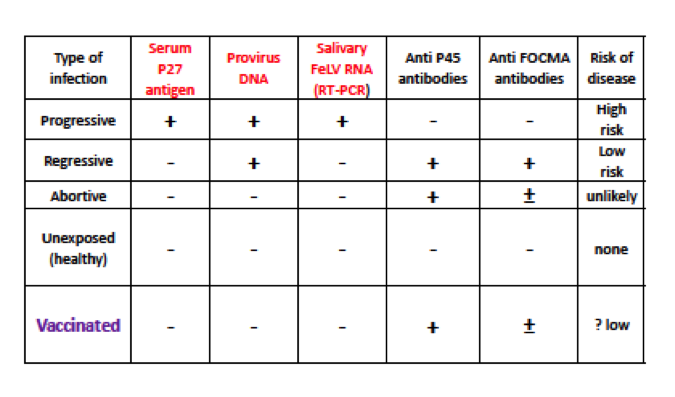
- Diagnosis of vraemic immuno-tolerant cats depends on detection of P27virus antigen in blood or serum
- In practice test kits are available online – lots of virus in circulation (+ve test)
- Abortive – can miss the virus particle in blood – but doesn’t mean it isn’t there
- Provirus DNA – virus RNA as well (virus going around in blood)
- Anti FOCMA antibodies – antibodies to an oncogenic antigen
- Prevention
- Testing and removal of infected animal from Catteries
- Vaccines
- Cell culture derivedgp70, FOCM Aantigen
- (Leucocell2/quantum cat FeLV)
- E. coli expressed Subunit vaccine (Leucogen)
- Recombinant canary pox virus
- Non core vaccine
- Not a high risk for the majority of cats.
- Often used to keep under control at breathing level
- Not a high risk for the majority of cats.
Equine infectious anaemia (swamp fever)
- Targets Monocyte macrophages.
- Frequently asymptomatic (avirulent strains)
- More virus than disease
- Virulent strains (acute viraemia, thrombocytopaenia and anaemia)
- Cyclical pathology (immune escape mutations)
- Partial recovery with time
- Notifiable in UK
- Antibodies in blood (Coggins test)
- Chinese attenuated vaccine
- Gives a +ve coggins test result
- Need to carry out serological tests to prove clearance of disease
- May be important for import/export certificate
PCR test for pro-viral DNA in lymphocytes
- Amplification of LTR region (avoids endogenous retrovirus false positive results)
- PCR able to detect 1 copy of viral DNA, but this requires an infected lymphocyte to be in the initial sample, so false –ve results do occur
- Promoters at either end are lost in endogenous so look for these in exogenous
- Not every lymphocyte is infected – lung cells are initial target
Lentivirus infections
- Feline Immunodeficiency Virus
- 3-6% of healthy cats positive for antibodies 30% of sick cats
- Disease burden associated with virus (indirect association with other pathologies)
- A widespread and common virus of cats, which was first identified in 1987.
- Related to HIV and similar in pathogenesis
- Takes a long time for infection to develop
- High incidence of FIV and FeLV tend to go together
- 3-6% of healthy cats positive for antibodies 30% of sick cats
- Lifelong infection: virus is present in blood and saliva
- Cats become infected usually by fighting, but can be trans-placental or at parturition (viraemic mother)
- Env binds to receptors CD134 and CXCR4
- Not many cells have CD134
- CXCR4 is a chemokine receptor (lots of cells have it) – T helper cells
- Virus infects activated feline CD4+ T-cells
- Acute phase I (2 months) → viraema, infection spreads to more CD4+ cells.
- Fever, malaise and lymphadenopathy
- Initial immune response, antibody production, and cytotoxic CD8 T-cells which reduce the number of infected CD4+ cells with apparent clinical recovery.
- Phase II. may last several years
- Mutations of the virus allow it to escape from CD8+ T-cell control
- CD4+ T-cells containing provirus cannot be eliminated
- Gradual decline of CD4+ T-cells follow recurrent cycles of expansion, immune elimination, mutation and escape of virus infected cells.
- Central role of CD4+ T-helper cells in immune responses and progressive drop in CD4+
- Reduced IL2 production
- Major CD4+ growth factor
- Reduced antibody production,
- Reduced blastogenesis
- Reduced Cell Mediated Immunity
- Phase III: onset of symptoms due to immunosuppression associated with opportunistic infections
- High frequency of fevers, gingivitis and mouth ulcers.
- Common presenting signs
- Reduced IL2 production
- Strong association with FeLV infection, and B-cell lymphomas
- Unsure of which causes the other, as occur at the same time
- Neurological symptoms can also occur in rare cases were infection of astrocytes and microglial cells predominates
- Diagnosis
- Depends of the detection of antibodies to virus
- Treatment
- Azidothymidine, which inhibits reverse transcriptase is partially effective.
- Several experimental vaccines but none effective virus persists in the presence of antibody and cytotoxic response.
- Feline Immunodeficiency Virus
Maedi/Visna in sheep
- (Similar pathogenesis to FIV)
- Maedi, infection of Monocytes and monocyte precursors, new virus is produced when monocytes become activated
- Lifelong infection with late onset of disease symptoms, chronic inflammation/fibrosis induced by infected monocytes
- Symptoms of breathlessness, consolidation of lungs and enlarged bronchial lymph nodes, fibrosis and hyperplasia of alveolar septae with monocyte infiltration
- Visna chronic demyelinating encephalitis with monocyte in filtration caused by a distinct subtype of virus. (Rare)
- Virus persists in the presence of antibody and cellular immune response possibly due to constant mutation and generation of escape variants.
- Transmission from mother to lambs in colostrum milk.
- Transmission of virus by aerosol from symptomatic sheep or from blood by biting insects.
- Control by eradication
- Removal of symptomatic sheep
- Removal of seropositive sheep
- Removal of PCR +ve sheep
- This hasn’t been engaged with with too much enthusiasm – would need to remove a lot of sheep to get the problem under control
Caprine Arthritis/encephalitis
- Similar virus to maedi/visna, but lesions occur as a proliferative synovitis with lymphocyte plasma cell and monocyte infiltration.
- Spread in colostrum and milk. (kids develop neurological symptoms)
- Several small ruminant lentivirus have been identified and sub-typed according to serology and host range.
- Some are found mainly in sheep (Maedi/visna) others are mainly goat (Caprine arthritis)
- Some occur equally in sheep and goats.
- They vary in their pathogenicity.
Ovine pulmonary adenomatosis (Jaagsiekte)
- Worldwide disease of sheep (except Australasia)
- Both sheep and goats contain several (27) related endogenous retroviruses (JSRV) in their genome
- Develop tumours in the lungs
- Neoplastic transformation of type II alveolar cells and non-cilliated bronchial epithelial cells
- Nodular adenomas/adenocarcinomas secrete large amounts of surfactant containing fluid.
- Metastasis to L.N.
- The disease is spread by the respiratory route, disease incidence can be as high as 20% following introduction of virus.
- But more commonly 1-5% of sheep in endemic flocks become affected.
- Clinical signs include breathlessness, lung sounds, large amount of pulmonary fluid only seen in some cases
- Tip sheep up on front legs, and fluid will pour out of their nose
- The JSRV genome does not contain a conventional oncogene.
- JSRV env protein binds to Hyaluronidase 2 (Hyal2)
- A hyaluronidase enzyme on the surface of many types of cell
- Including type 2 pneumocytes.
- JSRV env protein has transforming properties when transfected into a wide range of cells through binding to Phosphatidylinositol 3-kinases (a cell signalling pathway).
- Only exogenous protein affects the signalling pathway
- enJSRV env protein does not bind PI3_Kinase and lacks transforming properties.
- But enJSRV Env is expressed in the placenta and foetus where it has an important function in the development of the multinucleated trophoblast cells essential for implantation.
- Nearly identical, but without env gene
- Cells normally stop growing when they touch each other (e.g. skin), these will keep going
- Affected sheep have no antibody response to JSRV env protein as the expression of endogenous retrovirus causes the sheep to be immuno-tolerant to the jaagseike virus.
- Not all infected sheep develop lesions
- In infected flocks many more sheep have JSRV in peripheral blood lymphocytes than have lung lesions (despite lack of antibody response).
- Endogenous proteins block replication of exogenous
- Initial infection of a cells by JSRV is much more efficient in dividing cells.
- Type 2 pneumocytes are actively dividing in lambs in which JSRV is more likely to establish lung infection leading to the development of lung lesions.
- In Older sheep intercurrent disease with lung damage leads to pneumocyte division as part of the healing process which increases the risk of tumour development
- Co-expression of mutant GAG proteins from some endogenous retroviruses disrupts assembly and exit of mature wild type virus; hence act to limit JSRV spread within the lung and prevent development of tumours.
- Susceptible when healing
- ↑ risk of replication, develop tumour
- Virus is a trigger/amplifier of effects – usually another factor that triggers formation of a tumour
- JSRV env protein binds to Hyaluronidase 2 (Hyal2)
Images and files in the map









Information
- Description
- BVSc3 AD2 (Viruses) Mind Map on Retrovirus, created by Jess Pope on 21/12/2016.
- Created by
- Jess Pope
- Views
- 0
- History
-
- Created by Jess Pope · Dec 21, 2016
- Copied by Jess Pope · Dec 21, 2016
- Copied by Jess Pope · Dec 21, 2016
- Copied by Jess Pope · Dec 21, 2016
- Copied by Jess Pope · Dec 21, 2016
- Rating