
Notes
CNS physiology - Vision
Content of the notes (6)
Page 1
Eye anatomy
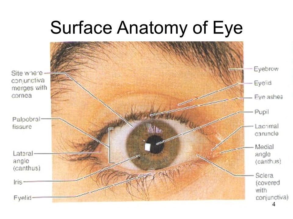
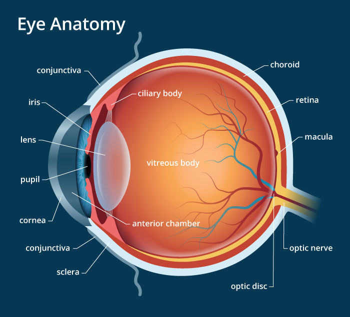
Iris
Pigmented smooth muscle
More pigment = darker eye colour (Brown, black)
Less pigment = lighter eye colour (Blue, green)
Blue = least pigmented
What is gluacoma?
Build up of excessive aqueous humour
Fluid becomes trapped in anterior eye compartment - drainage system blocked (can be due to Iris/Corneal junction being too narrow)
Use anticholinesterase drugs to treat (link pharma lecture Cholinergic drugs pg. 3 section iv)
Vitreous humour
Nutrients to retina
Structural support - maintains shape of eyeball
Choroid
Melanin-containing region
Absorbs light
Absent in albinism (lack of melanin) - causes poor eyesight due to light 'ping-ponging' around retina but not actually being absorbed (See vision anatomy)
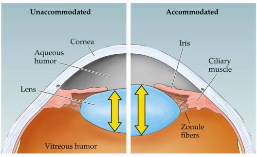
i) Lens accommodation
Focuses light onto retina
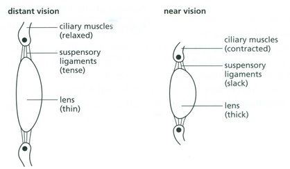
Unaccommodated:
Cillary muscles contracted
Zonule fibres relaxed
Lens becomes thick & round
Allows us to see things close-by
Accommodated:
Cillary muscles relaxed
Zonule fibres contracted
Lens becomes thin & flat
Allows us to see things far away
Light is focused by cornea & lens onto fovea in retina
Remember image projecting onto retina is inverted (Link vision anatomy lecture)
Emmetropia = 20:20 vision
What is the fovea?
Centre of the macula
Point where cone density is highest -approx 30'000 (only cones found in fovea!)
Acute vision (sharpest & most detailed images) arises from here
Macular degeneration can lead to damage to fovea
Link to vision anatomy lecture
Disorders of lens accommodation
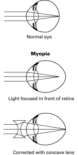
Myopia:
Short-sightedness
Light rays converge before reach retina
Results in fuzzy image when viewing long distance
Treated using concave lenses
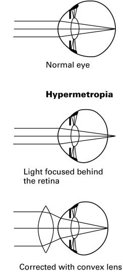
Hypermetropia:
Long-sightedness
Light rays converge 'after' retina
Results in fuzzy image when viewing close up
Treated using convex lenses
Presyopia:
Long-sightedness that occurs as we age
Page 2
Rods & cones
i) GF of rods & cones
Rods
Responsible for night vision
Very light sensitive (low threshold)
Contain high levels of photopigment
Low accuracy - more focused on general details, not specifics
High signal amplification - which converges & summates at bipolar cells in retina
Slow adapting
Cones
Responsible for day vision
Light insensitive (high threshold)
High accuracy - focused on fine details
Concentrated in fovea
Low signal amplification - little convergence
Fast adapting
3 types of cones:
Red (H)
Green (M)
Blue (L)
Light stimulation causes rods & cones to hyperpolarise!
ii) Rhodopsin
Pigment contained in retina
Opsin + retinal (a.k.a. retinaldehyde)
How does rhodopsin work?
11-cis retinal (unactivated rhodopsin) is floating about when no light is striking retina
When light hits, 11-cis retinal is converted into trans retinal (any kind) - it is now activated
This causes opsin & retinal to split
Metarhodopsin II (activated rhodopsin) is formed!
The metarho excites electrical change in photoreceptors (in our case, rods & cones)
Hyperpolarisation in Rs & Cs occurs & a signal is transduced!
Rods & cones contain diff isoforms of opsin (but both contain rhodopsin!)
Rods = Scotopsins
Cones = Photopsins

iii) Dark current vs light current
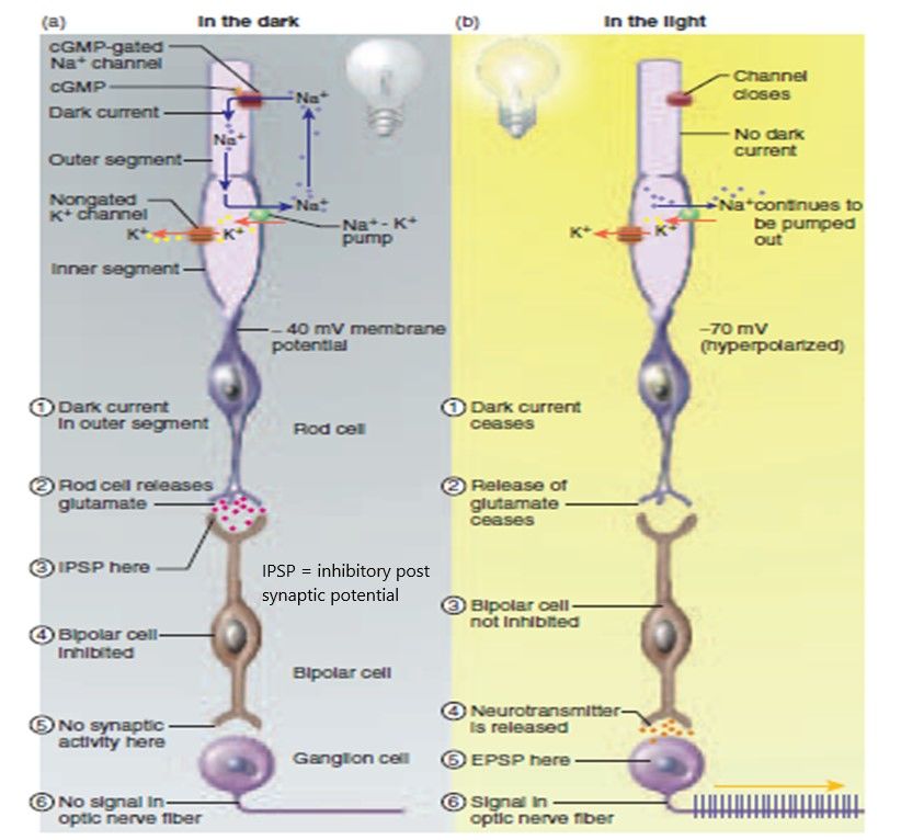
In the dark...
Sodium ion influx into outer rod segment
cGMP splints sodium channels open
K+ efflux from inner rod segment
Creates MP of -40mV
In the light...
Sodium ion influx stopped
cGMP levels drop - due to metarhodopsin II activating cGMP phosphodiesterase
K+ efflux remains
Creates hyperpolarisation
iv) Glutamate release
Glutamate = neurotransmitter used by rods & cones
Released in the dark!
Not released in the light!
Glutamate receptors located on bipolar cells
Page 3
Bipolar & ganglion cells
i) Bipolar cells
2 types of glutamate receptors on bipolar cells - Ionotropic & Metabotropic
Ionotropic
AMPA & kainate
Glutamate binding activates ligand gated sodium channels
Bipolar cell is depolarised
Signal is sent to off-centre ganglion cells
Metabotropic
mGluR6
Glutamate binding leads to closure of cGMP-gated sodium channels
Bipolar cell is hyperpolarised
Signal is sent to on-centre ganglion cells
Equal & opposite responses to the light signal
ii) On vs off centre
Contrast of visual info
Allows us to detect stimuli against a background
How does it work?
Half of the cells check for stimuli brighter than the background
Other half check for stimuli darker than the background
http://www.sumanasinc.com/webcontent/animations/content/receptivefields.html - video explaining on-centre off-centre vision
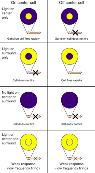
iii) Ganglion cells & parallel pathways
Bipolar cells synapse with ionotropic receptors on ganglion cells (AMPA, Kainate & NMDA)
Glutamate = neurotransmitter used
Depolarisation in ganglion cells & AP generated
3 types of ganglion cell:
P cells
M cells
K ganglion cells
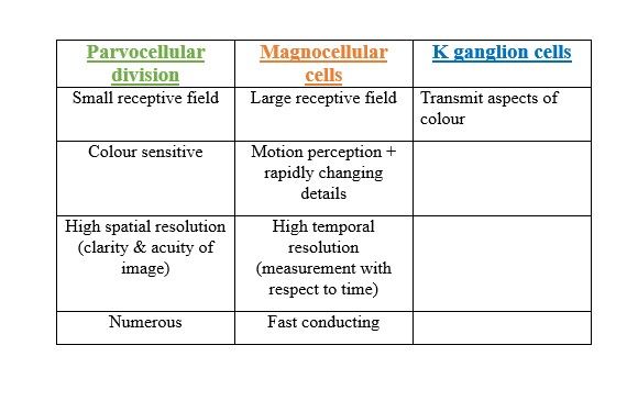
Page 4
Visual pathway beyond the retina
i) Image transmission from retina to cortex
Optic chiasm
Fibres from nasal half each eye decussate
Allows depth perception
(See Visual anatomy lecture)
Pretectum
Blinking response
Superior colliculus
Head & eye movements
Suprachiasmatic NHypo
Circadian rythms (Melanin secretion, sleepiness due to darkness falling)
Lateral geniculate nucleus
90% ganglion fibres head here
Cerebral cortex
Perception of vision
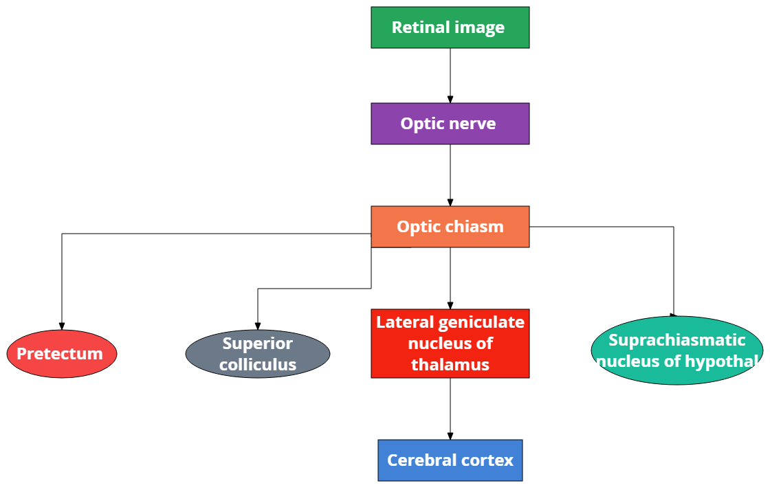
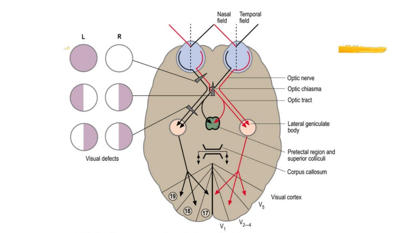
Page 5
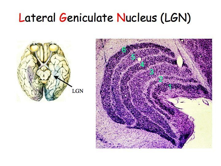
Lateral geniculate nucleus
90% of retinal axons synapse here
50% foveal representation
Sends signal on to cerebral cortex
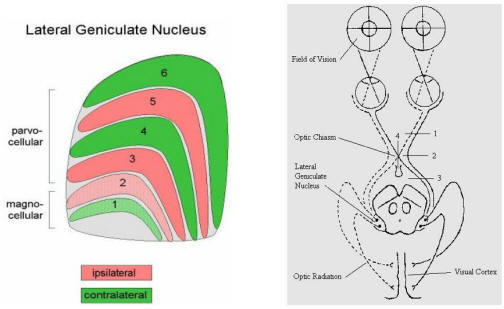
Composed of 6 cell layers of cell bodies separated by intralaminar layers:
Layers 1+2 = Magnocellular layer - M cells input
Layers 3+6 = Parvocellular layer - P cells input
Layers 1,4+6 = Contralateral nasal hemiretina
Layers 2,3+5 = Ipsilateral temporal hemiretina
Page 6
Primary visual cortex
Primary visual cortex comprised 6 layers of cells
LGN inputs enter in Layer 4 (Internal granular layer) + layers 2 & 3
Comprised of 4 sublayers:
4A
4B
4C alpha
4C beta
Axons M cells terminate 4C aplha - Motion information
Axons P cells terminate AC beta - Detail information
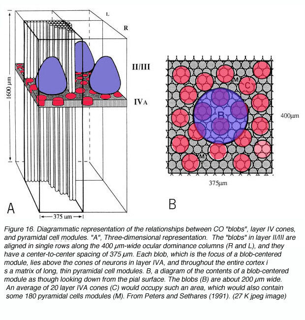
Axons intralaminar region LGN terminate layers 2 & 3 (Blobs) - Colour information
What are blobs?
Structures within visual cortex that 'put the colour on the picture'
Resemble K ganglion cells (they are specifically targeted by K-ganglion cells!)
Used in colour perception, colour discrimination + the learning & memory of the colour of objects
How do they work?
Each pink circle below represents a 'pixel' coming from the retina
The blob combines these 'pixels' together to create an image - to which it adds colour
i) Visual cortical pathways
These pathways form the basis of 'learning from & reacting emotionally to what we see'
Magnocellular pathway - Motion & change
V1 - V2 -V3 -MT
Dorsal pathway
Connects with posterior parietal cortex
V3 = orientation
MT = object movement
What does Magnocellular pathway transmit?
Processes P-channel info about object location & M-channel info about object movement
Gives us our sense of:
Spatial orientation
Binocular fusion/depth perception
Location, movement, direction & velocity of objects in space
Concerned with the 'WHERE' of the stimulus!!
Parvocellular pathway - Form & detail
V1 - V2 - V4
Ventral pathway
Connects with inferior temporal cortex
V4 = colour & form
What does Parvocellular pathway transmit?
P-channel info about object colour & form
Allows us to:
Recognise objects & colours
Read text
Learn & remember visual objects (words & their meanings)
Concerned with the 'WHAT' of the stimulus!!
Information
- Description
- Central Nervous System (CNS Physiology) Note on CNS physiology - Vision , created by Niamh McLoughlin on 05/11/2017.
- Created by
- Niamh McLoughlin · Nov 2017
- Updated
- Nov 9, 2017
- Views
- 16
- Rating