
Flashcards
Histo
Cards in this deck (40)
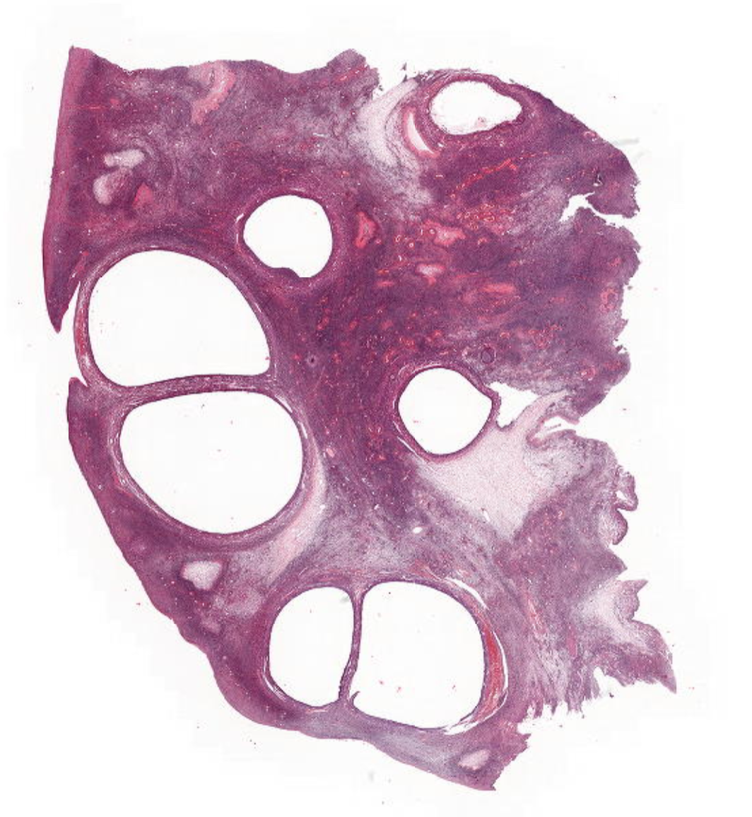
- 1—PCOS
1) primordial follicles
2)follicles and not cysts
3)crisscross stroma - 2—Serous Cystadeoma (Ovary)
1)corpus albicans
2)2 cysts needed for diagnosis
3)flattened ciliated cuboidal epithelium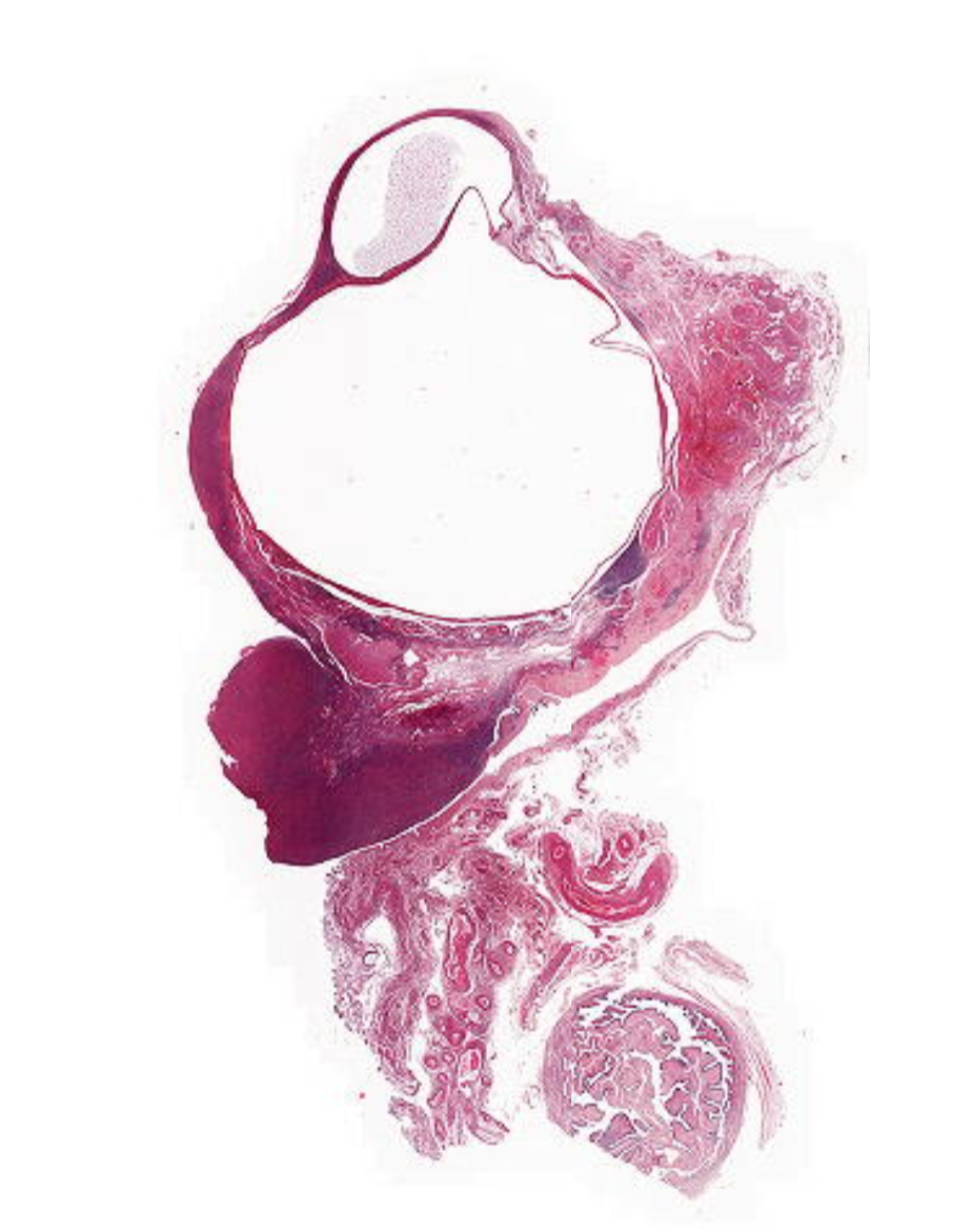
- 3—mucinous cystadenoma (ovary)
1)mucinous
2)fallopian tube remnants
3)multilocular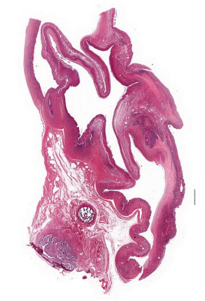
- 4—Serous Cystadenoma with Atypia
(serous borderline tumor)
1)no invasion
2)papillary/arborising
3)hobnailing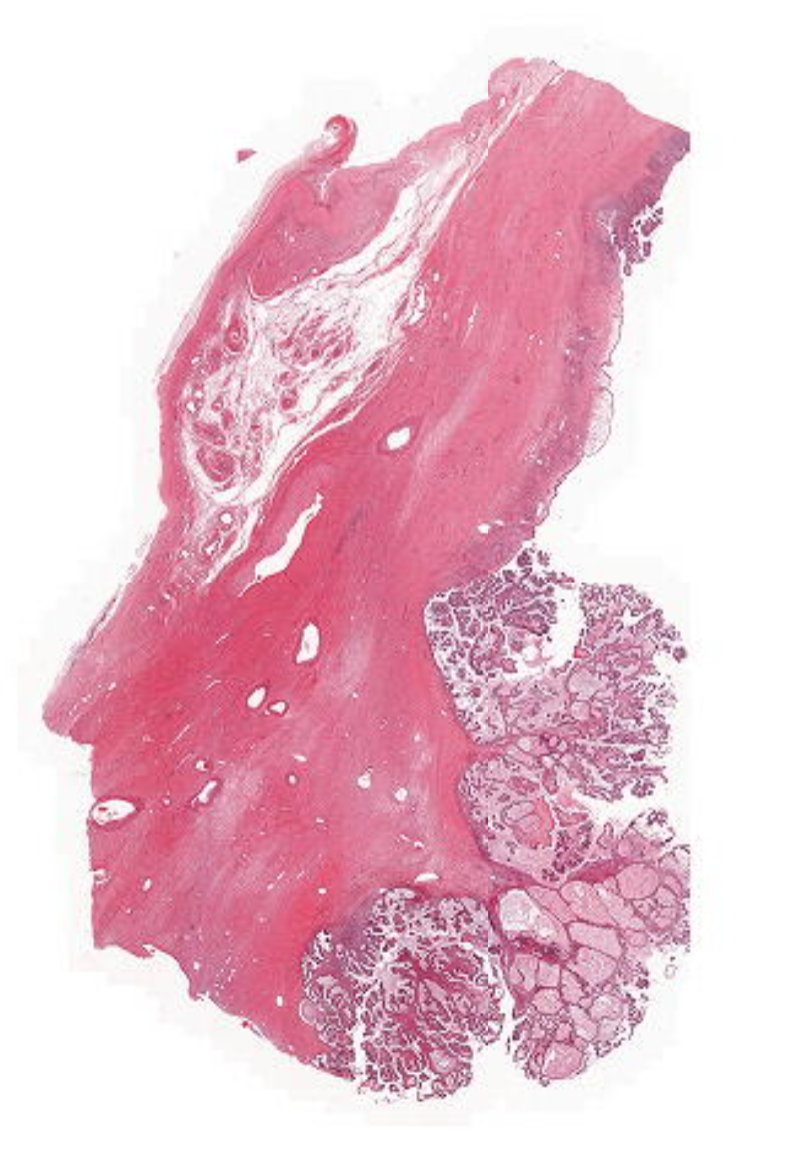
- 5—Serous High Grade Adenocarcinoma
(ovary)
1)papillary
2)hobnailing
3)pseudostratified epithelium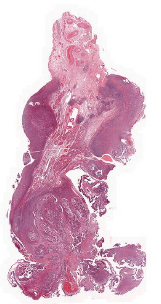
- 6—Brenner Tumour
(ovary) FUNEC (mnemonic)
3 main parts
1)extensive fibrosis
2)neoplastic endothelium
3)calcifications (blue)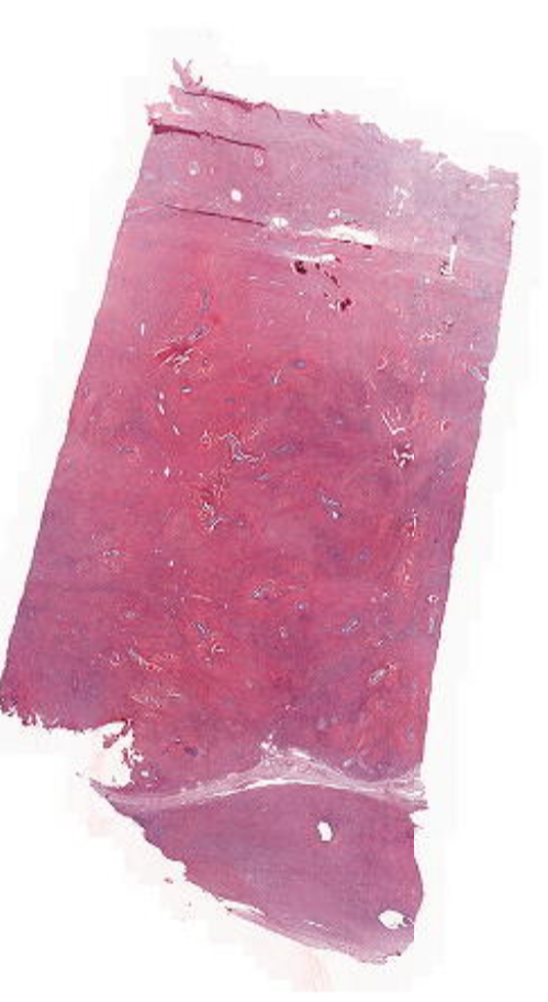
- 7—Mature Teratoma (ovary)
1)hair
2)nerve
3)eye (retina with pigment) = black
4) stomach/gastro goblet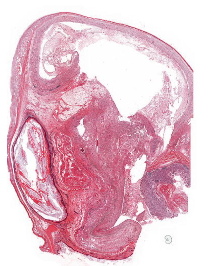
- 8—Immature Teratoma (ovary)
1)neural
2) cartilage
3) tooth
4) intestinal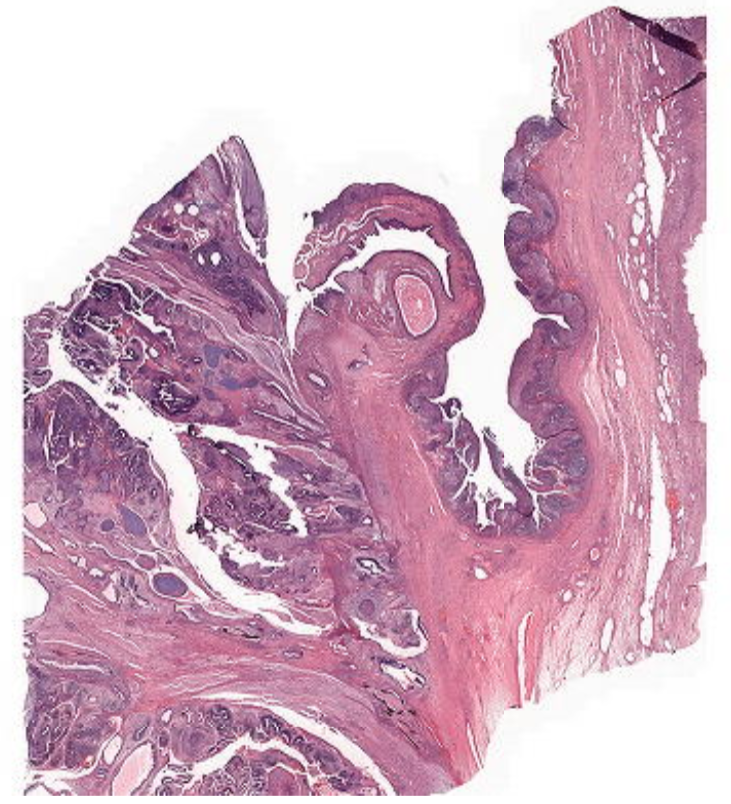
- 9—Dysgerminoma (ovary)
1)equivalent to a seminoma in men
2)tumour cells
3) fibrosis around vessels
4) lymphocytes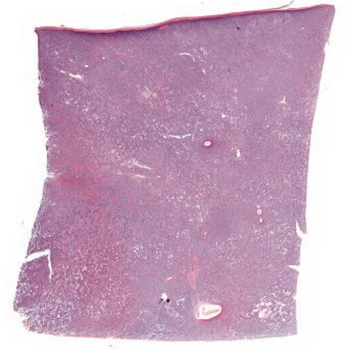
- 10—Granulosa Cell Tumour (ovary)
1) corpus albicans
2)call exner bodies = pseudorosettes
3)coffee bean nuclei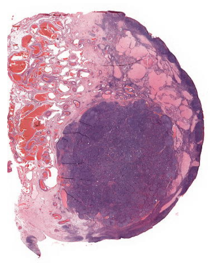
- 11—Ovary with Corpus Luteum Cyst
1)large cyst with fibrin blood and proteinaceous material
2)primordial follicles
3) granulosa layer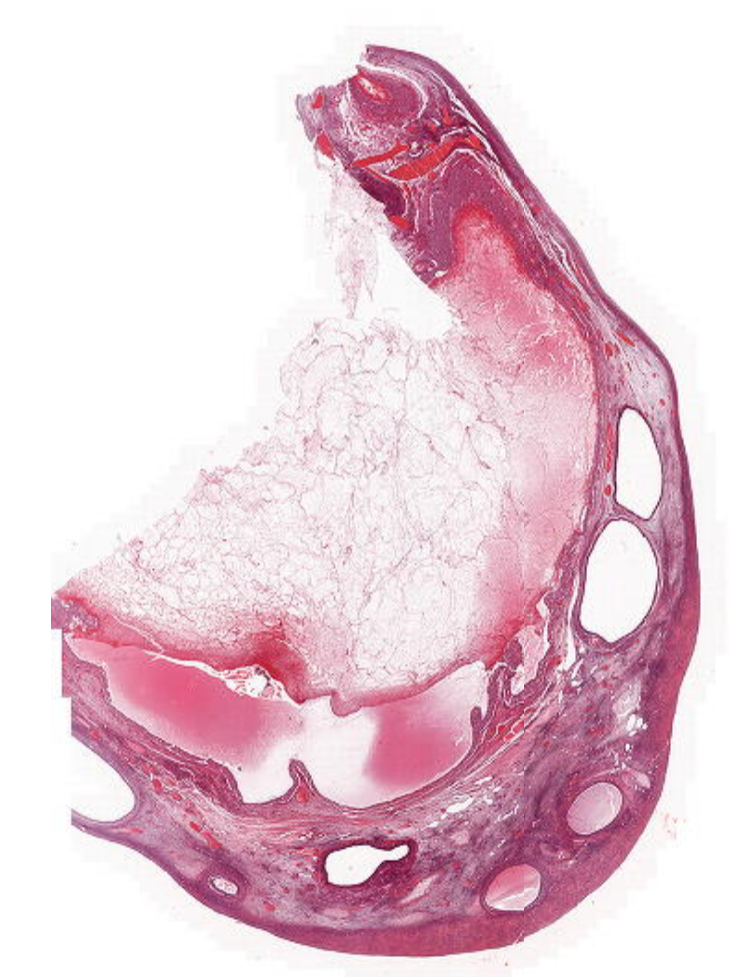
- 12—ovary with mucinous cystadenoma with atypia
1)cribriform growth
2)mucin = protein
3)talk about regular cancer markers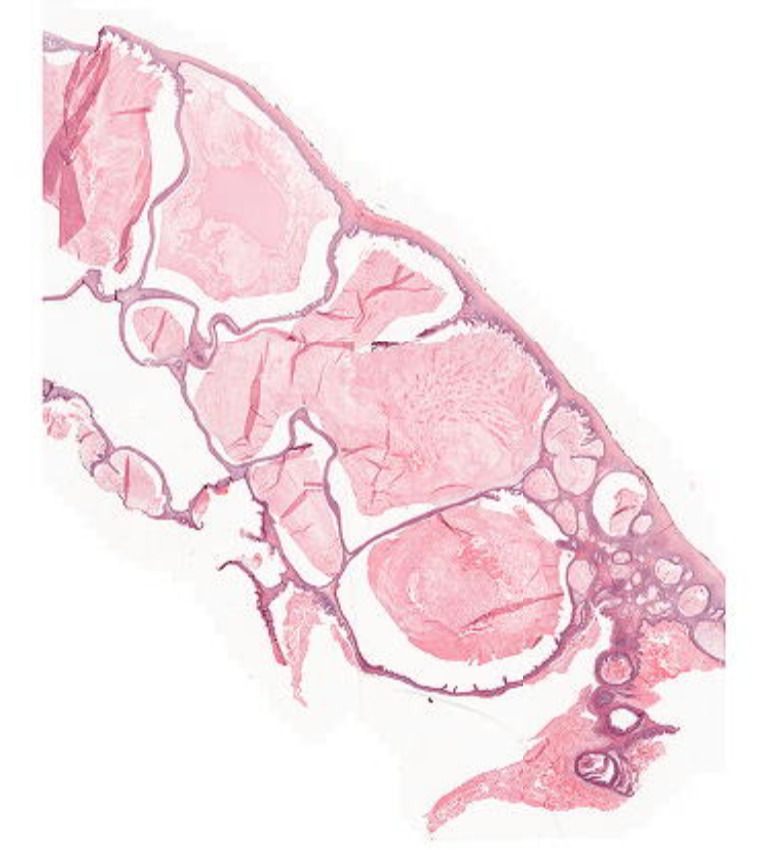
- 13—Ovary with Mucinous Adenocarcinoma Well Differentiated
1)focal stromal invasion
2)cribriform growth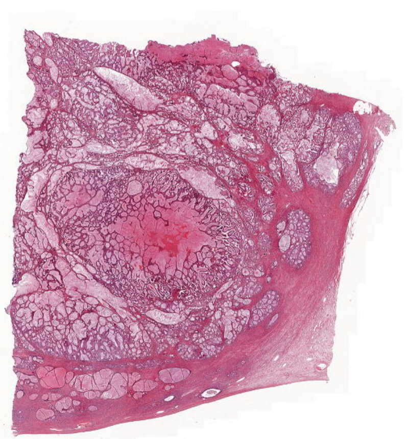
- 14—Ovary with Leydig Cell Tumour
1)corpus albicans
2)alternating criss/cross stroma
3)reinke crystals (can't see)
4)bi/tri nucleated cells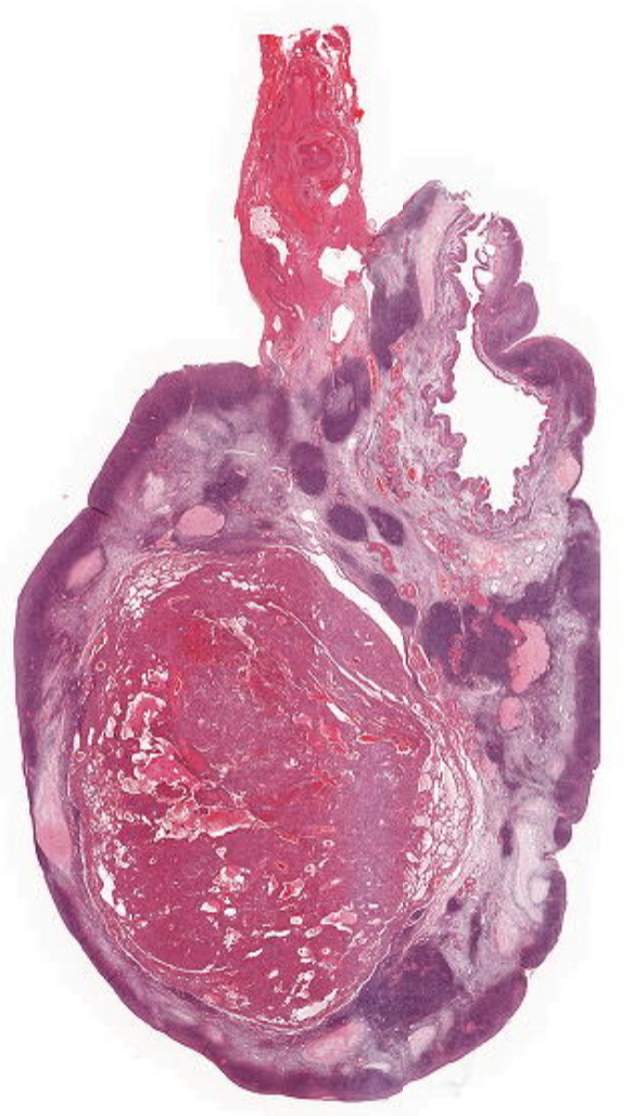
- 15—Tubal Pregnancy
1)Immature neural tissue (neural ectodermal tissue)
2)cyto/syncitiotrophoblasts
3)placental tissues
4)fallopian tube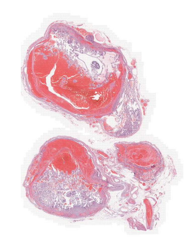
- 16—Molar Pregnancy
1)complete mole = no normal villi
2)syncitiotrophoblasts
3)BLOOD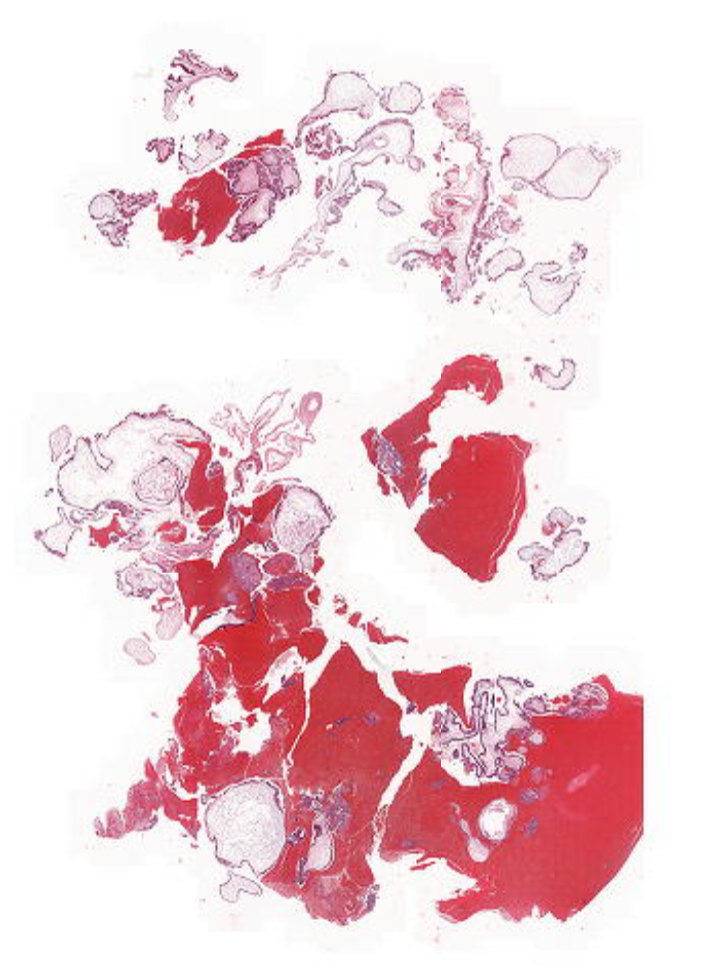
- 17—choriocarcinoma of the uterus
1)haemorrhage
2)necrosis
3)MITOSIS
4)talk about hall marks of cancer
5) trophoblasts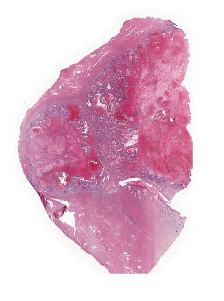
- 18—Immature Placenta with Villitis due to Toxoplasma
1)Immature placental tissue with increased trophoblastic proliferation is seen. Many villi show fibrosis. Small necroses with neutrophilic infiltration can be seen in scattered villi. Parasites are difficult to find, but are present as small cysts with basophilic (blue) dots.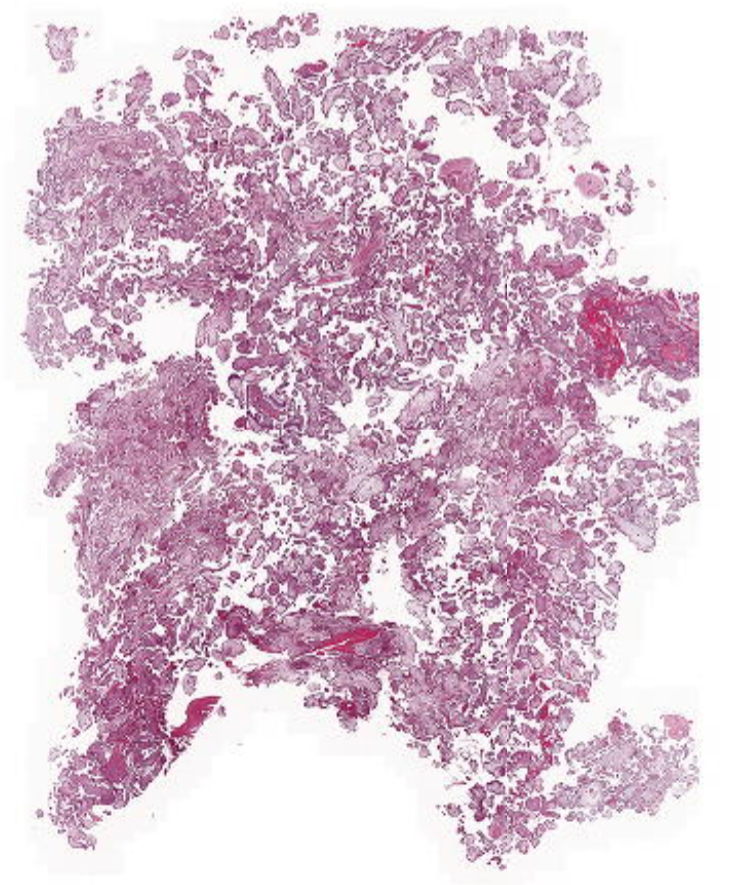
- 19—Placenta with Infarction
1)individual cell ghosts
2)necrosis
3)intervillous nuclei
4) top is foetal side with necrosis
5) debris
6) bottom is fibrotic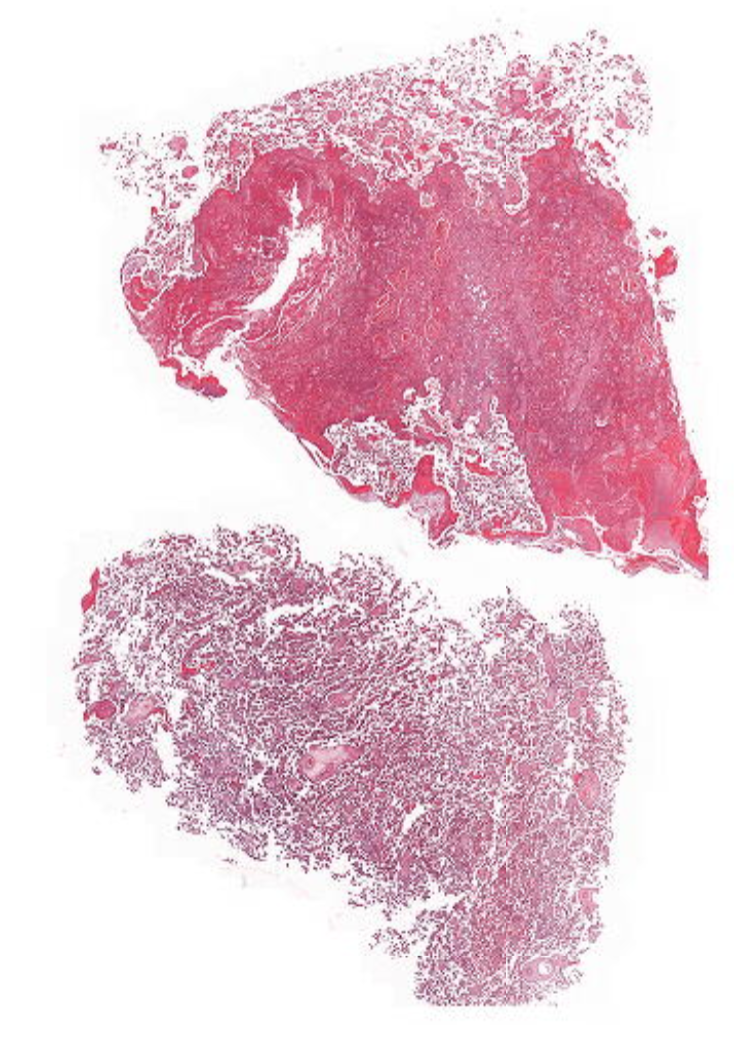
- 20—Testicular Atrophy
1)Tubules with concentric fibrosis have some degree of viability; surrounded by Sertoli cells
2) Seminiferous tubules of reduced caliber and with reduced number of germ cells
3)fibrosis around tubules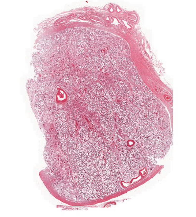
- 21—Testis with non-seminoma,
differentiated as a yolk sac tumour
1)NECROSIS and atrophy
2)Schiller-Duval bodies are tumour cells surrounding blood vessels
3)Intratubular germ cell neoplasia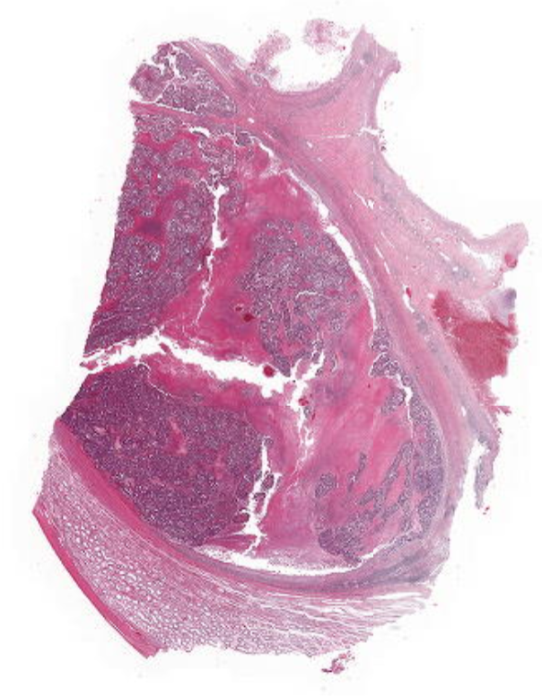
- 22—Testis with Seminoma
1)Intratubular germ cell neoplasia
2)adjacent seminiferous atrophy (looks like an elongated collapsed gland)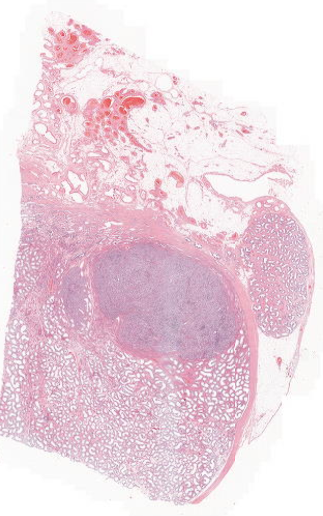
- 23—Neuroblastoma in Adrenal Gland
1)Tumor nuclei are slightly pleomorphic; small blue round cells
2)rosettes
3)eosinophilic matrix of neuropil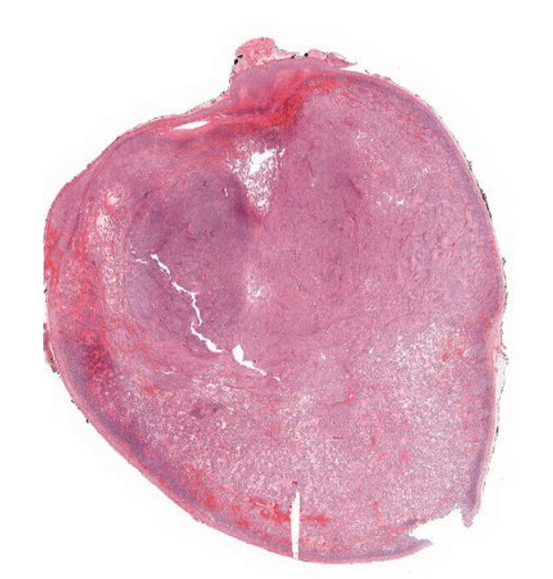
- 24—Embryonal Rhabdomyosarcoma
(in the nose)
Pieces of polypoid (botryoid) tissue covered by squamous epithelium
o Lots of haemorrhage
o Ulcerations in squamous epithelium
Tumor appears paucicellular
o Hypocellular myxoid areas and several vessels
o Increased cellularity around vessels and along the surface (cambium layer)
o Spindle cells with eosinophilic extensions
o Cross striations found if more mature tumour
An inflammatory reaction with granulocytes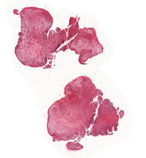
- 25—Wilm's Tumour
(Nephroblastoma)
Sheets of epithelial cells with distinct cytoplasm and gland-like structures (pseudo-rosettes)
o Areas of blastematous differentiation with cells with a high nuclear/cytoplasmic ratio
o Areas of mesenchymal differentiation with strands of fibroblast like cells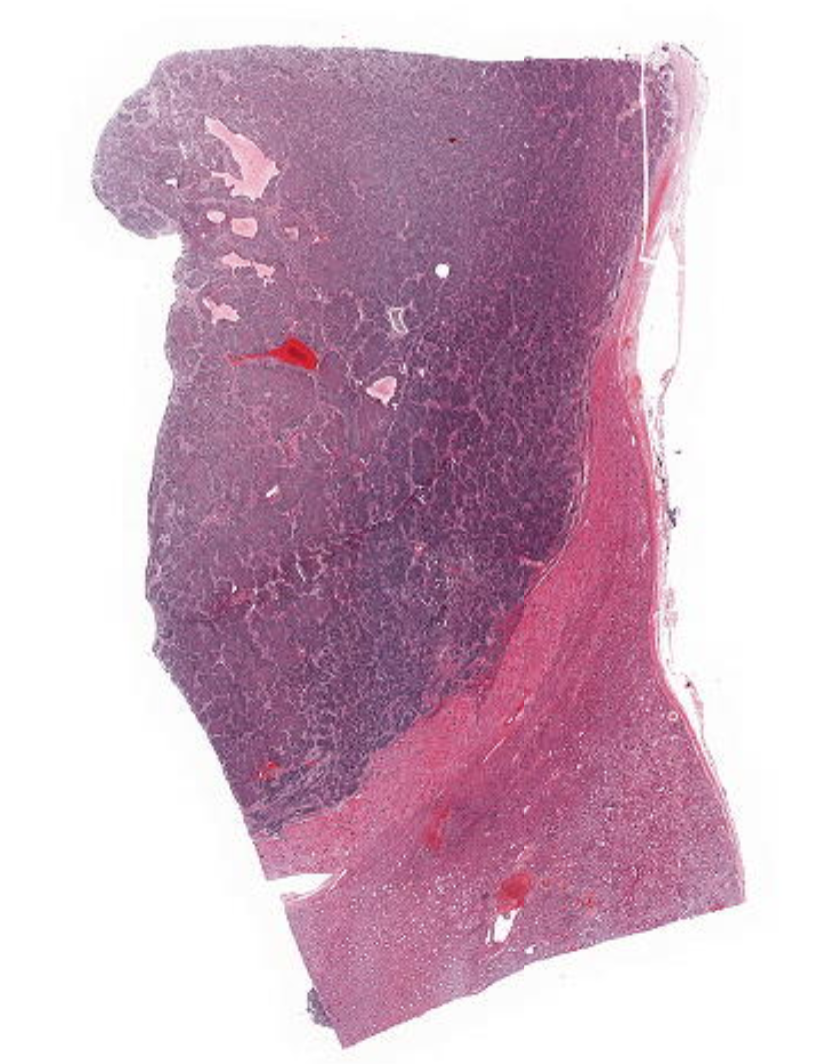
- 26—Immature Lung RDS
Only few of the alveoli contain air, which appears as dilated, cystic areas
o Alveoli contain eosinophilic fibrin, which is compressed like "hyaline membranes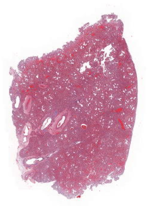
- 27—Cystic Renal Dysplasia
1) multiple cysts with flattened epithelium and fibrosis
2)Normal tubules and glomeruli are seen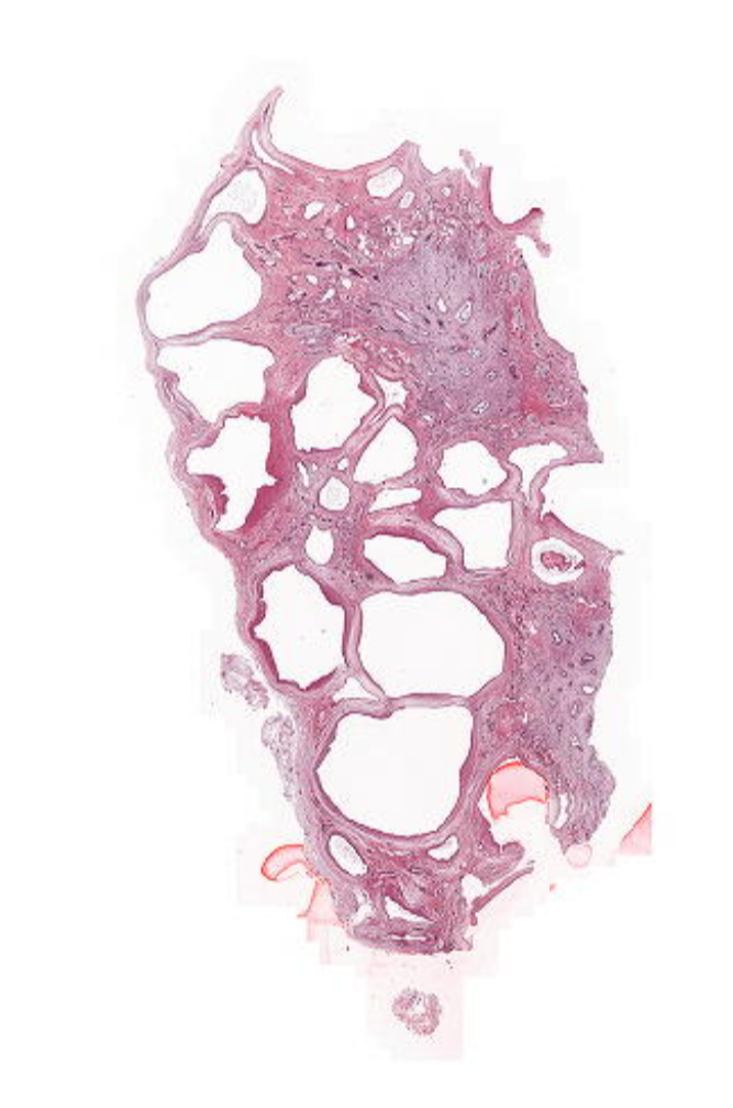
- 28—HSIL high grade intraepithelial lesion of the uterine cervix
1)Transformation zone
o Squamous epithelium disorganised, atypical, dysplastic some keratinisation through layer
o Mitosis seen at all layers – should only be seen basally normally
o Downgrowth of dysplastic epithelium into cervical glands
Lesion in limited to basement membrane (in situ CIN III lesion)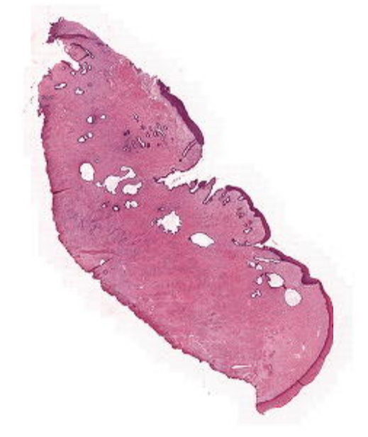
- 29—Condyloma (LSIL CIN1) of the cervix
1)thickened, hyperplastic squamous epithelium
2)vaculositation of cells
3)koilocytes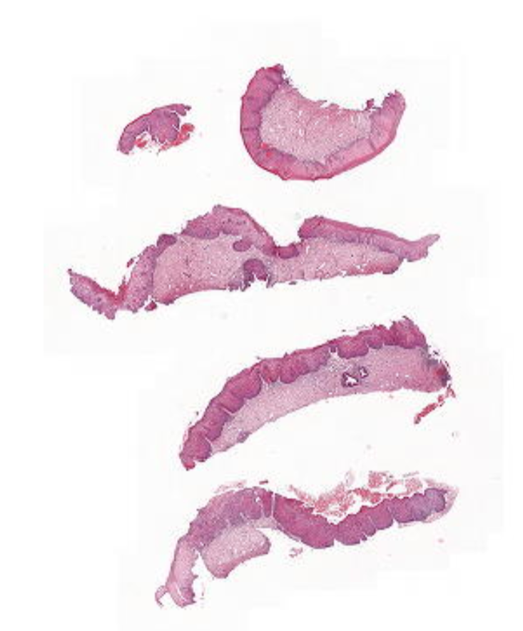
- 30—Squamous Cell Carcinoma of the Uterine Cervix
1)conisation
2)Thickened, atypical squamous epithelium
o Cells with ‘empty’ nuclei where chromatin more clumped
o Invasion into cervical stroma, able to visualise chromatin and red keratin pearls
o Abnormal keratinisation seen on surface and in infiltrating areas
o Vascular invasion not seen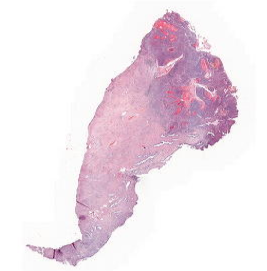
- 31—adenocarcinoma of the uterine cervix in situ and invasive
1)Atypical pseudostratified and hyperchromatic nuclei in glands, several mitotic figures
o Goblet cells and mucinous vacuoles in glands indicate intestinal metaplasia → highly suggestive of neoplasia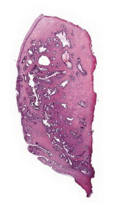
- 32—hyperplastic endometrium without atypia
1)Curettage
2)Small risk of progression to carcinoma, treated with progesterone until menopause
3)MITOSIS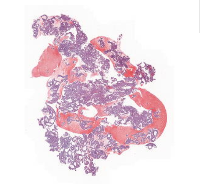
- 33—intramucosal well differentiated endometroid adenocarcinoma (type 1)
1)Areas of cribiform growth (formation of round glands with ‘smooth’ lining)
o Micro-abscesses (clusters of neutrophils) in lumen of glands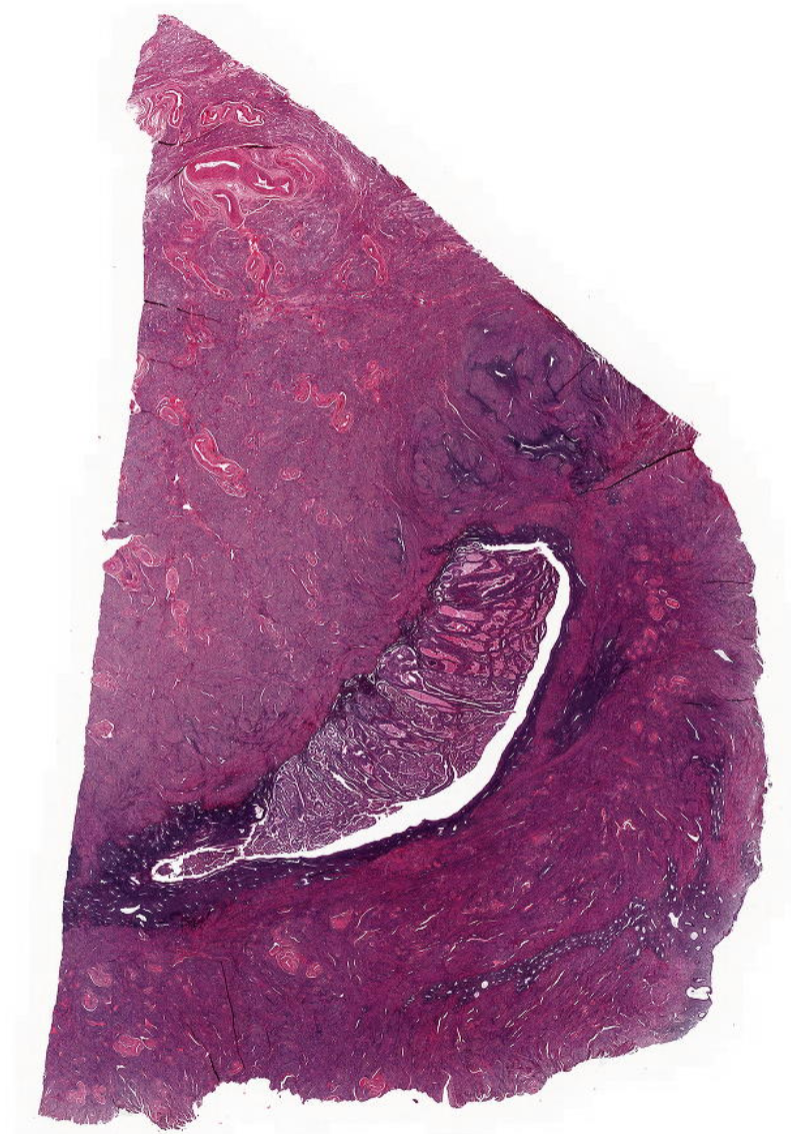
- 34—High grade adenocarcinoma
Serous type 2 (endo)
1)50% of wall
2)scallop/hobnail
3)talk about normal cancer markers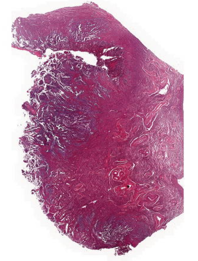
- 35—Serous Endometrial Intraepithelial Carcinoma (SEIC)
Polypoid lesion with stalk with no uterus
o Areas of large cysts (L) and normal glands (upper R)
o In ‘normal’ area, stroma is rich in vessels and eosinophilic, typical of polyps
o Cells lining cysts are flattened, monomorphic, and cuboidal (as opposed to columnar)
On (lower R) side, serous endometrial intraepithelial carcinoma
o Some glands have half normal epithelium and half serous, scalloping carcinoma cells
o Not invasive tumour as remains on surface of glands
o Large nucleoli, loss of polarisation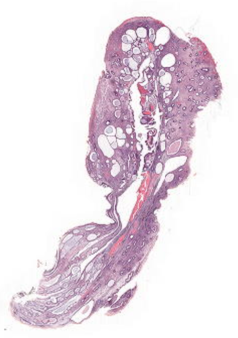
- 36—Fat connective tissue with islands of endometrial tissue and fibrosis/chronic inflammation
o Dilated glands filled with blood → pain
o Glands show columnar, partly flattened, cuboidal epithelium
Endometrial stroma with small vessels and haemorrhage around glandsEndometriosis
ENDO=both glands and stroma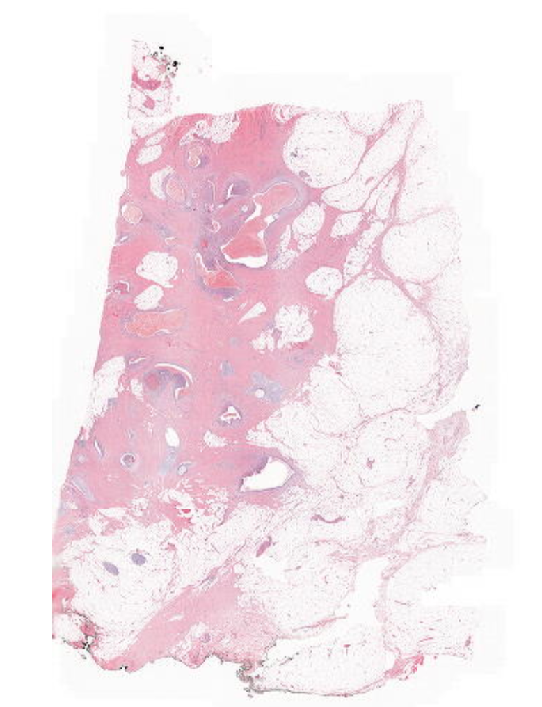
- 37—Adenomyosis
Border between endometrium and myometrium is irregular
o Deep extension of endometrium into myometrium including both glands and stroma
o Nil hyperplasia, atypia, or mitotic figures
Generates pain and irregular bleeding if gland is open to surface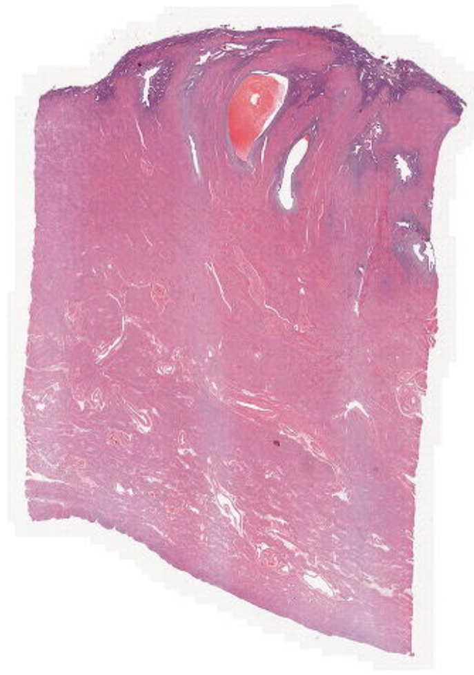
- 38—Uterus with Clear Cell Adenocarcinoma Type 2
Endometrial polyp with development of adenocarcinoma
o Polyp: dilated glands with normal gland tissue, eosinophilic stroma on (top R)
o Overtly atypical nuclei and clear cytoplasm
o Cells have distinct cell borders and line glandular spaces with hobnailing (also seen in serous carcinoma)
High grade tumour with marked atypia with hyperchromasia, large nucleoli
o High grade of pleomorphism and attempt to form glands
o Eosinophilic extracellular material characteristic of clear cell carcinoma
o Invasion through to myometrium (smooth muscle shown)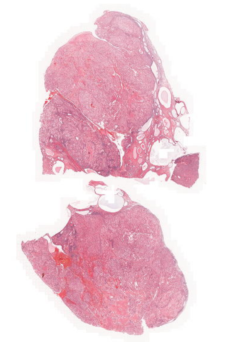
- 39—Uterus with Leiomyoma
Section through well-localised leiomyoma
o Smooth muscle fibre bundles, partly in ‘criss-cross’ pattern, elongated nuclei
o Normal myometrium with less compact strands of muscle fibres seen in periphery
No atypia of myocytes in the myoma, no necrosis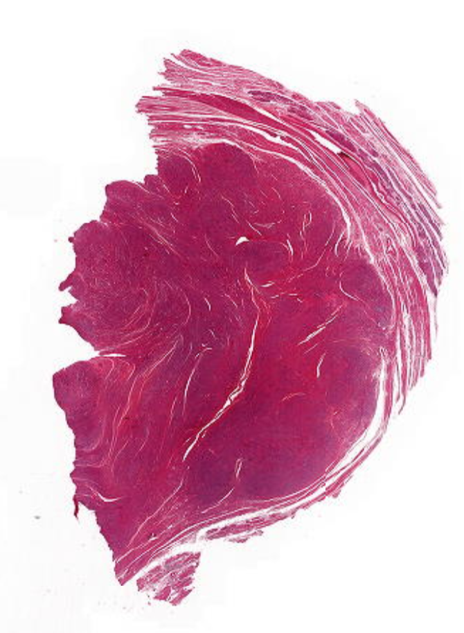
- 40—Uterus with Leiomyosarcoma
Section through overtly malignant tumour consisting of spindle cells
o High-grade atypia, multiple mitotic figures
Heterogenous leiomyosarcoma
o Relatively well-circumscribed at upper border
o Large area of necrosis in middle, loss of cells → most important hallmark of cancer
o Bundles of spindle cells poorly arranged; no bundles of cells
o Nuclei are atypical, course chromatin/clearing of nucleus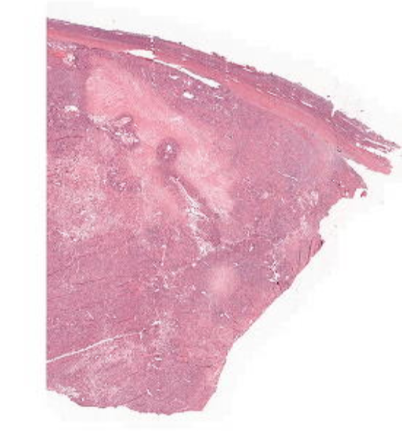
Information
- Description
- Public Health Flashcards on Histo, created by Benjamin Challis on 14/12/2018.
- Created by
- Benjamin Challis
- Views
- 77
- Rating