
Fichas
Surgery
Fichas del conjunto (55)
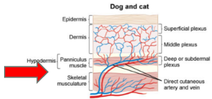
- 1Draw the layers of the skin. Why are these layers important?-Want to undermine skin below cutaneous trunci/platysma muscle -> avoid dermal necrosis
-Panniculus muscle allows more flexibility of dermis
-Direct cutaneous vessels can be damaged by shearing/crushing injuries -> skin necrosis 48-72 hrs later - 2Outline the phases of wound healing*Same phases for trauma vs surgery
1. Inflammation
-Platelets & neutrophils
-Remain in wound until trigger is gone
-"Clean up" crew
2. Tissue formation/regeneration
-Macrophages, fibroblasts
-"Architects"
-Angiogenesis
-Granulation tissue fills wounds
-Sign of minimal bacterial infection/necrosis
**Absence of granulation tissue by 3-5 d (dogs) or 5-6 d (cats) means there is a problem! -> infection, poor perfusion, excess necrotic tissue, etc.
3. Remodeling (aka secondary intention healing)
-Myofibroblasts, epithelial cells
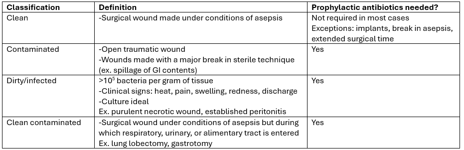
-Wounds heal from the edges in - 3Outline wound classifications
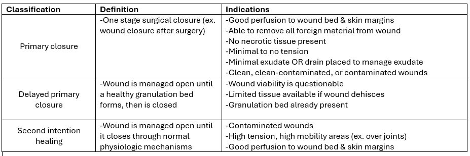
- 4Outline wound closure methods for small animals
- 5List patient and wound factors that inhibit healingPATIENT FACTORS
-Diabetes mellitus/HAC/endocrinopathy
-FIV
-Steroids (duration & dose-dependent)
-Chemotherapy
-Severe anemia
-Poor nutritional status
-Hypotension/hypothermia under GA
-Length of time under GA
-Being a cat
WOUND FACTORS
-High tension, high mobility
-Pressure points
-Radiation site
-Highly productive wounds
-Excessive suture material in wound bed
-Residual foreign material/necrotic tissue - 6Describe the types, functions and indications for drains1. Types of drains
a. Active
-Active suction
-Fully closed system
-Close wound over drain
b. Passive
-Relies on gravity
-Wound fluid wicks out around drain
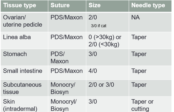
2. Functions
-Eliminate dead space
-Remove excess fluid
3. Indications
Place during surgical closure of healthy wounds where:
-Wound is exudative
-Dead space can't be managed by other means
-To manage a deep abscess - 7Describe the principles of drain usage and placementNOT a substitute for open wound mgmt
-Should not exit through primary incision
-Exit point must be ventral for passive drains & amenable to bandaging
-ONLY closed suction drains for abdomen
-Avoid in oncologic surgery
-Use active instead of passive when possible
-Remove the drain ASAP (2-4 days)
-Use the small number and size of drain possible
-Diameter of exit point should be 1.5-2x the drain diameter
-Don't fenestrate drain
-Don't suture drain at both ends
-Avoid if concerns about skin tension & tissue viability - 8Define surgical asepsis and surgical site infections (SSI)Surgical asepsis = complete absence of contamination by pathogenic organisms
-Prevents wound contamination
SSI = any infection of a surgical site
-Can occur 1 yr+ after surgery
-Purulent drainage
-Dehiscence of incision sites
-Abscess or other evidence of ifxn
-Heat, redness, pain, localized swelling - 9Outline the factors/risks that can cause surgical site infections1. Microbial pathogens
-Ifxn develops if >10^6 bacteria/gram of tissue
a. Duration -> ifxn rate doubles for every hr of surgical time
b. Type of procedure (dirty/contaminated > clean-contaminated > clean)
-Ortho procedures have high ifxn rates
2. Local wound environment
-If skin already infected, only perform sx if necessary -> otherwise clean & wait for ifxn to subside
3. Host defense mechanisms
a. Patient factors
b. Duration of anesthesia & surgery - 10Describe prevention of surgical infection at the patient level1. Skin health
-Skin over surgical site should be evaluated prior to & during hair removal
-Distant site ifxns are linked to ↑ SSIs
-Elective procedures should NOT be performed if multiple skin lesions present
2. Skin prep
-Remove transient microorganisms & reduce resident flora
-Clip only w/in 4 hrs of sx -> ↓ incidence of SSIs
-Avoid rashes & skin abrasions from shaving/clipping
-Clean w/ non-medicated soaps BEFORE applying antiseptics -> antiseptics are not active in dirty environments
-Scrub, paint, or spray antiseptics
-Antiseptic contact time varies + use a single-step process
-Alcohol-based products or chlorhex + alcohol have better efficacy - 11Describe prevention of surgical infection at the environmental level1. Draping
-Blood & fluid resistant
-Lint-free, antistatic, able to maintain isothermic enviro. relative to patient
-Should not be flammable
-Single-use disposable = less risk of SSI
-Can use self-adhesive drapes, but should only use ones infused w/ antimicrobial agents - 12Describe prevention of surgical infection at the personnel level-Do laundry & change soiled attire!
-Keep scrubs covered when not in OR
-Clean cell phones
1. Scrubs
-Tuck tops into pants
-No undershirts showing
2. Presurgical hand prep
-Transient microbiota = most common cause of SSI
-Resident microbiota = less pathogenic
-Use waterless products! -> alcohol-based hand rubs are superior
-Trim fingernails to 2 mm or less
3. Gowning & gloving
-Barrier b/w sterile & non-sterile areas
-Significant decrease of bacterial load in operating room
-Disposable gowns less permeable to bacteria
-Up to 45% incidence of glove perforations -> use colored undergloves & change gloves worn during draping
-Open, closed, and assisted gloving
-Assisted gloving is the most sterile - 13Describe the anatomy and pathophysiology of herniasCOMPONENTS/ANATOMY
-Epithelial-lined sac
-Hernia ring
-Contents
a. Internal hernia
-Does not involve abdominal wall (ex. mesenteric, diaphragmatic)
b. External hernia
-Hernia protrudes through a defect in the abdominal wall (ex. scrotal, prepubic)
PATHOPHYSIOLOGY
-Loss of domain -> things in improper places
-Incarceration -> adhesions, impedance of normal function
-Vascular strangulation (surgical emergency!) - 14Describe the surgical management of herniasGOALS OF SURGERY
1. Stabilize the patient
2. Release herniated structures & return them to normal locations
3. Repair the defect
EMERGENCY SURGERY
1. Suspicion for/clinical signs evident of GI entrapment or intestinal necrosis
2. Septic abdomen
3. Intestinal adhesions
4. Diaphragm rupture - 15Describe umbilical hernias in small animals-Common, congenital & heritable
-May have concurrent cardiac defects, incomplete xiphoid fusion, cryptorchidism, or PPDH
-Clinical signs rare
-May spontaneously resolve by 6 mos old
1. If soft, non-painful, reducible:
a. Small ring -> close at neutering
b. Large ring -> monitor & close at neutering
2. If firm, painful, assoc. w/ GI signs
-Surgical emergency
SURGICAL APPROACH
-Separate incision for males
-Incorporate into spay incision for F
-Careful incision over hernia
-Reduce or excise fat to expose hernia ring -> close w/ PDS - 16Describe inguinal hernias in small animalsANATOMY
-Inguinal canal contains: 1. vaginal process/ring; 2. genitfemoral n.; 3. external pudendal vessels; 4. +/- spermatic cord
-70-90% female, L>R, estrus & pregnancy are risk factors
DIAGNOSIS
-Palpation
-Ultrasound/radiographs
SURGERY
-As soon as diagnosed -> emergent if concern for organ entrapment
-Can approach via caudal abdominal incision or over the hernia
-ID & reduce herniated structures from the inside
-Check viability of reduced tissue
-Close inguinal ring -> careful of normal structures! - 17Describe scrotal hernias in small animals-Variation of an inguinal hernia -> stuff ends up in scrotum rather than inguinal rings
-Rare & often concurrent strangulation
-Chondrodystrophoids & brachycephalics
SURGERY
1. Reducible
-Prescrotal incision parallel to midline
-Reduce & close external inguinal ring
2. Not reducible
-Caudal midline laparotomy +/- approach inguinal rings
-Reduce hernia contents & evaluate viability - 18Describe abdominal/thoracic wall ruptures in small animalsETIOLOGY & PRESENTATION
-Polytrauma
ANATOMY: 1. prepubic tendon rupture; 2. dorsolateral; 3. paracostal; 4. intercostal
-Rule out concurrent pneumothorax, pulmonary contusions, v-tach, uro/hemoabdomen, or bile peritonitis
DIAGNOSIS
-Palpation
-"Skyline" radiographs/ultrasound
-Surgical exploration
SURGERY
**Stabilize cardiopulmonary fxn 1st!
1. Abdomen
-Approach via midline laparotomy
-Care for aorta/ureters, etc.
-2/0 PDS, simple interrupted pattern
-Use pubis periosteum to anchor sutures or pre-drill holes in pubic brim
2. Thoracic
-Circumcostal sutures w/ 0 or 2/0 PDS
-Must place a chest tube
-Be prepared to perform PPV or lung lobectomy - 19Describe the etiology and pathophysiology of diaphragm ruptures in small animalsETIOLOGY & PATHOPHYSIOLOGY
-Can be congenital or trauma-based
-Commonly rupture @ central tendon & pars costalis
-Thoracic & abdominal pressures equalize
-Loss of domain -> ↓ tidal volume, atelectasis, VQ mismatch, impaired venous return
-Assess for concurrent trauma (ex. pleural effusion, pneumothorax, strangulation)
a. Gastric herniation = SX EMERGENCY -> intubation, PPV, orogastric decompression, & sx
b. Liver lobe/splenic torsion/strangulation
-High volume pleural effusion
-↓ effective circulating volume & hypovolemic shock
-Thoracocentesis prior to anesthesia - 20Describe clinical signs, diagnosis, and treatment of diaphragm ruptures in small animalsCLINICAL SIGNS
-Dyspnea
-↓ lung sounds
-Asynchronous respiratory patterns
DIAGNOSIS
-Radiographs/CT/ultrasound
TREATMENT
-Stabilize & surgery
-O2, IV fluids, & opioid analgesics
a. Surgery
-Immediate PPV & head up position
-Midline laparotomy
-Reduce organs
-Place small bore chest tube under palpation before closing
POSTOP: may remain O2 dependent; chest tube should be aspirated q4hrs
-IV fluids, opioids, early feeding
COMPLICATIONS: chronic hernias; intrathoracic adhesions; re-expansion pulmonary edema; abdominal compartment syndrome - 21Describe peritoneopericardial diaphragmatic hernias (PPDH)-Congenital
-Pericardial sac connects abdominal cavity
-Asymptomatic until GI obstruction or cardiac tamponade
-Often concurrent w/ umbilical hernia & xiphoid defects
DX: ultrasound, radiographs
SURGERY
-Cranial midline laparotomy
-Evacuate pericardial sac
-Usu. do not need chest tube/PPV unless thorax compromised
-Chronically herniated liver lobes may be necrotic -> liver lobectomy
-Close w/ 0-3/0 PDS in ford interlocking pattern - 22Outline the classifications of hernias1. Location
-Internal vs external
2.Hernia contents
-ex. enterocoele, cystocoele, hepatocoele
3. Status
a. Reducible
-Contents can be returned to normal position by manipulation
b. Irreducible/incarcerated
-Cannot be manipulated to normal position, but vascular supply is intact
c. Strangulated
-Blood supply to contents is compromised
4. Etiology
a. Congenital
b. acquired
c. Iatrogenic (ex. incisional) - 23Describe the etiology and diagnosis of umbilical hernias in large animals-Mostly congenital, +/- heritable? (cattle & swine)
-QH predisposed, F>M
-Infection rare in foals; more common in cattle
DIAGNOSIS
1. Physical exam
-Fluctuant swelling at umbilicus at or shortly after birth
-Rarely irreducible or strangulated
2. Ultrasound
-Indicated if suspected ifxn or irreducible - 24Describe the treatment of umbilical hernias in large animals1. Conservative treatment
a. Manual reduction
-Useful for small (<2 fingers) hernias w/ no infection
-Owner examines & reduces 1-2x/day
-Hernia ring may close spontaneously
b. Abdominal corset
-Works in calves but not foals
c. Ring scarification
d. Strangulation
-Induce necrosis & sloughing of hernial sac
-Hernias <5 cm, reducible, non-infected
2. Surgery
-Large reducible hernias
-Small hernias that did not respond to conservative tx
-Strangulated or irreducible hernias
-Repair @ 3-4 months (after weaning)
a. Umbilical hernorrhaphy
-Open vs closed
b. Umbilical resection
-If infected, patent urachus, and/or unresponsive to conservative therapy - 25Describe ventral hernias in large animals-Iatrogenic from incisions
-Blunt trauma (getting kicked)
-Usually large, so strangulation less common
REPAIR
1. Traumatic
a. Immediate (w/in 8-12 hrs)
-Required if strangulated
-Tissue damage & severe inflamm. unfavorable for healing
b. Delayed (after 6-8 wks)
-Inflamm. subsided
2. Postoperative
-Repair delayed until inflamm. subsides & wound is not infected
REPAIR METHODS: re-closure of fascial & muscle layers; mesh herniorrhaphy for large defects/weak hernial ring - 26Describe prepubic tendon rupture in large animals-Usu. late term mares, esp. drafts
-Rare in cattle
-Uni or bilateral
-Usu. preceded by severe ventral edema & can be associated w/ avulsion of ventral musculature
DIAGNOSIS/TREATMENT
1. Pre-rupture
-Caudoventral abdominal edema
-Pain on palpation
-Reluctance to move pelvic limbs
TX: strict rest & control of edema; consider induced parturition
2. Post-rupture
-Sagging of caudoventral abdomen
-Disruption of prepubic tendon on rectal palpation or ultrasound
-Rear limb discomfort
TX: strict rest; induced foaling or terminal C-section; poor px for mare - 27Describe the etiology & presentation of inguinal/scrotal hernias in neonatal large animalsINGUINAL
-Congenital or acquired
-Rare in females
a. Direct (peritoneal defect)
-Contents pass through body wall & into inguinal canal
-Contents are outside of vaginal tunic
-Acquired >>>> congenital
b. Indirect
-Content enters inguinal canal via enlarged vaginal ring
-Contents are inside vaginal tunic
-Congenital or acquired
SCROTAL
-Inherited
-Common in foals, esp. SB & drafts -> may also have ruptured vaginal tunic
-Common in piglets & lambs
DX: fluctuant scrotal swelling
-preputial edema
-mild colic - 28Describe the treatment of inguinal/scrotal hernias in neonatal large animals1. Conservative
-Small reducible hernias in foals
-Reduce at least twice daily
-Monitor for strangulation
-Recommend primary closure castration when gelded
2. Surgical
-Piglets & lambs
-Foals that are colicky, have very large hernias. or hernia failed to respond to conservative tx
-Primary closure castration - 29Describe acquired inguinal/scrotal hernias in stallionsETIOLOGY
-After breeding activity or strenuous exercise
-Standardbreds predisposed
DIAGNOSIS
-Acute, severe colic -> strangulated contents
-Hard, cool testicle on affected side
-Rectal palpation
TREATMENT
**Surgical emergency**
-Can sometimes be manually reduced if treated quickly
-Herniorrhaphy w/ castration
-Resection of damaged intestines
- +/- Laparoscopy if testicle not damaged - 30Describe postcastration hernia in stallions-Herniation of bowel or omentum after castration
-Most likely in SBs
-Herniated bowel often strangulates -> acute colic
TREATMENT
**Surgical emergency**
1. Omental hernia
-Ligate & amputate
2. Bowel hernia
-Clean & reduce bowel into abdomen
-Close inguinal canal
-Can also close scrotum
PREVENTION
-Twist & tack spermatic cord prophylactically
-Prophylactic tacking via open castration - 31Describe acquired inguinal/scrotal hernias in beef bulls-Associated w/ breeding activity
-Strangulation rare, but fertility impaired
DIAGNOSIS
-Fluctuant scrotal swelling
-Rectal palpation
-Ultrasound
TREATMENT
-Standing flank celiotomy or lateral recumbency - 32Outline the basics of wound management1. Wear gloves
-Barrier nursing -> avoid MRSA colonization
2. Triage according to concurrent injuries
3. Apply sterile water-soluble gel to wound
4. Clip local area generously
5. Lavage
-Gel & hair out of wound
-Wound bed
-Tap water vs 35 mL syringe/18G needle
6. +/- Debride necrotic tissue
7. +/- Forage exposed bony surfaces
-Drill perforations into medullary cavity
-Promotes formation of granulation bed on exposed bony surfaces - 33Describe indications for and goals of bandagingINDICATIONS
-Support/protect surgical incision
-Manage contaminated traumatic wound until suitable for delayed primary closure
-Manage wound for closure by second intention
GOALS
-Maintain optimal environment for wound healing
a. Mechanically stable
b. Moist
c. Protected
3 layers: contact, absorptive, & outer - 34Describe the contact layer of a bandageLayer directly contacting wound
1. No granulation tissue
a. Minimal/moderately contaminated wound prior to granulation tissue
-Alginate dressings
-Tegaderm
-Soluble, non-traumatic, & moist
*Change q 2-4 days
b. Heavily contaminated/necrotic/ infected wound prior to granulation tissue
-Honey-soaked sterile gauze
-Saline-soaked lap sponges
-Sugar under lap sponges
-Debridement dressings
*Change q6-24 hrs
2. Post-granulation tissue formation
-Non-debridement dressing
-Hydrocolloid/hydrogel under non-adherent dressng
-Foam dressings - 35Describe the absorptive and outer layers of a bandageABSORPTIVE
-Prevents exudate from pooling on wound surfaces
-Redistributes pressure of bandage over entire wound area
-Common error = not using enough!
-Cotton, gauze, etc.
OUTER
-Controls inward pressure of bandage
-Vetwrap, elasticon, etc. - 36Outline common bandage complications~60% incidence
-Insufficient padding
-Too long b/w bandage changes
-Bandage allowed to become wet
-Bandage too tight
-Poor technique (ex. toes left out)
-Poor owner communication
-Poor staff training
-Traumatic/desiccating/moving contact layer
-Excess tension -> poor availability of surrounding tissues
-Poor wound vascularity (ex. bony surfaces) - 37Describe the basics of surgical wound closure-Atraumatic technique (ex. use DeBakey forceps)
-Use stay sutures when manipulating skin
-Undermine skin & use walking sutures to address tension
-Assess surrounding skin mobility to determine direction of closure
-Distal limbs require free grafts or releasing incisions -> skin can be mobilized & repositioned to close wounds over pressure points - 38Describe local flap techniquesLocal flaps rely on local perfusion -> no specific arterial supply
1. Advancement
-Pay attention to width of base relative to length of flap -> length can't be more than 1.5x larger than width
2. Rotational
-No set rules
-Use whatever skin is available
3. Transposition
4. Flank/elbow fold flap - 39Describe axial pattern flap techniquesHave specific arterial supply & named for supplying vessel
1. Caudal superficial epigastric
-Can close large wounds on thigh
2. Caudal auricular
3. Superficial brachial
etc. etc. - 40Describe free grafts-Can be used to manage chronic distal extremity wounds that won't heal on their own
-Requires patent blood supply in area to be grafted onto -> better to graft onto granulation tissue
Plasmatic imbibition -> inosculation -> revascularization in 5 days
-Trim fat off prior to grafting
-Can mesh free graft to reduce tension & improve surface area
-Punch grafts can be used on high mobility areas - 41List causes of graft/flap dehiscenceFREQUENT
-Excess tension
-Wound closed prematurely
-Inappropriate surgical technique
INFREQUENT
-Wound infection - 42What are the recommended ages for spaying and neutering dogs and cats?SPAY
Dogs <20 kg at full grown -> by 5-6 months
Dogs >20 kg at full grown -> no specific recommendations
Cats -> by 5 months old
NEUTER
Cats -> by 5 months
Dogs <20 kg at full grown -> by 5-6 months
Dogs >20 kg at full grown -> b/w 9-15 months
Cryptorchid neuter -> by 12 months - 43Outline the process of breaking the suspensory ligament in a dog1. Caudomedial traction on the proper ligament to pull the suspensory ligament taut
2. Palpate for most lateral component of suspensory & follow it dorsally
3. Isolate b/w thumb & forefinger
4. Flex the wrist to break at point of attachment on body wall
5. Controlled rupture as far distally from origin of vascular pedicle as possible - 44Outline the process of ligating the ovarian pedicle1. Break a window in an avascular area of the broad ligament
TWO CLAMP TECHNIQUE
-Minimal fat in pedicle -> no crush bed needed
2. Concave face of Carmaults towards ovary
3. Long tails on ligatures
4. Minimum 1 cm stump
-2/0 PDS or Maxon
THREE CLAMP TECHNIQUE
-Fatty/thick pedicle needing crush bed
-Cut b/w 2nd & 3rd clamps - 45Outline the general guidelines for suture size for a canine OVH
- 46Briefly describe the types of knots that can be used in a canine OVH1. Aberdeen knot
-Easiest knot to bury at the end of an intradermal pattern
2. Miller's knot
-Good for fatty pedicles/uterine body
3. Transfixion knot
-Encircle uterine vessel on one side, compress on the other - 47Outline the process of breaking the suspensory ligament in a cat1. Hold ovary by the proper ligament in the left hand
2. Push the body wall down w/ the 3rd finger & fan out broad ligament, vessels, & suspensory ligament
3. Capture suspensory ligament in a hemostat
4. Push down on hemostat to tear suspensory (keep ovary fixed in place!) - 48Describe the surgical differences when dealing with a pyometra vs an OVH-Long midline incision
-Fully exteriorize uterus
-Gentle handling!! Avoid rupturing uterus
-3-clamp ligation technique for ovarian pedicles
-Uterine vessels prominent -> ligate individually w/ Miller's
-Don't oversew uterine stump
-Lavage abdomen w/ saline prior to closure - 49List common complications of spays and how to prevent them1. Hemorrhage
-Gentle tissue handling
-Secure ligatures -> big stumps, long tails, tight knots
-Check internal margin of body wall for bleeding before closure
2. Incisional complications
-Clear SQ fat to visualize external rectus sheath
-Ensure anchor & finish knots are TIGHT -> 6 & 7 throws, respectively
-Quilting pattern in SQ tissues
-Appropriate analgesia +/- abx
3. Ovarian remnant syndrome
-Adequate midline incision
-Fully rupture suspensory ligament
-Palpate ovary location prior to clamp placement
-Double-check ovary resection
4. Ureteral injury
-Be careful!!
-Empty bladder during preop skin prep
-Avoid excessively caudal ligations of uterine body
-Do not include fat in uterine body ligature - 50Outline the process of a closed castration in a dog1. Push testicle cranially & to midline
2. Incise until vaginal tunic exposed (but don't enter!)
3. Push testis out through the incision
4. Break ligament of the tail of the epididymis
5. Break down fat around the spermatic cord
6. Use a crush bed for ligature & 3-clamp technique
7. Leave at least 1 cm stump
8. Do not close inguinal canals & lift tissue clear of urethra while closing SQ tissues - 51Outline the process of an open castration in a dog1. Incise vaginal tunic to expose surface of the testis
2. Extend incision proximally to expose vasculature
3. Ligate vasculature directly
4. Ligate vaginal tunic separately
*Otherwise very similar process - 52Outline the process of a feline neuter1. Pluck or clip fur + sterile skin prep
2. Scrotal incision -> leave vaginal tunic intact
3. Exteriorize testis
4. Strip fascia to isolate spermatic cord
5. Cord tie with an overhand knot & cut
6. Testicular stumps tucked back inside scrotum
7. Scrotal incisions left open - 53Describe the anatomy of a cryptorchid testicle-Not covered by vaginal tunic
-Often small
-Anchored by 3 structures:
1. Testicular artery & vein
-Course dorsally to vena cava & aorta
2. Vestigial gubernaculum
-Tethers testis to internal inguinal ring
3. Spermatic cord
-Crosses ureter & inserts on the prostate - 54Outline the process for a cryptorchid neuter in a dog1. Clip prepuce away from surgeon before draping
2. Skin incision from just in front of prepuce to mid-prepuce, off midline
3. Incise SQ tissue,, preputialis muscle & ligate preputial vessels
4. ID linea alba & enter abdomen on midline
5. Check to either side of urinary bladder
6. Find vas deferens where it crosses ureter
7. Try to pull testicle back through inguinal ring -> may need to enlarge ring
8. Remember to close ring if enlarged & repair preputialis muscle when closing the abdomen - 55Describe neuter complications and how to prevent them1. Hemorrhage
a. Scrotal from vaginal tunic or SQ vessels
b. Spermatic cord from ligature slippage
2. Urethral laceration/ligation
-Urine extravasation -> tissue necrosis
-Urinary obstruction
3. Inadvertent prostatectomy
-Complication of cryptorchid neuter
-Complete urinary obstruction + uroabdomen
PREVENTION
-Atraumatic tissue handling
-Thorough hemostasis
-Fully tighten ligatures
- +/- Prophylactic scrotal ablation in large mature dogs
-Ensure testis protects urethra during prescrotal incision
-No spay hooks!
-Caudal abdominal midline approach ONLY
Información
- Descripción
- NAVLE Veterinary Medicine Fichas sobre Surgery, creado por Danielle Keerbs el 06/10/2024.
- Creado por
- Danielle Keerbs · oct 2024
- Actualizado
- 9 oct 2024
- Visitas
- 1
- Valoración