
Mapa mental
The Heart
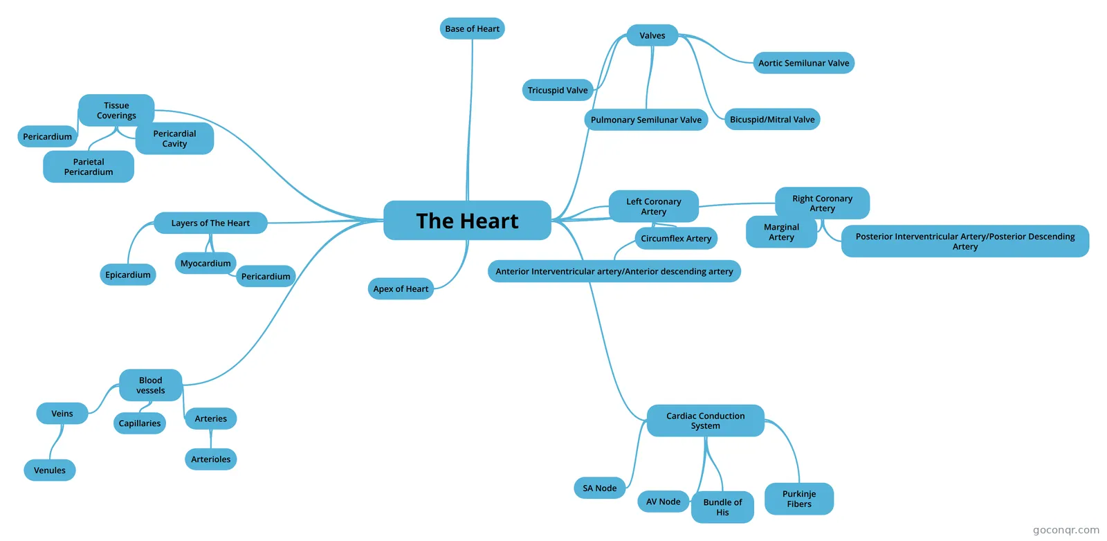
Mind map: The heart
Contenido del mapa
The Heart
- Coronary Anatomy: - Heart weights between 250-350 grams (less than a pound). - The heart represents about 1/200 or 0.5% of the body weight, yet it requires about 1/20 or 5% of the body's blood supply. - The only tissue layer that can adequately received oxygen and nutrients from the blood flowing within the chambers is the endocardium. - The thick myocardium and epicardium must rely on alternative supplies of blood to maintain their function. Examples of types of heart attack: - transmural - subendocardial
Layers of The Heart
- Epicardium
- - Corresponds to the visceral pericardium. –Functions as an outer protective layer –It is a serous membrane that consists of connective tissue covered by epithelium, and includes blood capillaries, lymph capillaries, and nerve fibers. –The deeper portion often contains fat, particularly along the paths of larger blood vessels.
- Myocardium
- –The thickest layer of tissue in the heart. –Composed of cardiac muscle tissue responsible for forcing the blood out of the heart chambers. –The fibers are arranged in planes, separated by connective tissues that are richly supplied with blood capillaries, lymph capillaries and nerve fibers.
- Pericardium
- –Inner layer and is lined with a thin layer of endothelium. •Forms the innermost lining of the walls of the chambers as well as the heart valves. •This layer joins with the endothelial linings of the blood vessels as they leave and enter the heart. •Also contains connective tissue that contains some specialized cardiac muscle fibers called Purkinje fibers.
- Epicardium
Base of Heart
- •The base of the heart consists mainly of the LA, part of the RA, and parts of the proximal portion of the large veins that enter the heart posteriorly. •The base is located superiorly and near the right sternal border at the level of second and third ribs.
Apex of Heart
- •Located inferiorly and to the left of the base at the level of the fifth inter-costal space. •Approx.2/3 of the mass of the heart is to the left of the mid-sternal border. •Contraction of the heart can be palpated at the apex (between 5th and 6th ribs). –This is referred to as the PMI (Point of maximal intensity)
Valves
- Tricuspid Valve
- RA -> RV: Tricuspid valve - Has three leaflets or cusps - Strong, fibrous strings (chordae tendineae) are attached to the cusps on the ventricular side
- •These strings originate from small mounds of cardiac muscle tissue (the papillary muscles) that project inward from the walls of the ventricle. •When the tricuspid valve closes, the chordae tendineae and papillary muscles prevent the cusps from swinging back into the atrium.
- •When the muscular wall of the RV contracts, the blood inside its chamber is put under increasing pressure, and the tricuspid valve closes passively •As a result the only exit is through the pulmonary trunk, which divides to form the left and right pulmonary arteries.
- Pulmonary Semilunar Valve
- •At the base of this trunk is a pulmonary valve (pulmonary semilunar valve). –This consists of three cusps. It opens as the right ventricle contracts. However, when the ventricular muscles relax, the blood begins to back up in the pulmonary trunk. •This causes the pulmonary valve to close, thus preventing a return flow into the ventricular chamber.
- Bicuspid/Mitral Valve
- •The LA receives the blood from the lungs through four pulmonary veins – two from the R and two from the L lung. •Blood passes from the LA into the LV through the atrio-ventricular orifice, which is guarded by a valve. •This valve consists of two leaflets, and is named the bicuspid or mitral valve. (it is shaped like a miter, a turban like headdress). •Like the tricuspid valve, the cusps of the bicuspid valve are prevented from swinging back by the papillary muscles and the chordae tendineae.
- Aortic Semilunar Valve
- •When the LV contracts, the bicuspid valve closes and blood exits through the aortic valve (aortic semilunar valve). –This valve also consists of 3 cusps.
- Tricuspid Valve
Tissue Coverings
- Pericardium
- Consists of an outer fibrous bas, the fibrous pericardium. - This surrounds a more delicate, double-layerd sac. - the inner layer of this sac is the visceral pericardium or Epicardium. This covers the heart.
- Parietal Pericardium
- At the base of the heart, the visceral pericardium turns back upon itself to become the parietal pericardium.
- The parietal pericardium, in turn, forms the inner lining of the fibrous pericardium. - the fibrous pericardium is a tough, protective sac composed largely of white fibrous connective tissue. It is attached to the central portion of the diaphragm, behind the sternum, the vertebral column, and the large blood vessels emerging from the heart.
- Pericardial Cavity
- Between the parietal and visceral layers of the pericardium (pericardium and epicardium) is a potential space, the pericardial cavity, that contains a small amount of serious fluid. - this fluid reduces friction between the pericardial membranes as the heart moves within them.
- Pericardium
Blood vessels
- Arteries
- Can be classified as elastic or muscular according to their size and function.
- Large arteries such as the aorta or those of the pulmonary trunk are called elastic arteries. •The tunica media of these vessels is thick and contains many elastic fibers. The elastic nature of these arteries helps maintain pressure within the vessels.
- Other smaller arteries distribute blood throughout the body. These arteries are called muscular arteries. Their tunica media contains primarily smooth muscle fibers. Muscular arteries are less distensible than elastic arteries
- Arterioles
- •Very small microscopic arteries that deliver blood to the capillaries. •Their lumens are smaller than 0.5 mm •Their tunica media is largely composed of smooth muscle with scattered elastic fibers. •These play a major role in regulating blood flow to the capillaries because of their ability to constrict or dilate. •Also, changes in arteriole diameter can affect systemic BP
- Capillaries
- •Microscopic vessels that connect the arterioles with the venules. •Form dense networks that branch throughout all tisues. •Average capillary is 1 mm in length and 0.01 mm in diameter. –This is just large enough for a single RBC to pass though. •They have extremely thin walls made of a single layer of cells and a basement membrane. •Capillaries do NOT have a tunica media or tunica adventitia. –This is what allows for the exchange of materials between blood and the tissue cells.
- Veins
- •Receive blood from the venules and have the same three tissue layers as arteries. •However, the tunica media of the veinsis thinner than that in the arteries. •Generally, the veins are thinner and more compliant than arteries, and act as blood reservoirs.
- •The walls of some veins, such as those in the legs, contain one-way valves that help maintain venous return to the heart by preventing retrograde blood flow even under relatively low pressures. •The valves in the veins are made up of folds of tunica intima (endothelium) and are similar in nature to the semilunar valves of the heart.
- Venules
- •Form from capillaries •Consist mainly of tunica intima and tunica adventitia •Collect blood from the capillaries
- Arteries
Cardiac Conduction System
- SA Node
- –Pacemaker –Located just below the epicardium in the posterior wall of the RA, below the opening of the superior vena cava. –The membranes of the nodal cells are in contact with each other, and these cells have the ability to excite themselves (spontaneous depolarization). This activity is rhythmic.
- AV Node
- •After the impulse travels from the SA node into the atrial syncytium, the R and L atria contract almost simultaneously. •Instead of passing directly into the ventricular syncytium, the impulse passes along fibers of the conduction system that are continuous with atrial muscle fibers. These conducting fibers lead to a mass of specialized cardiac muscle tissue called the AV node. –This node is located on the floor of the RA near the septum and just beneath the endocardium. It provides the only normal conduction pathway between the atrial and ventricular syncytia.
- •The fibers that conduct the cardiac impulse into the AV node (junctional fibers) have very small diameters, and because small fibers conduct impulses slowly, they cause the impulse to be delayed. •The impulse is delayed still more as it travels through the AV node, and this delay allows time for the atria to empty and the ventricles to fill with blood. •Once the impulse reaches the distal side of the AV node, it passes into a group of large fibers that make up the AV bundle (Bundle of His), and the impulse moves rapidly through them.
- Bundle of His
- •The AV bundle enters the upper part of the interventricular septum, and divides into right and left branches that lie just beneath the endocardium •About halfway down the septum, the branches give rise to enlarged Purkinje fibers.
- Purkinje Fibers
- •These fibers spread from the inter ventricular septum into the papillary muscles, which project inward from the ventricular walls, and then continue downward to the apex of the heart. •There they curve around the tips of the ventricles and pass upward over the lateral walls of these chambers. •Along the way, these fibers give off many small branches, which become continuous with cardiac muscle fibers. •The muscle fibers in the ventricular walls are arranged in irregular whorls, so that when they are stimulated by the impulses on the Purkinje fibers, the ventricular walls contract with a twisting motion. –This action squeezes or wrings the blood out of the ventricular chambers and forces it into the arteries.
- SA Node
Right Coronary Artery
- Courses to the right side of the heart and divides into two branches: marginal artery & posterior inter ventricular artery.
- Marginal Artery
- Serves the myocardium of the lateral part of the right side of the heart.
- Posterior Interventricular Artery/Posterior Descending Artery
- runs to the heart apex and supplies the posterior walls.
Left Coronary Artery
- Runs towards the left side of the heart and then divides into its major branches: anterior inter ventricular artery and circumflex artery.
- Anterior Interventricular artery/Anterior descending artery
- Supplies blood to the inter ventricular septum and anterior walls of both ventricles.
- Circumflex Artery
- Supplies the left atrium and the posterior walls of the left ventricle.
Información
- Descripción
- 1 KNR 307 Mapa Mental sobre The Heart, creado por Ashley Akiyama el 06/03/2016.
- Creado por
- Ashley Akiyama
- Visitas
- 2
- Valoración
Más sobre Heart Anatomy and Function