
Mapa mental
Attention
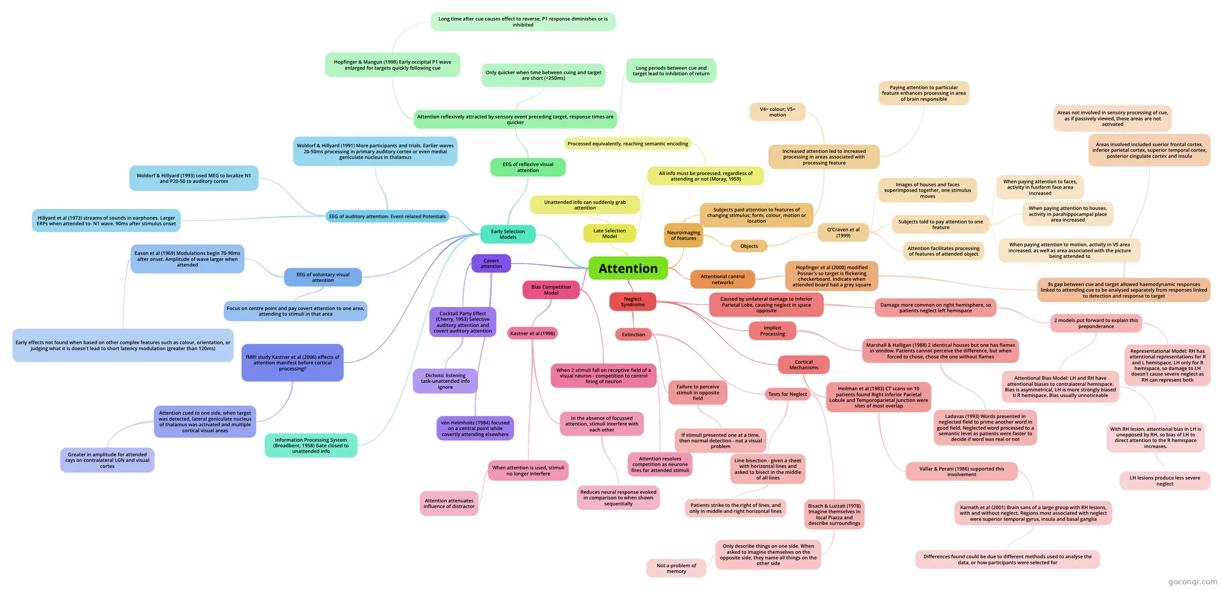
Mind map: Attention
Contenido del mapa
Attention
Early Selection Models
- Information Processing System (Broadbent, 1958) Gate closed to unattended info
- EEG of auditory attention- Event related Potentials
- Hillyard et al (1973) streams of sounds in earphones. Larger ERPs when attended to- N1 wave. 90ms after stimulus onset
- Woldorf & Hillyard (1991) More participants and trials. Earlier waves 20-50ms processing in primary auditory cortex or even medial geniculate nucleus in thalamus
- Woldorf & Hillyard (1993) used MEG to localize N1 and P20-50 to auditory cortex
- EEG of voluntary visual attention
- Focus on centre point and pay covert attention to one area, attending to stimuli in that area
- Eason et al (1969) Modulations begin 70-90ms after onset. Amplitude of wave larger when attended
- Early effects not found when based on other complex features such as colour, orientation, or judging what it is doesn't lead to short latency modulation (greater than 120ms)
- EEG of reflexive visual attention
- Attention reflexively attracted by sensory event preceding target, response times are quicker
- Only quicker when time between cuing and target are short (<250ms)
- Long periods between cue and target lead to inhibition of return
- Hopfinger & Mangun (1998) Early occipital P1 wave enlarged for targets quickly following cue
- Long time after cue causes effect to reverse, P1 response diminishes or is inhibited
- Attention reflexively attracted by sensory event preceding target, response times are quicker
- fMRI study Kastner et al (2006) effects of attention manifest before cortical processing?
- Attention cued to one side, when target was detected, lateral geniculate nucleus of thalamus was activated and multiple cortical visual areas
- Greater in amplitude for attended rays on contralateral LGN and visual cortex
- Attention cued to one side, when target was detected, lateral geniculate nucleus of thalamus was activated and multiple cortical visual areas
Covert attention
- von Helmholtz (1984) focused on a central point while covertly attending elsewhere
- Cocktail Party Effect (Cherry, 1953) Selective auditory attention and covert auditory attention
- Dichotic listening task-unattended info ignore
Late Selection Model
- Unattended info can suddenly grab attention
- All info must be processed, regardless of attending or not (Moray, 1959)
- Processed equivalently, reaching semantic encoding
Neuroimaging of features
- Subjects paid attention to features of changing stimulus; form, colour, motion or location
- Increased attention led to increased processing in areas associated with processing feature
- V4= colour; V5= motion
- Paying attention to particular feature enhances processing in area of brain responsible
- Increased attention led to increased processing in areas associated with processing feature
- Objects
- O'Craven et al (1999)
- Images of houses and faces superimposed together, one stimulus moves
- Subjects told to pay attention to one feature
- When paying attention to faces, activity in fusiform face area increased
- When paying attention to houses, activity in parahippocampal place area increased
- When paying attention to motion, activity in V5 area increased, as well as area associated with the picture being attended to
- Attention facilitates processing of features of attended object
- O'Craven et al (1999)
- Subjects paid attention to features of changing stimulus; form, colour, motion or location
Bias Competition Model
- When 2 stimuli fall on receptive field of a visual neuron - competition to control firing of neuron
- Attention resolves competition as neurone fires for attended stimuli
- Kastner et al (1998)
- In the absence of focussed attention, stimuli interfere with each other
- Reduces neural response evoked in comparison to when shown sequentially
- When attention is used, stimuli no longer interfere
- Attention attenuates influence of distractor
- In the absence of focussed attention, stimuli interfere with each other
- When 2 stimuli fall on receptive field of a visual neuron - competition to control firing of neuron
Attentional control networks
- Hopfinger et al (2000) modified Posner's so target is flickering checkerboard. Indicate when attended board had a grey square
- 8s gap between cue and target allowed haemodynamic responses linked to attending cue to be analysed separately from responses linked to detection and response to target
- Areas involved included suerior frontal cortex, inferior parietal cortex, superior temporal cortex, posterior cingulate cortex and insula
- Areas not involved in sensory processing of cue, as if passively viewed, these areas are not activated
- Areas involved included suerior frontal cortex, inferior parietal cortex, superior temporal cortex, posterior cingulate cortex and insula
- 8s gap between cue and target allowed haemodynamic responses linked to attending cue to be analysed separately from responses linked to detection and response to target
- Hopfinger et al (2000) modified Posner's so target is flickering checkerboard. Indicate when attended board had a grey square
Neglect Syndrome
- Caused by unilateral damage to Inferior Parietal Lobe, causing neglect in space opposite
- Damage more common on right hemisphere, so patients neglect left hemispace
- 2 models put forward to explain this preponderance
- Representational Model: RH has attentional representations for R and L hemispace, LH only for R hemispace, so damage to LH doesn't cause severe neglect as RH can represent both
- Attentional Bias Model: LH and RH have attentional biases to contralateral hemispace. Bias is asymmetrical, LH is more strongly biased ti R hemispace. Bias usually unnoticeable
- With RH lesion, attentional bias in LH is unopposed by RH, so bias of LH to direct attention to the R hemispace increases.
- LH lesions produce less severe neglect
- With RH lesion, attentional bias in LH is unopposed by RH, so bias of LH to direct attention to the R hemispace increases.
- 2 models put forward to explain this preponderance
- Damage more common on right hemisphere, so patients neglect left hemispace
- Extinction
- Failure to perceive stimuli in opposite field
- If stimuli presented one at a time, then normal detection - not a visual problem
- Tests for Neglect
- Line bisection - given a sheet with horizontal lines and asked to bisect in the middle of all lines
- Patients strike to the right of lines, and only in middle and right horizontal lines
- Bisach & Luzzati (1978) Imagine themselves in local Piazza and describe surroundings
- Only describe things on one side. When asked to imagine themselves on the opposite side, they name all things on the other side
- Not a problem of memory
- Only describe things on one side. When asked to imagine themselves on the opposite side, they name all things on the other side
- Line bisection - given a sheet with horizontal lines and asked to bisect in the middle of all lines
- Failure to perceive stimuli in opposite field
- Implicit Processing
- Marshall & Halligan (1988) 2 identical houses but one has flames in window. Patients cannot perceive the difference, but when forced to chose, chose the one without flames
- Ladavas (1993) Words presented in neglected field to prime another word in good field. Neglected word processed to a semantic level as patients were faster to decide if word was real or not
- Marshall & Halligan (1988) 2 identical houses but one has flames in window. Patients cannot perceive the difference, but when forced to chose, chose the one without flames
- Cortical Mechanisms
- Heilman et al (1983) CT scans on 10 patients found Right Inferior Parietal Lobule and Temporoparietal Junction were sites of most overlap
- Vallar & Perani (1986) supported this involvement
- Karnath et al (2001) Brain sans of a large group with RH lesions, with and without neglect. Regions most associated with neglect were superior temporal gyrus, insula and basal ganglia
- Differences found could be due to different methods used to analyse the data, or how participants were selected for
- Karnath et al (2001) Brain sans of a large group with RH lesions, with and without neglect. Regions most associated with neglect were superior temporal gyrus, insula and basal ganglia
- Vallar & Perani (1986) supported this involvement
- Heilman et al (1983) CT scans on 10 patients found Right Inferior Parietal Lobule and Temporoparietal Junction were sites of most overlap
- Caused by unilateral damage to Inferior Parietal Lobe, causing neglect in space opposite
Información
- Descripción
- Biological Mapa Mental sobre Attention, creado por eloisefeeney27 el 30/04/2014.
- Creado por
- eloisefeeney27
- Visitas
- 16
- Valoración