
Mapa mental
Development of Nervous System
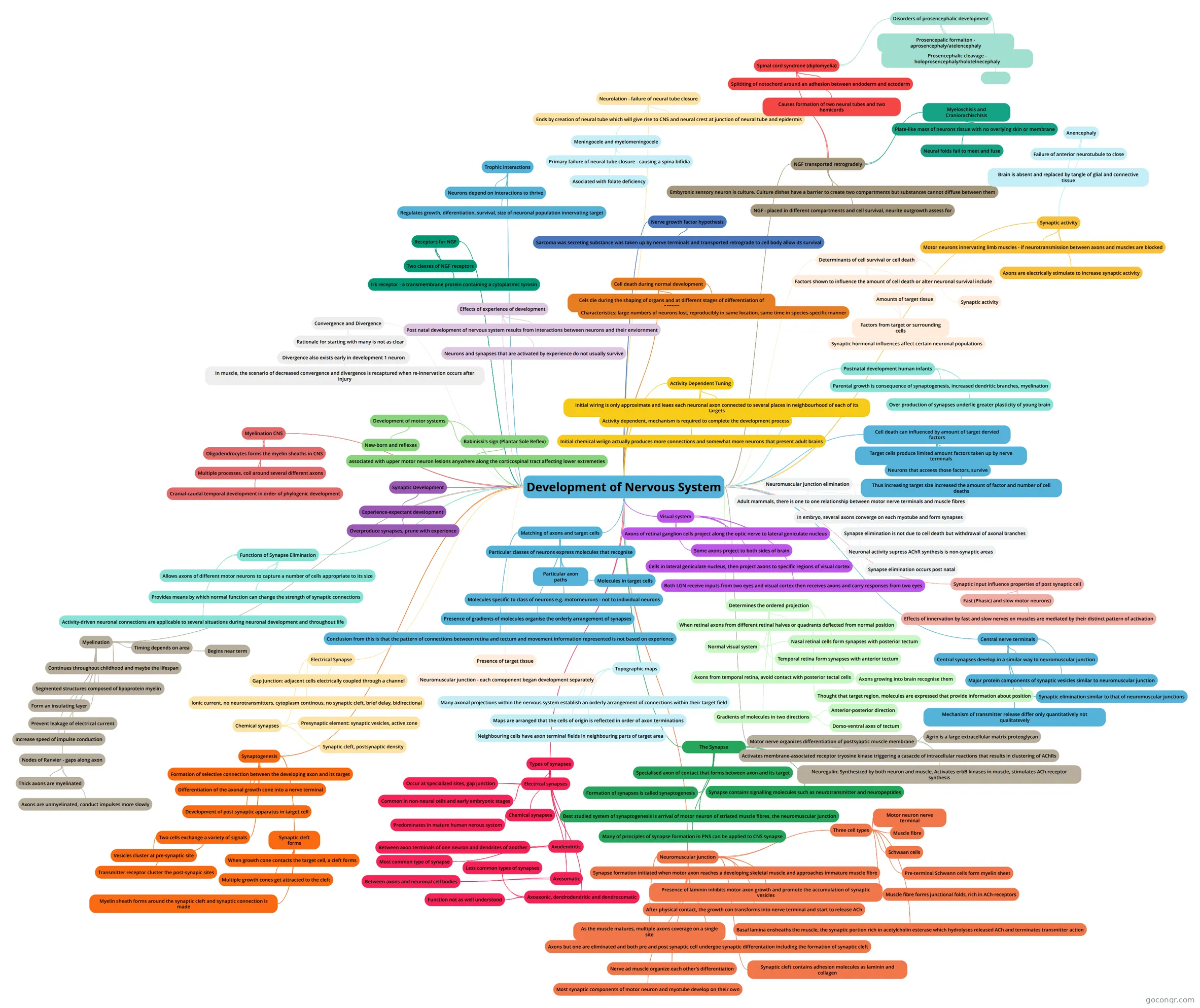
Mind map: Development of nervous system
Contenido del mapa
Development of Nervous System
Matching of axons and target cells
- Particular classes of neurons express molecules that recognise
- Particular axon paths
- Molecules in target cells
- Molecules specific to class of neurons e.g. motorneurons - not to individual neurons
- Presence of gradients of molecules organise the orderly arrangement of synapses
- Conclusion from this is that the pattern of connections between retina and tectum and movement information represented is not based on experience
- Particular classes of neurons express molecules that recognise
Presence of target tissue
- Neuromuscular junction - each comoponent began development separately
Topographic maps
- Many axonal projections within the nervous system establish an orderly arrangement of connections within their target field
- Maps are arranged that the cells of origin is reflected in order of axon terminations
- Neighbouring cells have axon terminal fields in neighbouring parts of target area
Visual system
- Axons of retinal ganglion cells project along the optic nerve to lateral geniculate nucleus
- Some axons project to both sides of brain
- Cells in lateral geniculate nucleus, then project axons to specific regions of visual cortex
- Both LGN receive inputs from two eyes and visual cortex then receives axons and carry responses from two eyes
Determines the ordered projection
- When retinal axons from different retinal halves or quadrants deflected from normal position
- Normal visual system
- Nasal retinal cells form synapses with posterior tectum
- Temporal retina form synapses with anterior tectum
- Axons from temporal retina, avoid contact with posterior tectal cells
- Thought that target region, molecules are expressed that provide information about position
- Axons growing into brain recognise them
- Gradients of molecules in two directions
- Anterior-posterior direction
- Dorso-ventral axes of tectum
The Synapse
- Specialised axon of contact that forms between axon and its target
- Formation of synapses is called synaptogenesis
- Synapse contains signalling molecules such as neurotransmitter and neuropeptides
- Best studied system of synaptogenesis is arrival of motor neuron of striated muscle fibres, the neuromuscular junction
- Many of principles of synapse formation in PNS can be applied to CNS synapse
Types of synapses
- Electrical synapses
- Occur at specialized sites, gap junction
- Common in non-neural cells and early embryonic stages
- Chemical synapses
- Predominates in mature human nerous system
- Axodendritic
- Between axon terminals of one neuron and dendrites of another
- Most common type of synapse
- Axosomatic
- Between axons and neuronal cell bodies
- Axoaxonic, dendrodendritic and dendrosomatic
- Less common types of synapses
- Function not as well understood
- Electrical synapses
Electrical Synapse
- Gap Junction: adjacent cells electrically coupled through a channel
- Ionic current, no neurotransmitters, cytoplasm continous, no synaptic cleft, brief delay, bidirectional
- Chemical synapses
- Presynaptic element: synaptic vesicles, active zone
- Synaptic cleft, postsynaptic density
Synaptogenesis
- Formation of selective connection between the developing axon and its target
- Differentiation of the axonal growth cone into a nerve terminal
- Development of post synaptic apparatus in target cell
- Two cells exchange a variety of signals
- Vesicles cluster at pre-synaptic site
- Transmitter receptor cluster the post-synapic sites
- Synaptic cleft forms
- When growth cone contacts the target cell, a cleft forms
- Multiple growth cones get attracted to the cleft
- Myelin sheath forms around the synaptic cleft and synaptic connection is made
Neuromuscular junction
- Synapse formation initiated when motor axon reaches a developing skeletal muscle and approaches immature muscle fibre
- Presence of laminin inhibits motor axon growth and promote the accumulation of synaptic vesicles
- After physical contact, the growth con transforms into nerve terminal and start to release ACh
- As the muscle matures, multiple axons coverage on a single site
- Axons but one are eliminated and both pre and post synaptic cell undergoe synaptic differentation including the formation of synaptic cleft
- Three cell types
- Motor neuron nerve terminal
- Muscle fibre
- Schwaan cells
- Pre-terminal Schwann cells form myelin sheet
- Muscle fibre forms junctional folds, rich in ACh-receptors
- Basal lamina ensheaths the muscle, the synaptic portion rich in acetylcholin esterase which hydrolyses released ACh and terminates transmitter action
- Nerve ad muscle organize each other's differentiation
- Synaptic cleft contains adhesion molecules as laminin and collagen
- Most synaptic components of motor neuron and myotube develop on their own
Motor nerve organizes differentiation of postsyaptic muscle membrane
- Agrin is a large extracellular matrix proteoglycan
- Activates membrane-associated receptor tryosine kinase triggering a casacde of intracellular reactions that results in clustering of AChRs
- Neuregulin: Synthesized by both neuron and muscle, Activates erbB kinases in muscle, stimulates ACh receptor synthesis
Neuromuscular junction elimination
- Adult mammals, there is one to one relationship between motor nerve terminals and muscle fibres
- In embryo, several axons converge on each myotube and form synapses
- Synapse elimination is not due to cell death but withdrawal of axonal branches
- Neuronal activity supress AChR synthesis is non-synaptic areas
- Synapse elimination occurs post natal
Synaptic input influence properties of post synaptic cell
- Fast (Phasic) and slow motor neurons)
- Effects of innervation by fast and slow nerves on muscles are mediated by their distinct pattern of activation
Central nerve terminals
- Central synapses develop in a similar way to neuromuscular junction
- Major protein components of synaptic vesicles similar to neuromuscular junction
- Mechanism of transmitter release differ only quantitatively not qualitatevely
- Synaptic elimination similar to that of neuromuscular junctions
Activity Dependent Tuning
- Initial wiring is only approximate and leaes each neuronal axon connected to several places in neighbourhood of each of its targets
- Activity dependent, mechanism is required to complete the development process
- Initial chemical wriign actually produces more connections and somewhat more neurons that present adult brains
Postnatal development human infants
- Parental growth is consequence of synaptogenesis, increased dendritic branches, myelination
- Over production of synapses underlie greater plasticity of young brain
Synaptic Development
- Experience-expectant development
- Overproduce synapses, prune with experience
- Experience-expectant development
Functions of Synapse Elimination
- Allows axons of different motor neurons to capture a number of cells appropriate to its size
- Provides means by which normal function can change the strength of synaptic connections
- Activity-driven neuronal connections are applicable to several situations during neuronal development and throughout life
Myelination
- Begins near term
- Continues throughout childhood and maybe the lifespan
- Timing depends on area
- Segmented structures composed of lipoprotein myelin
- Form an insulating layer
- Prevent leakage of electrical current
- Increase speed of impulse conduction
- Nodes of Ranvier - gaps along axon
- Thick axons are myelinated
- Axons are unmyelinated, conduct impulses more slowly
Myelination CNS
- Oligodendrocytes forms the myelin sheaths in CNS
- Multiple processes, coil around several different axons
- Cranial-caudal temporal development in order of phylogenic development
Development of motor systems
- New-born and reflexes
- Babiniski's sign (Plantar Sole Reflex)
- associated with upper motor neuron lesions anywhere along the corticospinal tract affecting lower extremeties
Convergence and Divergence
- Rationale for starting with many is not as clear
- Divergence also exists early in development 1 neuron
- In muscle, the scenario of decreased convergence and divergence is recaptured when re-innervation occurs after injury
Effects of experience of development
- Post natal development of nervous system results from interactions between neurons and their enviornment
- Neurons and synapses that are activated by experience do not usually survive
Cell death during normal development
- Cels die during the shaping of organs and at different stages of differentiation of organs
- Characteristics: large numbers of neurons lost, reproducibly in same location, same time in species-specific manner
Determinants of cell survival or cell death
- Factors shown to influence the amount of cell death or alter neuronal survival include
- Amounts of target tissue
- Factors from target or surrounding cells
- Synaptic activity
- Synaptic hormonal influences affect certain neuronal populations
- Factors shown to influence the amount of cell death or alter neuronal survival include
Cell death can influenced by amount of target dervied factors
- Target cells produce limited amount factors taken up by nerve terminals
- Neurons that acceess those factors, survive
- Thus increasing target size increased the amount of factor and number of cell deaths
Synaptic activity
- Motor neurons innervating limb muscles - if neurotransmission between axons and muscles are blocked
- Axons are electrically stimulate to increase synaptic activity
- Anencephaly
- Failure of anterior neurotubule to close
- Brain is absent and replaced by tangle of glial and connective tissue
Nerve growth factor hypothesis
- Sarcoma was secreting substance was taken up by nerve terminals and transported retrograde to cell body allow its survival
NGF transported retrogradely
- Embyronic sensory neuron is culture. Culture dishes have a barrier to create two compartments but substances cannot diffuse between them
- NGF - placed in different compartments and cell survival, neurite outgrowth assess for
- Spinal cord syndrone (diplomyelia)
- Splititing of notochord around an adhesion between endoderm and ectoderm
- Causes formation of two neural tubes and two hemicords
- Disorders of prosencephalic development
- Prosencepalic formaiton - aprosencephaly/atelencephaly
- Prosencephalic cleavage - holoprosencephaly/holotelnecephaly
- Myeloschisis and Craniorachischisis
- Plate-like mass of neurons tissue with no overlying skin or membrane
- Neural folds fail to meet and fuse
Receptors for NGF
- Two classes of NGF receptors
- Irk receptor - a transmembrane protein containing a cytoplasmic tyrosin
Trophic interactions
- Neurons depend on interactions to thrive
- Regulates growth, diferentiation, survival, size of neuronal population innervating target
Neurolation - failure of neural tube closure
- Ends by creation of neural tube which will give rise to CNS and neural crest at junction of neural tube and epidermis
- Meningocele and myelomeningocele
- Primary failure of neural tube closure - causing a spina bifidia
- Asociated with folate deficiency
Información
- Descripción
- Universität Neuroscience Mapa Mental sobre Development of Nervous System, creado por Paras Chrysostomou el 21/05/2016.
- Creado por
- Paras Chrysostomou
- Visitas
- 0
- Valoración