
Flashcards
NERVOUS SYSTEM
Cards in this deck (166)
- 1Two types of motor disease1. ALS (Amyotrophic lateral sclerosis)
2. MS (Multiple Sclerosis) - 2ALSamyotrophic lateral sclerosis
This is a progressive degeneration of upper and lower motor neurons through protein aggregration, where the signals required for contraction/movement of muscles are disrupted - so there is an inability to initiate and control voluntary movement.
Extreme consequence of this is that it can lead to an inability to eat, move and eventually breathe.
Important to note that ALS doesn't affect brain function itself.
No known cure so mortality is 100% - 3upper motor neurones vs lower motor neuronesupper motor neurones originate in the cerebral cortex and travel down to the brain stem or spinal cord
lower motor neurones begin in the spinal cord and go on to innervate muscles and glands throughout the body - 4List a) Early symptoms of ALS b) Symptoms - weeks to months c) symptoms at far later stages d? Average life expectancya) Slight weakness in hand and limbs
b) twitching/cramping - muscles
loss of motor control - hands/legs
increased tripping/falling
persistent fatigue
slurred speech
c) difficulty breathing/swallowing- dysphasia/paralysis
d) 2-5 years after diagnosis - 5disease mechanism behind ALSExact cause = unknown
Genetic mutations + environmental factors have equal importance - 6how many genes have been associated with ALS20
- 7two factors which are the main drivers of symptoms in those with ALSneuronal dysfunction and cell death.
- 8protein aggregationdisordered proteins or mis-folded proteins aggregate (i.e., accumulate and clump together)
- 9dymyelinating disease and multiple sclerosisdymyelinating diseases - diseases where normal myelin is broken down for inappropriate reasons, often by an inflammatory process
e.g. multiple sclerosis - affects the conduction of electrical signalling of the CNS - 10myelinmixture of proteins and phospholipids forming a whitish insulating sheath around many nerve fibres, increasing the speed at which impulses are conducted.
- 11where does MS commonly developpatients 20-30
- 12symptoms of MS and life expectancyblurred vision, uncontrolled voluntary movement, loss of sensation and balance.
life expectancy slightly reduced - 13symptom progression with MSClinically isolated syndrome (CIS) describes the first episode that causes inflammation and damage to the nerves (myelin)
Relapsing remitting MS (RRMS) follows a predictable pattern where symptoms worsen and then improve.
Secondary progressive MS (SPMS)- This is the more aggressive form of the disease that RRMS can develop into if untreated. - 14clinical characteristics of MS and combined effectsFormation of lesions in CNS (known as plaques)
Inflammation due to autoimmune responses
destruction of myelin
Combined effects: disrupted signalling between neurons in the CNS which leads to symptoms - blurred vision, involuntary movement, loss of sensation, balance - 15reason why MS not solely be geneticScientists compared genetic information in three sets of identical twins. One of each pair had MS, and the other didn't — yet their genes proved essentially identical.
- 16excitable cells and communicationexcitable cells = muscle cells + neurones
uses their bioelectricity to communicate with downstream neurones, which then communicates with another etc
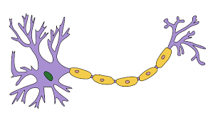
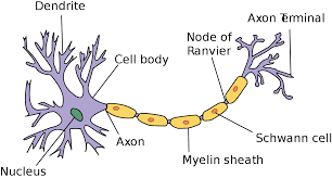
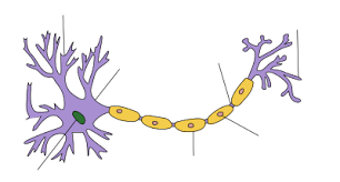
neurones sense stimuli (chemicals, pressure, pain), transmitting info in form of electrical impulses - from one neuron to the next or over longer distances to enable a result (contraction of a muscle fibre, release of messenger chemicals such as hormones and neurotransmitters) - 17anatomy of nueronedendrites -collects electrical signals from upstream neurones and brings this to the cell body (soma).
soma - integrates incoming signals and generates outgoing signal to axon which generates a process called neurotransmission
axon - passes electrical signal to downstream neurones via the dendrites of the downstream neurones. - 18—
- 19Law of Dynamic PolarizationInformation always flows from presynaptic to postsynaptic with regard to a specific synapse
(in other words, the neurons have a preferred direction for propagation of information) - 20specialised cell in nervous systemGlial cells - cells which do not produce electrical impulses but are still found in central and peripheral NS.
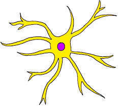
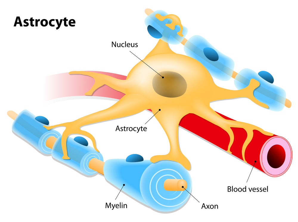
- types are schwann cells and astrocytes - 21astrocytesBranched glial cells which fill spaces between neurones in CNS
Prevents unwanted signals being received by neurons by ensuring the neurotransmitters are 'mopped' up and transferred directly from neuron A to B only. - 22swhann cellsspecialised glial cells which produce myelin in PNS
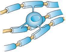
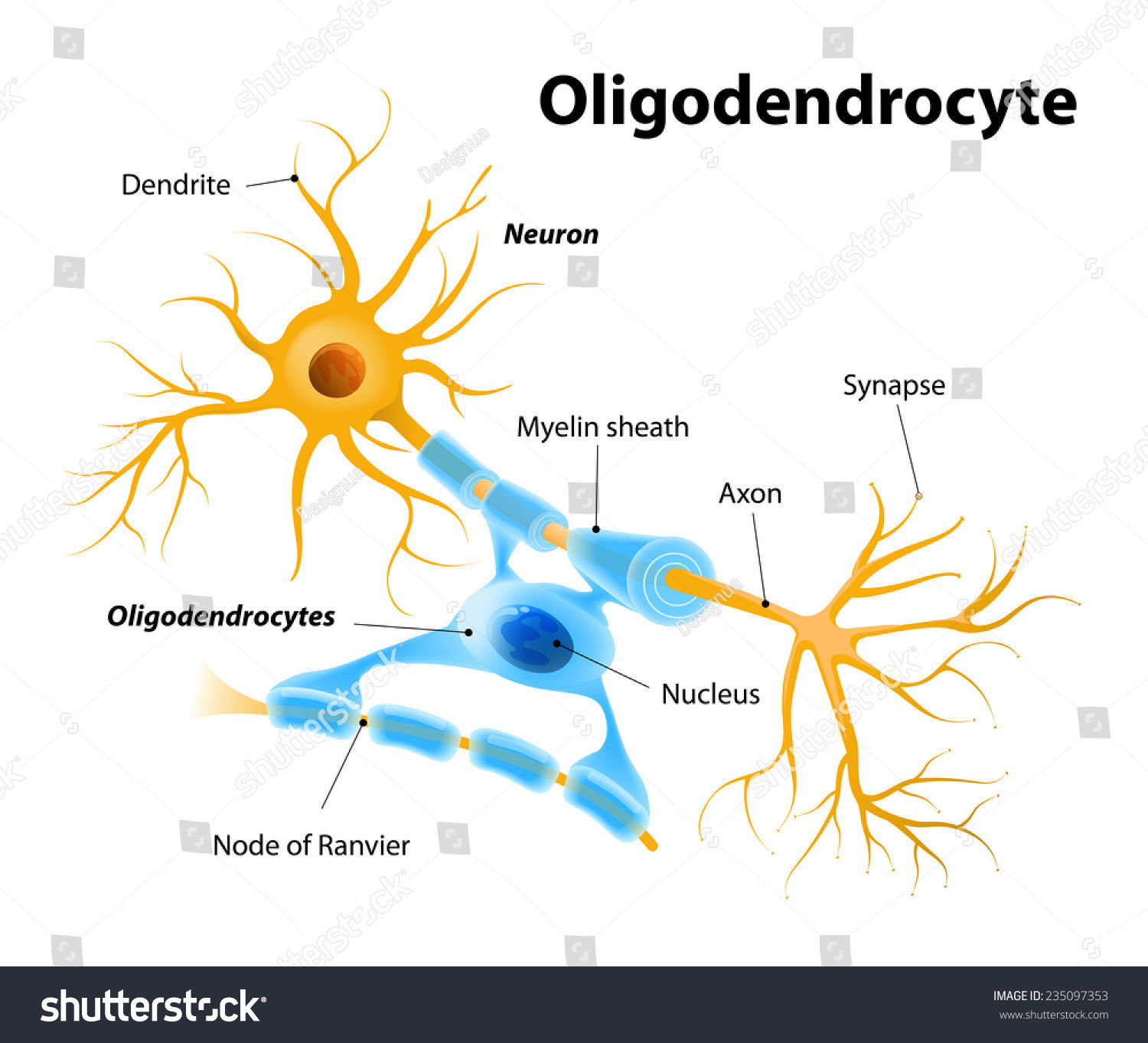
- 23Oligodendrocytestype of large glial cell found in the central nervous system which produces the myelin sheath to insulate axons
- 24—
- 25—
- 26—
- 27afferent axonssensory axons
take info from periphery to CNS
e.g. Afferent axons from skin sends info to brain/spinal cord (CNS) - info can be regarding temp, pressure - 28efferent axonsmotor axons which innervate muscles and so transfer info from CNS to muscles
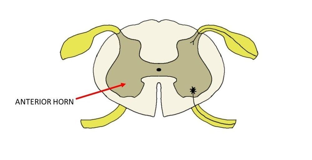
- 29location of efferent and afferent axonsDorsal root of spine - afferent axons
Vental root - efferent axons - 30—
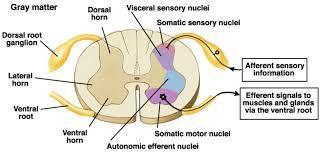
- 31brain vs spinal cordBrain concerned with cognition/memory/emotion/movement
Spinal cord concerned with reflective actions which are automatic - so short-latency responses to stimuli which does not require the brain (hand on hot surface) - 32electrical currents in terms of biologymovement of ions in solutions
- 33membranes of neurones and sodium/potassium concentrationsneurone is bound by a plasma membrane with phospholipid bilayer, a structure so thin that its fine detail can be revealed only by microscope.
Low chloride/sodium ion conc in cytoplasm (high potassium ions conc)
High chlorine/sodium ion conc in extracellular fluid (low potassium ion conc.)
so high potassium conc inside cell and high sodium conc outside cell - 34—diagram showing membrane
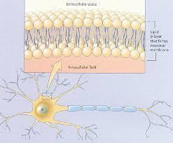
- 35major ion pump exampleSodium potassium ATPase
ATP converted to ADP and resulting phosphate group is used in removing 3 potassium ions from the cell, and then moves 2 sodium ions into the cell. - 36concentrations of Na and k ions relative to cytoplasm and extracellular fluidCytoplasm:
Na - 15mM
K - 100mM
Extracellular fluid
Na - 150mM
K - 5mM
Ratio out/in
K - 1:20
Na - 1:10 - 37resting membrane potentialelectrical charge of a neurone when it is not excited.
-65mV - 38type of channel which dictates the resting membrane potential and factors influencing this channelleak potassium channel - two pore domain potassium channel which is open at rest. They are not voltage dependent - enables movement of potassium ions in and out of cell to help set the membrane potential.
These can be regulated by external pH, oxygen tension and stretch of membrane itself. - 39creation of equilibrium potential across the membraneIn terms of the charges on the Na+ and K+, the ratio of their charges is 1:1. So no charge is accumulating inside/outside membrane
But the potassium leak channel means the ratios for the cations to anions differ inside and outside of the cell - so inside of membrane develops a net negative charge (K+ leaves)
Initially diffusion is dominant force in moving K+ ions across the membrane but as the inside of the membrane becomes more negatively charged, this negative electrical force pulls the K+ ions back through the potassium channel into the inside of the cell.
Eventually the electrical force which is pulling K+ into cell counterbalances the force of diffusion pushing K+ ions out of the cell and thus creates the equilibrium potential.
This is where electrical and diffusion forces are opposite in direction but equal in size - so there is no net movement of K+ ions through the channel despite the conc gradient.
Equilibrium potential for K+ is approx -80mV. - 40action potential- basic functional unit of neurone (neurone is basic unit of NS)
- mode through which a neurone transports electrical signals - 41depolarisationChange of membrane resting potential from -65mV to 0mV - this can result in a single action potential or multiple depending on degree of current injected and the duration of this.
- 42factor increasing action potential firing rateaction potential firing rate increases as the depolarising current increases.
- 43factors telling NS the nature of the external stimulinumber and frequency of action potentials
- 44threshold for the action potential-50 to -55 mV
- sufficient depolarisation needed to reach this threshold - 45last action potentialindicates that the flow of injected current has now been stopped or that the stimulus has been removed.
- 46control of skeletal musculatureaction potentials used for bidirectional signalling in long-term communication
- e.g. foot to brain
- so motor commands signalled from CNS to PNS (going to the foot) to move it BUT there is sensory and proprioceptive feedback from the muscles which will then propagate back up through the PNS to the CNS to inform the brain that the foot is moving.
So, these motor-sensory feedback loops enables control of the skeletal musculature.- - 47types of diseases occurring through disruption of action potentialsALS/MS/Alzheimers/locked-in-syndrome/Charcot-Marie tooth disease
- 48stages of action potential1 - Resting membrane potential sitting at -65mV due to flow of K+ ions through the leak potassium channel.
2. Rising phase - rapid depolarisation induced by opening of Na+ voltage-gated channels.
3. overshoot - Refers to that part of the action potential where the membrane potential is positive (inside with respect to the outside)
3. Falling phase - hyperpolarisation induced through opening of K+ voltage-gated channels.
4.undershoot - point at which hyperpolarisation results in a membrane potential that is slightly more negative than the resting potential.
5. Resting membrane potential is re-established - 49—
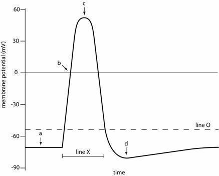
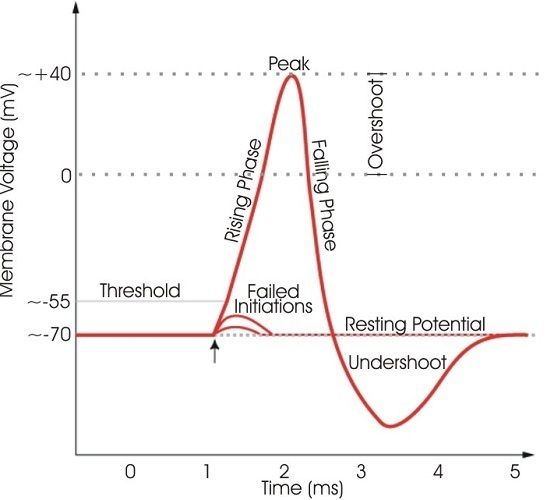
- 50opening/closing of voltage-gated Na+ channels during action potentialProtein channel is closed at rest (-65 to -70mV)
Protein opens when threshold is reached (-55mV)
Depolarisation results in a conformational change in the protein , opening a pore. Protein then acts as a selective filter to enable passage of Na+ ions.
The channel opens very fast to produce rapid depolarisation (this is the rising phase) -
The channel stays open for 1ms before inactivating/closing (channel is inactivated by a physical pore block) - 51opening/closing of voltage-gated K+ channels during action potentialrequire depolarisation to open.
Slow to open (1ms after depolarisation) unlike Na+ voltage dependent channels
So the delay in opening is why they get the name "delay rectifier potassium channels
When channel opens and there is an influx of potassium ions, the cell will begin to hyperpolarise (become more negative now that potassium is moving out of the cell)
channel will close when -65mV (resting membrane potential) is reached. - 52process of voltage gated channels when stimulus received - 1st halfinjected current is applied to/stimulus is received by lipid membrane, (e.g. neurotransmitter binds to ligand-gated sodium channel), and the conformational change in this sodium channel creates an initial influx of sodium ions because of the concentration gradient and electrical gradient, meaning there is a wave of initial depolarisation within the cell.
When this depolarisation occurs, it takes the membrane potential up from -65/70mV to -59mV (i.e. threshold potential is reached) this results in the opening of the Na+ voltage dependent channel via a conformational change. The influx of Na+ ions into the cell causes a further, more explosive and rapid depolarisation (rising phase) and essentially, the action potential is now being generated. The influx of Na+ lasts for approx 1ms before the channel is inactivated by a physical block.
with influx of Na+membrane moves rapidly toward 0 mV and then continues in a positive direction to a peak of +30 mV. The positive value at the peak of the action potential indicates that there is an excess of positive ions inside (overshoot) - 53process of voltage gated channels when stimulus received - 2nd halfThe initial depolarisation also means the delay rectifier potassium channel opens and the release of potassium ions from the cells essentially causes hyperpolarisation (inside of cell becomes more negative - falling phase) and when -65mV is reached the channel closes (i.e., it closes when the resting membrane potential is restored.)
However, due to the delay in closing of this potassium voltage-gated channel, there is an "undershoot' (a period of after-hyperpolarization) which means that the membrane potential is below the resting membrane potential. The resting membrane potential is restored through the leak potassium channel returning to its resting state. - 54refractory periodTime in which an excitable cell is unable to generate a subsequent action potential.
Absolute refractory period = This is when all sodium channels are open so ultimately there are none others available to generate action potentials.
Relative refractory period = period in which the cell is hyperpolarised - lower probability of action potential being generated. - 55factors influencing the action potential conduction velocity (or its firing rate)Resistance of plasma membrane to passage of ions
Diameter of axon dictates how fast action potential propagates (thin diameter means a higher resistance and this will restrict the conduction velocity. Whereas wide diameters means the axial resistance is low so less restriction in terms of conduction velocity - so thus action potential propagates further.)
Density of voltage dependent sodium channels - higher densities among the axis = higher probability of strong depolarisation so action potentials can be activated more rapidly. - 56saltatory conductionthe propagation of action potentials along myelinated axons
node-to-node propagation of the action potential - 57myelin sheath structurenot continues since there is a gaps in the axon known as the nodes of ranvier (gaps which are essentially high concentrations of sodium channels)
- 58specific location where action potential begins and subsequent propagationAP begins at the axon hillock (initial segment right next to the soma) which will then propagate along the axon underneath the myelin sheath and then depolarises the nodes of reverie which leads to explosive activation of the sodium channels thus generating another action potential around the node of ranvier.
This action potential is then propagating down the axon again, to the next node of ranvier to generate another action potential and so forth. - 59—
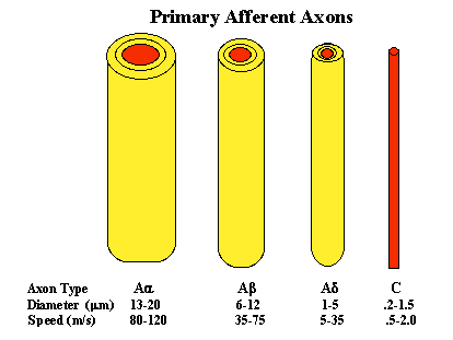
- 60different types of axonsAlpha-a axons - large diameter axons - so low axial resistance - higher generations of action potentials - have myelin sheaths. They propagate info from proprioreceptors
a-beta axons - relatively small in diameter - also contain myelin sheaths. They propgate info from mechanoreceptors on the skin
a-delta - receive info from free nerve endings present in the skin, and they transmit info regarding pain/temp/crude touch. Diameter is much smaller but still contain myelin sheath.
c-fibres - free nerve endings which are smaller in diameter and do not contain myelin sheath. Thus, velocity of conduction of the action potential is much slower - transmit info regarding pain/temp/sensation of itch - 61—diagram to show different axons
- 62propriorecetors vs mechanoreceptorsproprioreceptors - sensory receptor which receives stimuli from within the body, especially one that responds to position and movement.
mechanoreceptors -respond to touch, pressure, vibration, stretch, and itch - 63mechanosensiitve ion channelsgating depends on stretch of surrounding membrane (so they can respond to membrane tension/stretch by altering their conformation)
- 64receptors in the skinthermoreceptors
Meissner's corpuscles (senses touch)
nociceptors (sense pain)
Pacinian corpuscle (senses different pressure application to skin) - 65process when sensory receptors are activated within skin cellsaction potential within receptors in skin would begin in the dorsal root and propagate back from the periphery to the spinal cord where it can be processed in spinal cord or to brain (to assess whether input was tough/pressure.)
- 66Dermatomearea of the skin which contains primary afferents that transduce info from periphery to spinal cord.
- 67factors encoded in the action potential firing frequencyStimulus strength and duration.
stimulus onset/offset encoded in 1st/last action potentials.
Pressing stronger into hand (stronger pressure) would generate a far stronger action potential. - 68effects of damaged myelin sheath/ having unmeylinated axons in patientsLeads to slow generation of action potentials so info transmitted very slowly - so its fundamentally slowing the nerve impulses - so.....
loss of vision
bladder or bowel problems
unusual nerve pain
overall fatigue
with severe deterioration of myelin...
numbness
loss of reflexes and uncoordinated movements
poorly controlled blood pressure
blurred vision
dizziness
racing heart beat or palpitations
memory problems
pain - 69voltage/electrochemical potential across cellmembrane that exhibits a membrane potential is said to be polarized. That is, its membrane has a negative pole (the side on which there is an excess of negative ions) and a positive pole (the side on which there is an excess of positive ions).
the value −70 mV indicates that the potential difference has a magnitude of 70 mV and that the inside of the membrane is negative with respect to the outside surface - 70voltmeter and readingsrecorded as a red line on the screen of the voltmeter. Upward deviations of the red line are depolarizations (come from excitatory stimuli); downward deviations of the red line are hyperpolarizations (comes from inhibitory stimuli).
- 71specific ions passing across membraneIn the neuron's plasma membrane, channels for the transport of the major anions (negative particles) are either nonexistent or mostly closed.
For example, there are no channels to allow the exit of the large anionic protein molecules that dominate the intracellular fluid.
Chloride ions (Cl − ), the dominant extracellular anions, are likewise "trapped" on one side of the membrane because chloride ions are repelled by the protein anions inside the cell. This means that the only ions that can move efficiently across a neuron's membrane are the positive ions sodium and potassium. - 72local/graded potentialproduced when a ligand opens a ligand-gated channel in the dendrites, allowing ions to enter (or exit) the cell.
A slight shift away from the RMP (resting membrane potential) in a specific region of the plasma membrane. - 73another term for ligand-gated channelsstimulated-gated channels
- 74hyperpolarisationmovement away from 0 and usually below RMP.
- 75voltage-gated channels locationthere are many voltage-gated Na + channels and K + channels in the membrane of the neurones summation zone (axon hillock) and conduction zone (axon)
- 76threshold potential definitionthe minimum magnitude a voltage fluctuation in the summation or conduction zone must have to trigger the opening of a voltage-gated ion channel.
it is essentially a positive feedback mechanism to produce action potential - 77positive feedback vs negative feedbackPositive feedback occurs to increase the change or output: the result of a reaction is amplified to make it occur more quickly.
Negative feedback occurs to reduce the change or output: the result of a reaction is reduced to bring the system back to a stable state. - 78factor encoded in frequency of action potentialstrength of stimulus - not changing the magnitude of the action potentia
- 79reason for inability of action potential to move backwardsprevious segment of membrane remains in a refractory period too long to allow such restimulation. This is the mechanism responsible for the one-way movement of action potentials along axons.
(brief period during which a local area of an axon's membrane resists restimulation - For about 0.5 ms) - 80purpose of nodes of ranvierallow for ions to diffuse in and out of the neurone, propagating the electrical signal down the axon
- 81fastest and slowest nerve impulse transmission in humansthose that innervate the skeletal muscles conduct electrical impulses fastest
slowest fibres are those such as the sensory receptors of skin - 82nerve system and anaestheticsMany anaesthetics produce their effects by inhibiting the opening of sodium channels and thus blocking the initiation and conduction of nerve impulses.
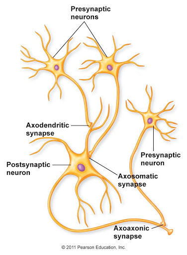
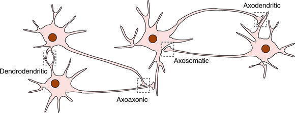
- 83synapsespecialised structure sitting between the axon tip of an upstream neuron and the dendrite of a downstream neuron
- 84types of synapseschemical and electrical
- 85electrical synapsesOccur where two cells are joined end-to-end by gap junctions.
Because the plasma membranes and cytoplasm are functionally continuous in this type of junction, an action potential can simply continue along the postsynaptic plasma membrane as if it belonged to the same cell.
Electrical synapses occur between cardiac muscle cells and between some types of smooth muscle cells. - 86chemical synapsesUse a chemical transmitter called a neurotransmitter to send a signal from the presynaptic cell to the postsynaptic cell.
.
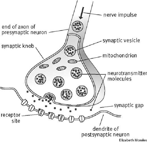
Three structures make up a chemical synapse:
1.A synaptic knob
2.A synaptic cleft
3.The plasma membrane of a postsynaptic neuron - 87synaptic knobend of the presynaptic neurone containing vesicles containing neurotransmitters
- 88synaptic cleftIt forms a junction between two or more neurones and helps nerve impulse pass from one neurone to the other.
synaptic cleft is not an empty space—it contains fluid, enzymes, adhesion molecules, and other substances. - 89—
- 90axodendritic synapsesone between the axon of one neurone and the dendrites of another
- 91axosomatic synapsesone between the axon of one neurone and the cell body/soma of another
- 92axoaxonic synapsesone neurone projecting its axon terminals onto another neurones axon
less common but offer a way for one neurone to inhibit or facilitate synaptic transmission by another neurone. - 93—
- 94reason for neurotransmittersAn action potential that has travelled the length of a neuron stops at its axon terminals. Action potentials cannot cross synaptic clefts.
Instead, neurotransmitters are released from the synaptic knob, cross the synaptic cleft, and bring about a response by the postsynaptic neuron. - 95excitatory vs. inhibitory neurotransmittersExcitatory neurotransmitters cause depolarization of the postsynaptic membrane (both K+ and Na+ channels opened)
Inhibitory neurotransmitters cause hyperpolarization of the postsynaptic membrane (opening k+ or Cl- channels) - 96mechanisms of synaptic transmission - part one1.When an action potential reaches a synaptic knob, voltage-gated calcium channels in its membrane open and allow calcium ions (Ca 2+ ) to diffuse into the knob rapidly. Many voltage-gated Ca 2+ channels are present in the membrane of the neurones output zone.
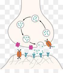
2. The increase in intracellular Ca 2+ concentration triggers molecular motors to pull neurotransmitter vesicles to the plasma membrane of the synaptic knob. Once there, the vesicles fuse with the membrane and release their neurotransmitter via exocytosis. Thousands of neurotransmitter molecules spurt out of the open vesicles into the synaptic cleft.
3. The released neurotransmitter molecules almost instantaneously diffuse across the narrow synaptic cleft and contact the postsynaptic neuron's plasma membrane. Here, neurotransmitters briefly bind to receptor molecules that are also gated channels. Binding of neurotransmitters triggers the channels to open. - 97mechanisms of synaptic transmission - part two4. The opening of ion channels in the postsynaptic membrane may produce a local potential called a postsynaptic potential. Excitatory neurotransmitters cause both Na + channels and K + channels to open. Because Na + rushes inward faster than K + rushes outward, there is a temporary depolarization called an excitatory postsynaptic potential (EPSP).
Inhibitory neurotransmitters cause K + channels and/or Cl − channels to open. If K + channels open, K + rushes outward; if Cl − channels open, Cl − rushes inward. Either event makes the inside of the membrane even more negative than at the resting potential. This temporary hyperpolarization is called an inhibitory postsynaptic potential (IPSP).
5. Once a neurotransmitter binds to its postsynaptic receptors, its action is quickly terminated. Several mechanisms bring this about. - 98neurotransmitters after useneurotransmitter's action inhibited when bound to post-synaptic receptors
Some neurotransmitter molecules are metabolized into inactive compounds by synaptic enzymes.
Other neurotransmitter molecules simply diffuse out of the synaptic cleft and are transported into nearby glial cells. The glial cells may release them again for reuptake by the presynaptic neurone, sometimes after breaking them down into another form. - 99summation - two typestemporal and spatial
- 100efficiency of excitatory neurotransmitter if released by one synaptic knobamount of excitatory neurotransmitter released by one knob is not enough to trigger an action potential. It may, however, facilitate initiation of an action potential by producing a local depolarization of the synaptic membrane—an EPSP.
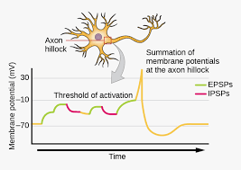
- 101difference between temporal and spatial summationSpatial summation occurs when multiple presynaptic neurones together release neurotransmitter (e.g. acetylcholine) and their local potentials may spread far enough to reach the axon hillock, where they may summate and subsequently
exceed the threshold of the postsynaptic neurone to cause action potential.
Temporal summation occurs when one presynaptic neurone releases neurotransmitter many times over a period of time to post-synaptic neurone to produce action potential. - 102Summationtemporal and spatial summation
Usually both excitatory and inhibitory transmitters are released at the same postsynaptic neuron.
The excitatory neurotransmitters produce local EPSPs, and the inhibitory neurotransmitters produce local IPSPs. Summation of these opposing local potentials occurs at the axon hillock, where many voltage-gated ion channels are located.
If the EPSPs predominate over the IPSPs enough to depolarize the membrane to the threshold potential, the voltage-gated channels will respond and produce an action potential. The action potential is then conducted without decrement along the axon's membrane.
If the IPSPs predominate over the EPSPs, the membrane will not reach the threshold potential. The voltage-gated channels at the axon hillock will not respond, and no action potential will be conducted along the axon. - 103serotonin and releaseserotonin - contributes to wellbeing and happiness
released at an axoaxonal synapse and triggers the axon terminal of a presynaptic neurone to block its potassium channels for up to several weeks.
Thus whenever there is an action potential arriving at that presynaptic neurone, it lasts longer (because potassium fails to rapidly repolarize the membrane.)
A prolonged action potential means that the presynaptic calcium channels stay open longer and trigger the release of more neurotransmitter than usual. - 104acetylcholineexcites skeletal muscle cells but inhibits cardiac muscle cells.
This illustrates that neurotransmitters can have inhibitory effects at some synapses and excitatory effects at other synapses.
It also shows that : their function is determined by the postsynaptic receptors, not by the neurotransmitters themselves. - 105ionotropic receptorsmembrane receptors that are part of an ion channel and thus act directly to change ion permeability when stimulated.
Can result in generation of action potential if influx encourages sufficient depolarisation. - 106metabotropic receptorsdo not form an ion channel pore but use signal transduction mechanisms, often G proteins, to activate a series of intracellular events using second messenger chemicals.
activation of a secondary messenger (secondary messenger cascade) promotes release of G-protein subunits (alpha subunits) - these bind with effector protein or modulate activity of other ion channels by binding with them.
Binding of this with neurotransmitter requires GDP since it is an energy-active process, GDP also causes translocation of G-protein which will then activate a membrane-bound enzyme.
This results in a cascade of activation of other downstream enzymes which triggers a cascade in itself
enzymes activated - e.g. phospholipase C and Adenylyl cyclase. - 107norepinephrineAKA NA
Binds to a metabotrophic receptor.
neurotransmitter norepinephrine binds to G protein-coupled receptors (GPCRs). Triggering the GPCR causes G proteins to activate the membrane-bound enzyme adenyl cyclase .
The adenyl cyclase removes phosphate groups from adenosine triphosphate (ATP) to form cyclic adenosine monophosphate (cAMP) .
Cyclic AMP is the "second messenger", triggering the activation of the protein kinase A enzyme that eventually causes Na + channels in the postsynaptic membrane to open. - 108metabotropic second-messenger mechanism compared to direct ionotropic mechanismmetabotropic second-messenger mechanism is usually slower and longer lasting than the more direct ionotropic mechanism
- 109secondary-messenger mechanismInvolves intracellular messengers, it can regulate other cellular processes, such as cytoskeleton movement, gene expression, and shuttling of proteins along the axonal transport system.
- 110structural classification of neurotransmitterssmall-molecule transmitters and large-molecule transmitters.
- 111small-molecule transmittersmolecules of a smaller size than those in the large-molecule category.
Small-molecule neurotransmitters are amino acids or are derived from individual amino acids
Small-molecule transmitters are subdivided into four main chemical classes:
Class I Acetylcholine
Class II Amines
Class III Amino acids
Class IV Other small molecules - 112large-molecule transmittersmade up of more than one amino acid—usually chains of 2 to 40 amino acids.
so effectively they are all neuropeptides—chains of several amino acids strung together by peptide bonds. - 113Class III Amino acids Class IV Other small moleculessmall-molecule transmitters
Class III - Amino Acids (contain an amine group, carboxylic acid group, and a specific R group)
Glutamate (glutamic acid, Glu) CNS, Excitatory; most common excitatory neurotransmitter in CNS
Gamma-aminobutyric acid (GABA) common inhibitory neurotransmitter in brain
Glycine (Gly)common inhibitory neurotransmitter in spinal cord
Class IV - other small molecules
Nitric oxide (NO)
Purines (contain adenine, a double-ring purine structure)
ATP
adenosine - 114Class I Acetylcholine Class II AminesClass I just ACh
Class II - monoamines - such as dopamine, NA, and adrenaline - which are all catecholamines and histamine/serotonin. - 115examples of larger-molecule transmittersNeuropeptides (chains of amino acids)
Endorphins
Dynorphin - 116link between depression and neurotransmittersa deficit of serotonin, dopamine, norepinephrine, or other amine neurotransmitters exists at synapses along particular pathways of the brain that affect one's mood.
- 117neuromodulatorAlthough neuropeptides may be secreted by a synaptic knob by themselves, some may be secreted along with a second, or even third, neurotransmitter.
In such cases, the neuropeptide is thought to act as a neuromodulator. A neuromodulator is a "co-transmitter" that regulates or modulates the effects of the neurotransmitter released along with it - 118convergence and divergenceoccurs when more than one presynaptic axon synapses with a single postsynaptic neuron.
Convergence allows information from several different pathways to be funneled into a single pathway.
For example, the pathways that innervate the skeletal muscles may originate in several different areas of the central nervous system. Because pathways from each of these motor control areas converge on a single motor neuron, each area has an opportunity to control the same muscle.
Divergence occurs when a single presynaptic axon synapses with many different postsynaptic neurones.
Divergence allows information from one pathway to be "split" or "copied" and sent to different destinations in the nervous system. For example, a single bit of visual information may be sent to many different areas of the brain for processing. - 119retrograde signallingthe transmission of chemical information from a postsynaptic to a presynaptic cell
(concept that signals can be sent "backward" at a synapse) - 120causations of nerve damagephysical injury can cause either local or widespread loss of sensation or motor control.
In some spinal cord injuries, paralysis may occur
Viral infections can also cause damage to nerves, as can autoimmune reactions triggered by viral infections
strokes and transient ischaemic attacks (TIAs), or mini-strokes, reduction of blood flow due to blockage or leakage can reduce glucose and oxygen delivery and damage nerve fibres - 121CVA and nerve damageCVA - cerebrovascular accidents
In a CVA, prolonged oxygen starvation can kill neurones, which then release stored excitatory neurotransmitters such as glutamate. These transmitters flood nearby cells, acting as excitotoxins that harm nearby neurones—thus spreading the loss of function caused by the CVA. - 122method of communication by excitable cellschemical neurotransmission
- 123reason for biological mechanism for inactivation of neurotransmitter actionensures rapid point-point (neuron to neuron) communication in the sense that it can end quickly)
- 124two most common amino acids in CNSGABA and glutamate
- 125synthesis and storage of neurotransmittersEnzymes required for synthesis of neurotransmitters, enzymes generated in cell soma, travel to pre-synaptic terminal, these enzymes then synthesise the neurotransmitters and package them into vesicles - vesicles then diffuse to post-synaptic neurones across the synaptic cleft, releasing the neurotransmitter for generation of bioelectricity at the downstream neurone.
Precursors then travel to pre-synaptic terminal to generate more neurotransmitters (through enzyme intervention) - 126role of calcium in neurotransmissionAction potential invades pre-synaptic terminal and thus membrane depolarises - this means that voltage-gated calcium channels open and the influx of calcium ions encourages vesicle fusion - so the vesicles can then release neurotransmitters into synaptic cleft for transmission.
- 127maintenance of neurotransmitter stock1. Docking - vesicle binds with a neurotransmitter before the action potential invades.
2. Ca2+ sensing - sensed Ca2+ triggers vesicle fusion.
3.. endocytosis - new vesicle membrane is pinched off the pre-synaptic terminal to give an empty vesicle.
4. loading - empty vesicle filled with neurotransmitter. - 128endocytosis vs exocytosisEndocytosis is the process of capturing a substance or particle from outside the cell by engulfing it with the cell membrane, and bringing it into the cell.
Exocytosis describes the process of vesicles fusing with the plasma membrane and releasing their contents to the outside of the cell. - 129examples of excitatory/inhibitory neurotransmittersdecrease excitability of post-synaptic neuron. GABA and glycine. Released from inhibitory synapses, e.g. GABA binds to Gaba receptors on post-synaptic neuron, opens chloride channels which produce hyperpolarisation of cell.
excitatory - increases excitability of post-synaptic neuron. Glutamate, noradrenaline, acetyl choline. Occurs through opening of ion channels to give depolarisation etc. - 130quantal hypothesis of neurotransmissionStimulation of post-synaptic axon will result in each vesicle producing a quantal response
- 131what does summation demonstrate and why is it necessaryneurons dont simply recieve input from one other neuron.
they can integrate input from tens to thousands of pre-synaptic neurons,
Summation of inputs necessary to reach threshold to generate action potential to propagate info. - 132summary of temporal and spatial summation in terms of EPSP'sspatial summation -this is summation of ESPS generated at different synapses.
temporal summation - summation of ESPS generated at the same synapse - results in far greater ESPS - 133inhibitory inputinitially the excitatory input would depolarise the dendrite of the neuron and the depolarisation would passively propagate to the soma.
Inhibitory input would then suppress the excitation - so reduce depolarisation (opening of chloride channels encourages hyperpolarisation) and prevent generation of AP. - 134cholinergic synaptic transmissioninvolves nicotinic receptors - ionotopic, nicotine and curarine binds- and muscarinic receptor - metabotopic, muscarine and atropine bind - both activated by acetyl choline.
Nicotine would activate ACh receptors in brain.
Curare - blocks muscle contractions, so can cause paralysis.
atropine - ophthalmic use, and dilates the pupils. - 135GABAergic synaptic transmissionethanol - affects level of inhibition, effects CNS (cognition) and PNS (motor function)
Barbiturates - mild sedation to full anaesthesia,
Neurosteroids - endogenous modulators, level of it alters during menstraul cycles.
All three of these can bind to the GABA Cl - ion channel - 136somatic systemcontrols contraction of skeletal muscle for voluntary movements
this is where PNS and CNS combine - 137Autonomic nervous system (ANS)Consists of sympathetic and parasympathetic system
1. sympathetic nervous system - includes pre-ganglionic fibres which attach onto post-ganglionic fibres. When activated in the periphery they can contract smooth muscle, cardiac muscle, and gland cells.
2. Parasympathetic nervous system - also containing pre-ganglionic fibres and post-ganglionic fibres
both parasympathetic and sympathetic nervous system work in concert to control contractions of smooth muscles, cardiac muscles - 138pre-ganglionic neurones and post-ganglionic neurones of sympathetic NSits a chain of sympathetic ganglion
pre-ganglionic neurons release acetyl choline and post-ganglionic neurons release noradrenaline.
so the sympathetic neurons would be located next to their targets and are involved in fight/flight responses. It achieves this fight/flight response by the binding of noradrenlaine with post-synaptic adrenoreceptors which will thus relax airways in the lungs, inhibit digestion, and accelerates heart rate.
Fast point-point communication - 139pre-ganglionic neurones vs post-ganglionic neuronespreganglionic neurons, originates in the brainstem or the spinal cord, and the postganglionic neurons, lies outside the central nervous system in collections of nerve cells called autonomic ganglia (so in the periphery.)
- 140Adrenaline - clinical useused to treat patients in cardiac arrest since it accelerates heart rate and increases peripheral resistance (the resistance of the arteries to blood flow)
- 141substances released in parasympathetic nervous systemThis releases acetylcholine.
controls actions which do not require immediate actions - digestion, metabolic functions and kidney/liver regulation.
Achieves so by....when acetyl choline binds to different post-synaptic acetylcholine receptors to give constriction of airways, stimulates digestion and slows hear rate.
parasympathetic neurones will travel large distances. - 142somatic motor systemsingle function: controls and commands all skeletal muscles in the body.
so this is under voluntary control and generates behaviour.
motor command generated in motor cortex - this electrical signal descends through brainsteam into the spinal cord which activates upper motor neurons of the spinal cord which will then continue on the signal to activate downstream motor neurons of the spinal cord which will then be projected onto the muscle fibres to enable contraction of certain body parts. - 143organisation of lower motor neuronesdistributed within ventral horn in a predictable way
the neurones innervating the axial muscles are medial to those innervating the distal muscles.
and the neurones innervating flexors are dorsal to those innervating extensors. - 144alpha motor unitsa single motor neurone - this synapses with many muscle fibres to ensure the speed of the contractile force is even.
alpha motor neurones - directly trigger the generation of force by muscles
then obviously, small motor neurones will innervate small muscles (large motor neurons innervate the larger muscles.)
however, the alpha motor nueron with all the muscle fibres it innervates it is classed as the motor unit. - 145motor neurone poolmore than one motor unit needed clearly since damage to one would mean a loss of contractile ability by the muscle. so this motor neuron pool is essentially a collection of alpha motor neurons which innervates a single muscle (e.g. biceps)
so many come together to form a neuron pool where contraction of muscles results from the individual and combined action of the motor units.
arrangement of this motor neurone pool means muscle activity can be maintained despite damage to a single motor neurone. - 146neuromuscular junction and receptor activationpoint of contact between a motor neurone and a skeletal muscle cell - so its a collection of pre-synaptic terminals which fan out and make contact with muscle fibres.
in the pre-synaptic terminals are pre-synaptic vesicles filled with neurotransmitter acetyl choline. when action potential invades the terminal there is depolarisation and calcium channel opens to give influx of calcium ions, the vesicles then diffuses into the membrane and releases the acetyl choline into the synaptic cleft which it will diffuse across to bind to and activate post-synaptic acetyl choline receptors on muscle fibres to initiate muscle contraction.
can activate two types of receptors
1. these receptors can be ionotropic - nicotinic actyl choline receptor. binding induces conformational change, which then opens the ion channel to allow influx of sodium ions - so then there is a depolarisation of post-synaptic membrane and thus gives an action potential.
2. can also bind to metabotropic receptors which will signal a protein cascade. - 147subsequent when the acetyl choline unbinds from acetyl choline post-synaptic receptoracetyl choline broken down by enzyme known as acetyl cholinesterase (AChE) into choline and acetic acid.
choline is then taken into pre-synaptic terminal via a choline transporter and choline is combined with acetyl- CoA to give acetyl choline.
then the acetyl choline is carried by a choline transporter to the vesicles where this neurotransmitter is packaged into vesciles, at the pre-synaptic terminal for re-release. - 148structure of a muscle fibrethere is the sarcolemma - an excitable cell membrane covering the muscle fibre.
myofibrils - contract in response to action potentials sweeping down the sarcolemma.
sacroplasmic reticulum - this is an extracellular stack that stores Ca2+ - Ca2+ released in an explosive fashion used in contraction process. - 149excitation-contraction couplingmuscle contraction occurs since thin (actin) filaments slide along the thick (myosin filaments)
So with the action potential generated along the sacrolemma, the release of Ca2+ by the sacrolplasmic reticulum - this calcium then binds to troponin which exposes myosin binding sites on the actin and the myosin then binds the troponin to the actin, and the myosin head pivots, sliding down the actin (i.e. muscle contraction)
Myosin will disengage at expense of ATP - 150specific stages for muscle contraction1. action potential occurs in motor neurone
2. Exocytosis of ACh (acetyl choline)
3. Post-synaptic depolarisation
4. Ca2+ release from sacroplasic reticulum
5. myosin filaments sliding against the actin
6. muscle contraction - 151when does relaxation of the muscle occurwhen ATP or Ca2+ levels drop.
- 152what does one action potential produceA single twitch - 5Hz
- 153what threshold is needed for full muscle contraction80Hz action potentials - rapid summation
(unfused tetanus)
there is however a partial relaxation of the muscle fibres between the twitches. - 154at 20Hz5Hz gives first twitch of muscle but the muscle doesn't relax back to baseline before the second stimulus arrives.
this results in summation of the twitches to create a larger force for muscle contractions.
(temporal summation) - 155at 100HzFused tetanus
rapid muscle contractions which is maintained for the duration of the stimulus - so no relaxation of muscles during duration - 156reflexinvoluntary, instantaneous movement in response to stimuli (does not require the brain)
- 157why is resting membrane potential -65 and not -80 mV?Due to presence of other ions such as Cl-
- 158local potentialAKA a grading potential
changes in membrane potential that vary in size depending upon the strength of the stimulus. - 159Explain why local potentials shrink as they spreadLocal potentials vary depending on the size of the stimulus
and their spread varies also, depending on the surface area
of the cell. If it is a small stimulus in a relatively small
neurone, the potential will travel further than the same
stimulus in a larger neurone simply because of the
difference in surface area and the resultant amount of leak
channels (through which the potential can be lost) - 160Explain the concepts of spatial and temporal summation of local potentialsSpatial summation refers to 2 stimuli acting on a cell at the
same time to produce either a stimulus with a combined
amplitude. Temporal summation refers to stimuli which
act in a series; one after the other to produce a larger
stimulus. If the second stimulus occurs before the first
one dies away, the result will be an increase in amplitude
of the stimuli which could then go on to create an action
potential - 1613 characteristics of graded potentialsThey are graded, which means the change in membrane voltage that occurs is proportional to the size of the stimulus. A stronger stimulus can open more ion channels. A stimulus that lasts for a long time can either open more ion channels or keep channels open for a longer time. In either case, more ions are able to cross the cell membrane, which produces a larger change in membrane voltage.
They are decremental, meaning that the signal grows weaker as it moves farther from the site of stimulation. Ion channels are opened at the site of stimulation and that is where ions move across the cell membrane. As a result, there is a high concentration of ions right around the ion channels. Once the ions cross the membrane, they diffuse away from the channel and there are fewer and fewer ions as they move away from the open channels. Fewer ions results in a smaller change in membrane potential.
They are reversible. If the stimulus comes to an end, the ion channels close and resting membrane potential is re-established before the signal travels very far. - 162excitatory and inhibitory local potentialslocal potentials can either excite the cell or inhibit the cell depending on what type of ion channel is opened.
If the stimulus opens a sodium channel, sodium enters the cell and depolarizes the membrane around the open channels.
If the stimulus opens a chloride channel/potassium channel, chloride ions enter the cell and make the local membrane potential more negative than the RMP (hyperpolarizes the cell).
Depolarization excites the cell (usually by ACh) and makes it more likely to send a signal to other cells (i.e. cause an action potential) Hyperpolarization inhibits the cell and makes it less likely to send a signal to other cells. - 163is action potential conduction decremental or nondecrementalnondecremental.
propagation of a nerve impulse along an axon in which the amplitude of the impulse is maintained as it progresses
(with decremental it gets gradually weaker) - 164why are local potentials often called graded potentialssince the magnitude of deviation from the RMP is proportional to the magnitude of the stimulus.
- 165Why are local potentials are called "local" nerve signalsbecause they are more or less isolated to a particular region of the plasma membrane. That is, local potentials do not spread all the way to the end of a neurones axon.
- 166reason for decremental conduction with graded potentialsdecline in strength occurs because as Na spreads out under the plasma membrane and depolarizes it, K+ flows out and reverses the effect of the Na+ inflow.
Information
- Description
- PLAB PROCESSES + SYSTEMS Flashcards on NERVOUS SYSTEM , created by Slim Shady on 19/11/2020.
- Created by
- Slim Shady
- Views
- 2
- Rating