
Flashcards
Lower Limb SWAY
Cards in this deck (90)
- 1What 3 ligaments maintain the hip joint?Pubofemoral ligament
Iliofemoral ligament
Ischiofemoral ligament - 2Describe the iliofemoral ligament.Lies anteroinferiorly.
Reinforces the anterior & inferior aspects of the joint capsule.
Originates from the iliopubic ramus, superior pubic ramus & obturator crest. - 3Describe the iliofemoral ligament.Thick, triangular ligament.
Lies on anterior & superior aspects of hip joint.
Blends with joint capsule.
Proximal attachments: between anterior inferior iliac spine & acetabular rim.
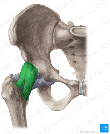
is the strongest ligament - prevents hyperextension of hip joint. - 4Describe the ischiofemoral ligament.Weakest of all 3 capsular ligaments.
Lies posteriorly & strengthens posterior aspect of joint capsule.
Attached medially to ischial bone below acetabulum.
Runs superolaterally around capsule.
Attaches to the base of greater trochanter, deep to iliofemoral ligament.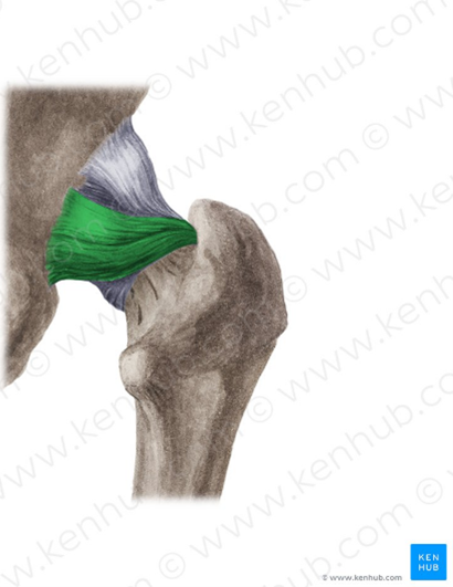
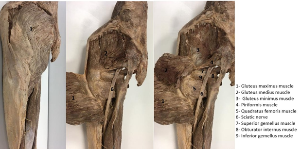
- 5What are the 3 muscles of the gluteal region?Gluteus maximus
Gluteus medius
Gluteus minimis - 6Describe the origin, insertion, nerve supply & actions of the Gluteus maximus.Origin: Lateroposterior surface of sacrum & coccyx, Gluteal surface of ilium.
Insertion: Iliotibial tract, Gluteal tuberosity of femur.
Nerve supply: Inferior gluteal nerve
Actions: Extension, abduction, adduction, lateral rotation of thigh.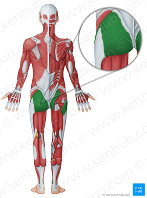
- 7Describe the origin, insertion, nerve supply & actions of the Gluteus medius.Origin: Gluteal surface of ilium.
Insertion: Lateral aspect of greater trochanter of femur.
Nerve supply: Superior gluteal nerve.
Actions: Abduction & medial rotation of thigh.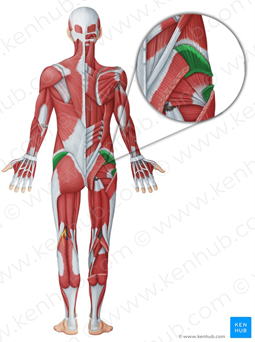
- 8Describe the origin, insertion, nerve supply & actions of the Gluteus minimis.Origin: Gluteal surface of ilium.
Insertion: Anterior aspect of greater trochanter of femur.
Nerve supply: Superior gluteal nerve.
Actions: Abduction, medial rotation of thigh.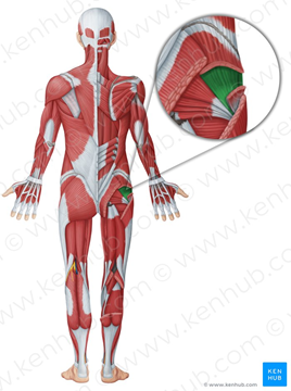
- 9What are the 6 deep gluteal muscles?Quadratus femoris
Piriformis
Obturator internus
Obturator externus
Superior gemellus
Inferior gemellus - 10Describe the origin, insertion, nerve supply & actions of the quadratus femoris.Origin: Ischial tuberosity
Insertion: intertrochantic crest of femur
Nerve supply: nerve to quadratus femoris
Actions: lateral rotation of thigh & stabilizes head of femur in acetabulum.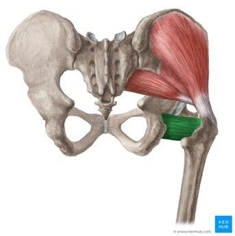
- 11Describe the origin, insertion, nerve supply & actions of the piriformis.Origin: anterior surface of sacrum & gluteal surface of ilium
Insertion: greater trochanter of femur
Nerve supply: Nerve to piriformis
Actions: lateral rotation & abduction of thigh, stabilises head of femur in acetabulum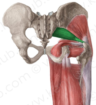
- 12Describe the origin, insertion, nerve supply & actions of the obturator internus.Origin: posterior surface of obturator membrane & bony boundaries of obturator foramen
Insertion: medial surface of greater trochanter of femur
Nerve supply: L5 & S1 nerves
Actions: lateral rotation of extended thigh, abduction of flexed thigh, stabilisation of hip joint.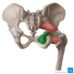
- 13Describe the origin, insertion, nerve supply & actions of the obturator externus.Origin: anterior surface of obturator membrane & bony boundaries of obturator foramen
Insertion: trochanteric fossa of femur
Nerve supply: Obturator nerve (L2-L4)
Actions: lateral rotation & abduction of thigh, stabilises head of femur in acetabulum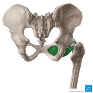
- 14Describe the origin, insertion, nerve supply & actions of the superior gemellus.Origin: ischial spine.
Insertion: medial surface of greater trochanter of femur
Nerve supply: L5-S1 nerves
Actions: lateral rotation & abduction of thigh, stabilises head of femur in acetabulum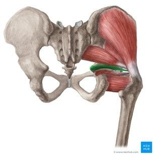
- 15Describe the origin, insertion, nerve supply & actions of the inferior gemellus.Origin: ischial tuberosity
Insertion: medial surface of greater trochanter of femur
Nerve supply: L4-S1
Actions: lateral rotation & abduction of thigh, stabilises head of femur in acetabulum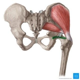
- 16—
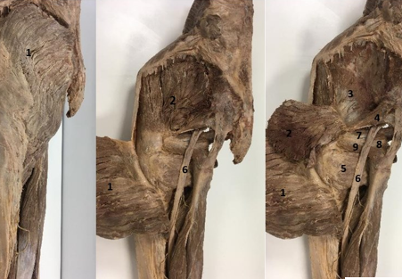
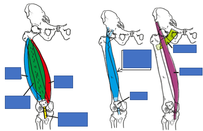
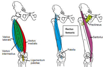
- 17What 5 muscles comprise the anterior compartment of the thigh? What is the action of each muscle?Rectus femoris - flexion of hip joint & extension of knee joint.
Vastus lateralis - extension of knee joint
Vastus intermedius - extension of knee joint
Vastus medialis - extension of knee joint
Sartorius - flexion of knee joint - 18What nerve innervates these muscles of the anterior compartment of the thigh?Femoral nerve (L2-L4)
- 19—
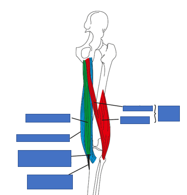
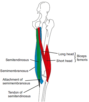
- 20Name the 3 muscles that comprise the posterior compartment of the thigh.Biceps femoris
Semitendinosus
Semimembranosus
(Hamstring muscles) - 21To what do all 3 muscles attach proximally?Ischial tuberosity
- 22What does each muscle attach to distally?Biceps femoris - head of fibula
Semitendinosus - medial condyle of tibia
Semimembranosus - medial shaft of tibia - 23All 3 muscles are innervated by what nerve?Sciatic nerve
- 24—
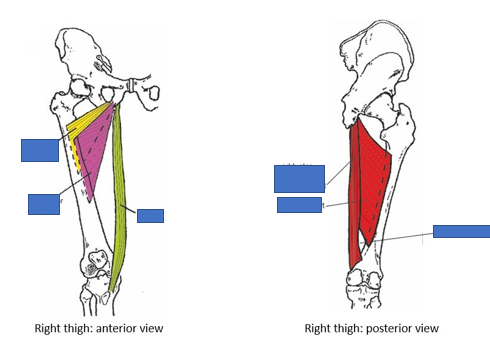
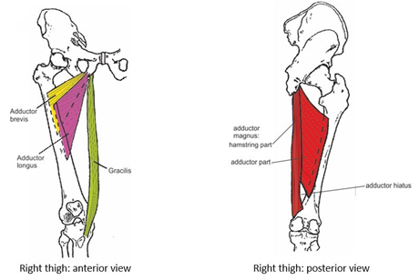
- 25Name the 5 muscles that comprise the medial compartment of the thighAdductor longus
Adductor brevis
Adductor magnus
Gracilis
Obturator externus - 26To what structure do all 5 muscles of the medial compartment of the thigh attach to proximally?Pubic bone
- 27What nerve innervates these 5 muscles? What is the exception?Obturator nerve
The hamstring part of the adductor magnus is innervated by the tibial nerve. - 28—
- 29Where is the femoral triangle located?Anterior to hip joint
In the superomedial third of thigh - 30What are the borders of the femoral triangle: a) Superior border? b) Medial border? c) Lateral border? d) Apex? e) Floor? f) Roof?a) Inguinal ligament
b) Adductor longus
c) Sartorius
d) Point at which sartorius & adductor longus meet
e) Adductor longus, pectineus, iliopsoas
f) Fascia lata - 31What are the contents of the femoral triangle?Femoral nerve (L2-L4)
Femoral artery
Femoral vein
Superficial & Deep inguinal lymph nodes - 32Describe the knee joint.Articulation: rounded condyles of femur, condyles of tibia.
Classification: synovial hinge joint.
Capsule: surrounds articular surfaces posteriorly & is absent anteriorly.
Ligaments: medial & lateral collateral ligaments, anterior & posterior cruciate ligaments
Synovial membrane: lines capsule
Nerve supply: femoral, obturator, common peroneal, tibial nerves.
Blood supply: branches from popliteal artery
Movements: flexion, extension, medial & lateral rotation - 33Name & describe the following ligament.Patellar ligament
Strong, thick fibrous band.
Distal continuation of quadriceps femoris tendon.
Apex of patella to tibial tuberosity.
Function: Stabilises the patella.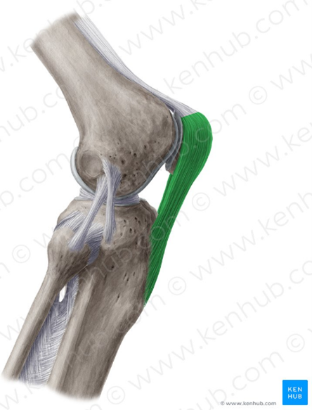
- 34Name & describe the following ligament.Lateral (fibular) Collateral ligament
Strong ligament
Origin: Lateral epicondyle of femur
Extends distally & attaches to lateral surface of head of fibula
Splits tendon of biceps femoris into 2.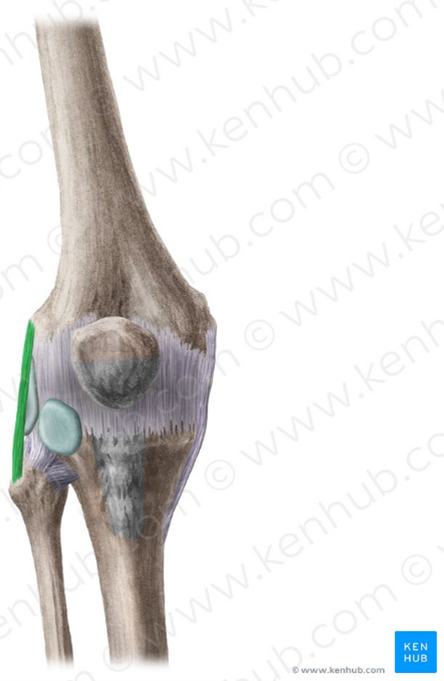
- 35Name & describe the following ligament.Medial (tibial) collateral ligament
Secures the knee joint
Prevents excessive lateral movement by restricting rotation of extended knee.
Superficial origin: proximal to medial epicondyle of femur.
Superficial insertion: medial epicondyle of tibia, medial shaft of tibia.
Deep origin: area of distal femur
Deep insertion: medial meniscus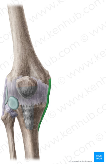
- 36Describe the anterior & posterior cruciate ligaments.Anterior:
Origin - anterior intercondylar area of tibia
Insertion - posterior part of medial surface of lateral femoral condyle.
Passes under the transverse ligament.
Prevents posterior rolling, displacement of femoral condyle during flexion & hyperextension of the knee joint.
Posterior:
Origin - Posterior intercondylar area of tibia
Insertion - anterior part of lateral surface of medial femoral epicondyle.
Twice as strong as anterior cruciate.
Prevents anterior rolling, displacement of the femoral condyle during flexion & prevents hyperflexion of knee joint.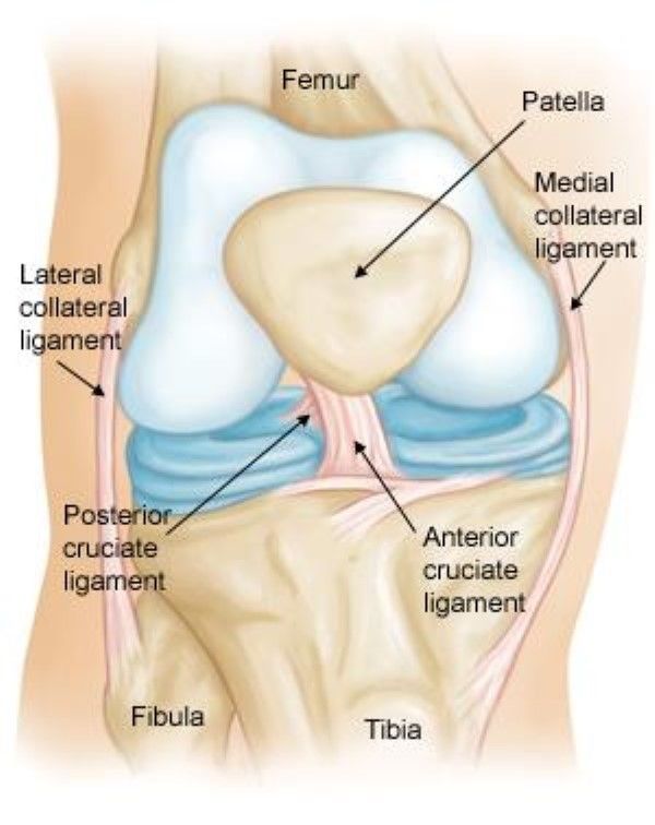
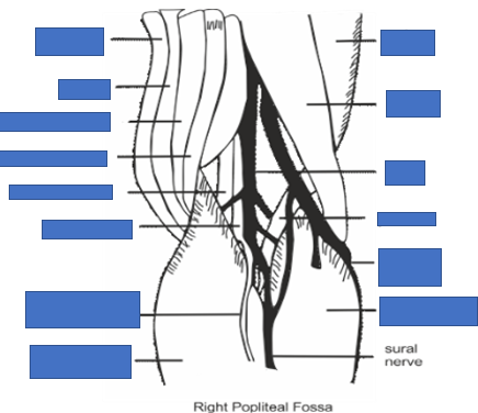
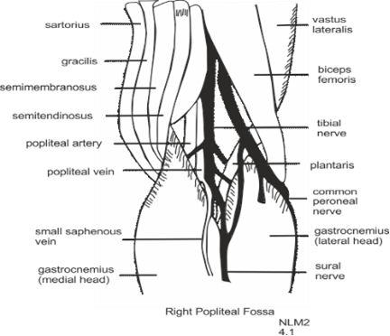
- 37State the location of the popliteal fossa.Posterior aspect of knee
- 38Describe the borders of the popliteal fossa: a) Lateral? b) Medial? c) Floor? d) Roof?a) Biceps femoris (above), Lateral head of gastrocnemius & plantaris (below).
b) Semimembranosus & Semitendinosus (above), Medial head of gastrocnemius (below)
c) Popliteal surface of femur, Capsule of knee joint, Posterior cruciate ligament, Popliteus
d) Skin, Superficial fascia, Deep fascia - 39Describe the contents of the popliteal fossa.Tibial nerve & Common peroneal nerve
Popliteal artery
Popliteal vein
Several lymph nodes - 40—
- 41What 4 muscles constitute the anterior compartment of the leg?Tibialis anterior
Extensor digitorum longus
Extensor hallucis longus
Peroneus tertius - 42Describe the origin, insertion, nerve supply & actions of the Tibialis anterior.Origin: lateral surface of tibia & interosseous membrane.
Insertion: medial cuneiform & base of metatarsal 1
Nerve supply: Deep fibular nerve
Actions: Dorsiflexion of foot at talocrural joint & Inversion of foot at subtalar joint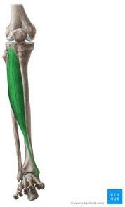
- 43Describe the origin, insertion, nerve supply & actions of the extensor digitorum longus.Origin: medial surface of fibula, lateral tibial condyle & interosseous membrane.
Insertion: distal & middle phalanges of digits 2-5.
Nerve supply: deep fibular nerve
Actions: Extension of digits 2-5 at metatarsophalangeal/interphalangeal joints, dorsiflexion of foot at talocrural joint & eversion of foot at subtalar joint.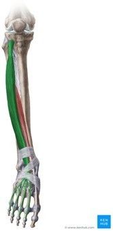
- 44Describe the origin, insertion, nerve supply & actions of the extensor hallucis longus.Origin: middle third of medial surface of fibula & interosseous membrane.
Insertion: base of distal phalanx of digit 1.
Nerve supply: Deep fibular nerve
Actions: Extension of digit 1 at metatarsophalangeal/interphalangeal joints, dorsiflexion of foot at talocrural joint.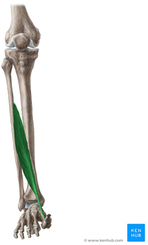
- 45Describe the origin, insertion, nerve supply & actions of the peroneus tertius.Origin: medial surface of fibula & interosseous membrane.
Insertion: dorsal surface of metatarsal 5.
Nerve supply: deep fibular nerve
Actions: Dorsiflexion of foot at talocrural joint & Eversion of foot at subtalar joint. - 46What 3 muscles comprise the superficial group of the posterior compartment of the leg?Gastrocnemius
Soleus
Plantaris - 47Describe the origin, insertion, nerve supply & actions of the gastrocnemius.Origin: Lateral femoral condyle (lateral head), Medial femoral condyle & popliteal surface of femoral shaft (medial head).
Insertion: posterior surface of calcaneus.
Nerve supply: Tibial nerve.
Actions: Plantarflexion of foot at talocrural joint & Flexion of leg at knee joint.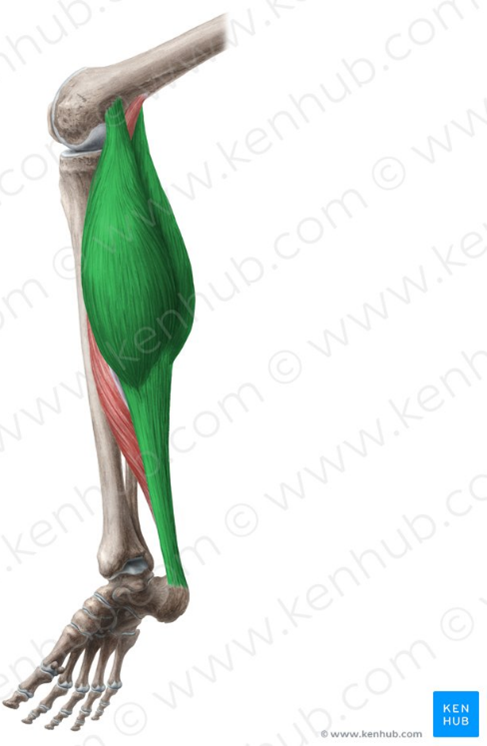
- 48Describe the origin, insertion, nerve supply & actions of the soleus.Origin: Soleal line, medial border of tibia, posterior border of fibula.
Insertion: Posterior surface of calcaneus.
Nerve supply: Tibial nerve.
Actions: Plantarflexion of foot at talocrural joint.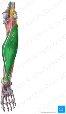
- 49Describe the origin, insertion, nerve supply & actions of the plantaris.Origin: Lateral supracondylar line of femur, Oblique popliteal ligament of knee.
Insertion: Posterior surface of calcaneus.
Nerve supply: Tibial nerve
Actions: Plantarflexion of foot at talocrural joint, Flexion of knee at knee joint.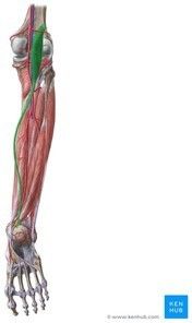
- 50What are the 4 deep muscles of the posterior compartment of the leg?Tibialis posterior
Flexor digitorum longus
Flexor hallucis longus
Popliteus - 51Describe the origin, insertion, nerve supply & actions of the tibialis posterior.Origin: Posterior surface of tibia, Posterior surface of fibula, Interosseous membrane.
Insertion: Tuberosity of navicular, All cuneiforms, Cuboid, Bases of metatarsals 2-4.
Nerve supply: Tibial nerve.
Actions: plantarflexion of foot at talocrural joint, inversion of foot at subtalar joint, supports medial longitudinal arch of foot.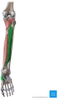
- 52Describe the origin, insertion, nerve supply & actions of the flexor digitorum longus.Origin: Posterior surface of tibia
Insertion: Bases of distal phalanges of digits 2-5
Nerve supply: Tibial nerve
Actions: Flexion of digits 2-5 at metatarsophalangeal/interphalangeal joints, Plantarflexion of foot at talocrural joint, Inversion of foot at subtalar joint.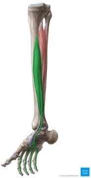
- 53Describe the origin, insertion, nerve supply & actions of the flexor hallucis longus.Origin: Posterior surface of fibula, Interosseous membrane.
Insertion: Base of distal phalanx of digit 1.
Nerve supply: Tibial nerve.
Actions: Flexion of digit 1 at metatarsophalangeal/interphalangeal joints, Plantarflexion of foot at talocrural joint, Inversion of foot at subtalar joint.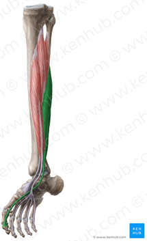
- 54Describe the origin, insertion, nerve supply & actions of the popliteus.Origin: Lateral condyle of femur.
Insertion: Posterior surface of tibia.
Nerve supply: Tibial nerve.
Actions: Unlocks knee joint & Knee joint stabilisation. - 55What are the 2 muscles of the lateral compartment of the leg?Peroneus longus
Peroneus brevis - 56Describe the origin, insertion, nerve supply & actions of the peroneus longus.Origin: Head of fibula & proximal 2/3 of lateral surface of fibula.
Insertion: Medial cuneiform bone & metatarsal 1.
Nerve supply: Superficial peroneal nerve.
Actions: Plantarflexion of foot at talocrural joint, Eversion of foot at subtalar joint, Supports longitudinal & transverse arches of foot. - 57Describe the origin, insertion, nerve supply & actions of the peroneus brevis.Origin: Distal 2/3 of lateral surface of fibula.
Insertion: Tuberosity of metatarsal 5.
Nerve supply: Superficial peroneal nerve.
Actions: Plantarflexion of foot at talocrural joint, Eversion of foot at subtalar joint. - 58Describe the tibiofibular joints.Proximal tibiofibular joint - synovial plane joint between head of fibula & lateral tibial condyle.
Distal tibiofibular joint - syndesmosis joint between fibular notch at inferior ends of tibia & fibula. - 59Describe & classify the following joints: a) Tarsometatarsal joints b) Intermetatarsal joints c) Metetarsophalangeal joints d) Interphalangeal jointsa) Plane synovial joints between cuboid, cuneiforms & bases of metatarsals.
b) Plane synovial joints between bases of lateral 4 metatarsals.
c) Synovial ellipsoid joints that permit limited flexion, extension, abduction & adduction.
d) Synovial hinge joints that permit limited flexion & extension of digits. - 60Describe the ankle joint.Includes 3 joints: talocrural joint, subtalar joint, inferior tibiofibular joint.
Articulates with lower end of tibia, 2 malleoli & body of talus.
Synovial hinge joint.
Allows plantarflexion & dorsiflexion.
Strengthened by collateral ligaments. - 61Describe the: a) Medial ligament. b) Lateral ligament.a) Strong, triangular bands. Radiate from medial malleolus to the talus, calcaneus & navicular.
b) Pass from lateral malleolus of fibula to the talus & calcaneus. - 62Describe the 3 arches of the feet. How are all 3 arches stabilised?Medial longitudinal arch
Lateral longitudinal arch
Transverse arch
Short interosseous ligaments - hold tarsals together.
Spring ligament - attached to calcaneus & navicular; helps to prevent the weight of the body driving the talus between the calcaneus & navicular.
Long & Short plantar ligaments.
Muscle tone - especially during movement when muscles function as dynamic ligaments. - 63Describe the medial longitudinal arch.On medial aspect of foot.
Formed by calcaneus, talus, navicular, cuneiforms & 3 metatarsals. - 64Describe the lateral longitudinal arch.Lower than the medial longitudinal arch.
Formed by calcaneus, cuboid & lateral 2 metatarsals. - 65Describe the transverse archIn the region of the bases of the metatarsals.
- 66What 3 muscles constitute layer 1?`Abductor hallucis
Flexor digitorum brevis
Abductor digiti minimi - 67Describe the origin, insertion, nerve supply & actions of the abductor hallucis.Origin: Medial process of calcaneal tuberosity, Flexor retinaculum, Plantar aponeurosis.
Insertion: Base of proximal phalanx of digit 1
Nerve supply: Medial plantar nerve
Actions: Abduction of digit 1 at metatarsophalangeal joint, Flexion of digit 1 at metatarsophalangeal joint, Support of longitudinal arch of foot.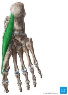
- 68Describe the origin, insertion, nerve supply & actions of the flexor digitorum brevis.Origin: medial process of calcaneal tuberosity, plantar aponeurosis.
Insertion: middle phalanges of digits 2-5.
Nerve supply: Medial plantar nerve.
Actions: Flexion of digits 2-5 at metatarsophalangeal joints, Supports longitudinal arch of foot.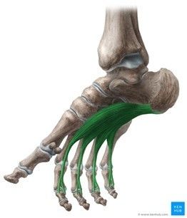
- 69Describe the origin, insertion, nerve supply & actions of the abductor digiti minimi.Origin: Calcaneal tuberosity & Plantar aponeurosis.
Insertion: Base of proximal phalanx of digit 5 & Metatarsal 5
Nerve supply: Lateral plantar nerve
Actions: Flexion & Abduction of digit 5 at metatarsophalangeal joint, Supports longitudinal arch of foot.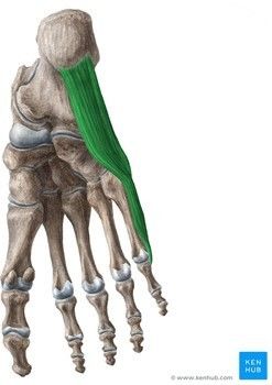
- 70What 2 muscles comprise the 2nd layer of the foot?Quadratus plantae
Lumbricals - 71Describe the origin, insertion, nerve supply & actions of the quadratus plantae.Origin: Medial surface of calcaneus, Lateral process of calcaneal tuberosity.
Insertion: Tendon of flexor digitorum longus.
Nerve supply: Lateral plantar nerve.
Actions: Flexion of digits 2-5 at metatarsophalangeal joints.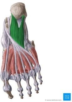
- 72Describe the origin, insertion, nerve supply & actions of the lumbricals.Origin: Tendons of flexor digitorum longus.
Insertion: Medial bases of proximal phalanges of digits 2-5, Extensor expansions of digits 2-5.
Nerve supply: Lumbrical 1 - medial plantar nerve, Lumbricals 2-4 - lateral plantar nerve
Actions: Flexion & Adduction of digits 2-5 at metatarsophalangeal joints, Extension of digits 2-5 at interphalangeal joints.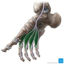
- 73What 3 muscles comprise the 3rd layer of the foot?Flexor hallucis brevis
Adductor hallucis
Flexor digiti minimi brevis - 74Describe the origin, insertion, nerve supply & actions of the flexor hallucis brevis.Origin: Tendon of tibialis anterior, Medial cuneiform, Lateral cuneiform, Cuboid.
Insertion: Base of proximal phalanx of digit 1
Nerve supply: Medial plantar nerve
Actions: Flexion of digit 1 at metatarsophalangeal joint, Supports longitudinal arch of the foot.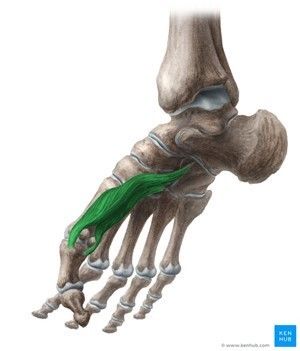
- 75Describe the origin, insertion, nerve supply & actions of the adductor hallucis.Origin: Bases of metatarsals 2-4, Cuboid, Lateral cuneiform.
Insertion: Base of proximal phalanx of digit 1
Nerve supply: Lateral plantar nerve
Actions: Flexion & Adduction of digit 1 at metatarsophalangeal joint, Supports longitudinal & transverse arches of the foot.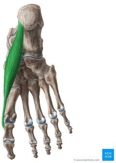
- 76Describe the origin, insertion, nerve supply & actions of the flexor digiti minimi brevis.Origin: Base of metatarsal 5, Long plantar ligament.
Insertion: Base of proximal phalanx of digit 5
Nerve supply: Lateral plantar nerve
Action: Flexion of digit 5 at metatarsophalangeal joint.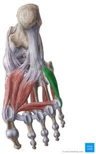
- 77What muscles comprise the 4th layer of the foot?Dorsal & Plantar interossei.
- 78Describe the origin, insertion, nerve supply & actions of the dorsal interossei.Origin: Opposing sides of metatarsals 1-5.
Insertion: 1 - medial base of proximal phalanx, 2-4 - lateral bases of proximal phalanges of digits 2-4..
Nerve supply: Lateral plantar nerve
Actions: Flexion & Abduction of digits 2-4 at metatarsophalangeal joints, Extension of digits 2-4 at interphalangeal joints.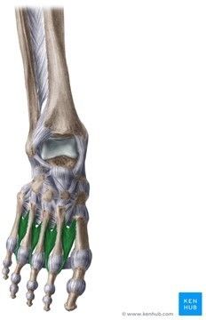
- 79Describe the origin, insertion, nerve supply & actions of the plantar interossei.Origin: Medial aspects of metatarsals 3-5.
Insertion: Medial bases of proximal phalanges of digits 3-5, Extensor expansions of digits 3-5.
Nerve supply: Lateral plantar nerve.
Actions: Flexion & Adduction of digits 3-5 at metatarsophalangeal joints, Extension of digits 3-5 at interphalangeal joints.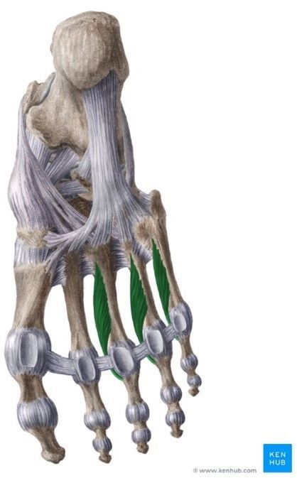
- 80—
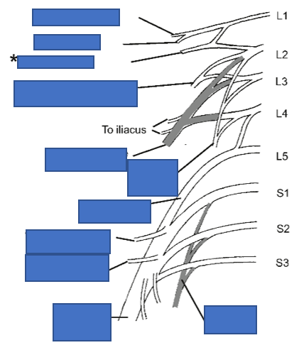
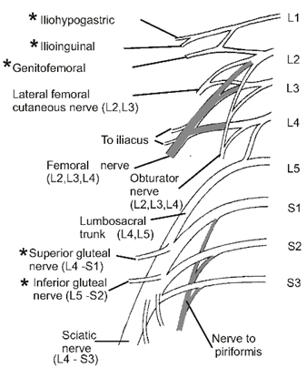
- 81Describe the lateral femoral cutaneous nerve (including its roots).Ventral rami of L2 & L3.
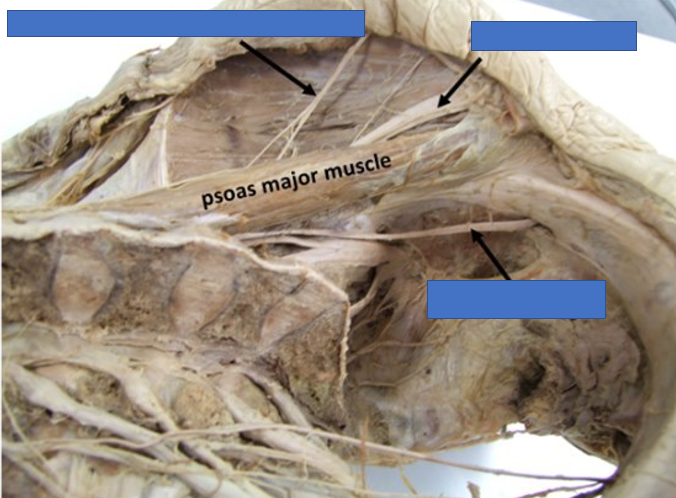
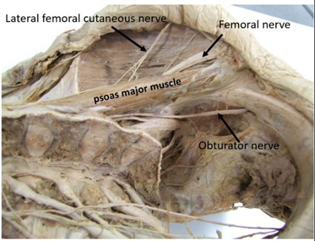
Emerges from lateral border of psoas major muscle.
Passes laterally under inguinal ligament.
Supplies skin overlying lateral thigh. - 82Describe the obturator nerve (including its roots).L2-L4
Emerges from medial border of psoas major muscle.
Passes through obturator foramen.
Enters medial side of thigh to supply adductor muscles & skin overlying medial aspect of the thigh. - 83Describe the femoral nerve (including its roots).L2-L4
Emerges from lateral aspect of psoas major muscle.
Enters thigh behind the inguinal ligament.
Lies lateral to femoral blood vessels in femoral triangle.
Innervates anterior thigh muscles & skin overlying anterior aspect of the thigh. - 84—
- 85Describe the superior & inferior gluteal nerves (including their roots).Superior gluteal nerve ==> L4-S1
Inferior gluteal nerve ==> L5-S2
Both arise from posterior divisions of sacral plexus.
Exit the pelvis via greater sciatic notch to supply gluteal muscles. - 86Describe the sciatic nerve (including its roots).L4-S3
Emerges from pelvis via greater sciatic notch.
Passes midway between greater trochanter & ischial tuberosity.
Supplies all leg & foot muscles, all joints in lower limb & most skin over leg & foot.
Branches into tibial nerve & common peroneal nerve. - 87Describe the following arteries in the lower limb: a) Superior & Inferior gluteal arteries b) External iliac artery c) Femoral artery d) Popliteal artery e) Anterior tibial artery f) Posterior tibial artery g) Medial & Lateral plantar arteriesa) Pass through greater sciatic foramen & supply gluteal muscles & hip joint.
b) Passes deep to inguinal ligament & continues as femoral artery.
c) Lateral to femoral vein in femoral triangle & passes through adductor hiatus to enter popliteal fossa as popliteal artery.
d) Descends through popliteal fossa, deep to tibial nerve & popliteal vein. Divides into anterior & posterior tibial arteries.
e) Descends along interosseous membrane & crosses ankle joint --> forms dorsalis pedis artery & lies lateral to tendon of extensor hallucis longus.
f) Descends in the deep posterior compartment of leg with the tibial nerve. Passes behind medial malleolus to enter the foot & divides into plantar arteries.
g) Form plantar arterial arch & gives off digital arteries to the digits. - 88—
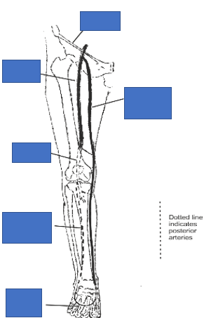
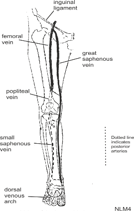
- 89Explain the process of intramembranous ossification.1. Mesenchymal cells cluster & differentiate into osteoblasts.
2. Ossification centres form.
3. Secreted osteoid traps osteoblasts & osteoblasts differentiate into osteocytes.
4. Trabecular matrix & periosteum form.
5. Compact bone develops superficial to trabecular bone.
6. Crowded blood vessels condense into red bone marrow. - 90Explain the process of endochonral oossification, using the picture as an aid.1. Mesenchymal cells differentiate into chondrocytes that produce a cartilage model.
2. Blood vessels on edge of cartilage model recruit osteoblasts into the bony collar.
3. Capillaries penetrate cartilage & deposit bone inside the cartilage model - forming primary ossification centre.
4. Cartilage & chondrocytes continue to grow at epiphyses whilst the medullary cavity expands & remodels.
5. Secondary ossification centres form at the epiphyses after birth.
6. Hyaline cartilage remains at epiphyseal plate & at joint surface as articular cartilage.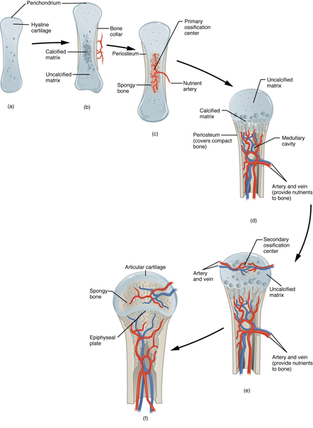
Information
- Description
- University - Medicine Locomotor Year 1 (Anatomy) Flashcards on Lower Limb SWAY, created by Jacob Coe on 14/03/2022.
- Created by
- Jacob Coe
- Views
- 0
- Rating