
Flashcards
Lower Limb Flashcards
Cards in this deck (53)
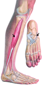
- 1- Head & upper 2/3rds of lateral Fibula -> base of 1st metatarsal & medial cuneiform - Superficial Peronal N (L5, S1, S2) - Lateral Compartment, Evertor of foot - Tendon post to lat malleolusPeroneus Longus
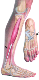
- 2- Inf 2/3rds of lateral fibula -> tuberosity on lateral side of base of 5th metatarsal - Superficial Peroneal N (L5, S1, S2) -Lateral Compartment, Evertor of foot - Tendon post to lat malleolusPeroneus Brevis
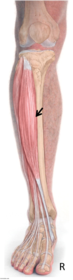
- 3- Lateral condyle of tibia, superior 1/2 of lateral tibial surface, interosseous membrane -> medial and inferior surfaces of medial cuneiform and base of 1st metatarsal - Deep Peroneal N (L4, L5) - Dorsiflexes ankle; inverts foot - Most medial of tendons coming into footTibialis Anterior
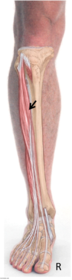
- 4- Lat condyle of tibia, superior 2/4 of medial surface of fibula, interosseus membrane -> middle and distal phalanges of lateral 4 digits - Deep Peroneal N (L4, L5) - extends lateral 4 digits; dorsiflexes ankleExtensor Digitorum Longus (EDL)
- 5- Inferior 1/3rd of anterior fibula and interosseus membrane -> dorsum of base of 5th metatarsal - Deep Peroneal N (L4, L5) - Dorsiflexes ankle; assists in eversion of footPeroneus Tertius
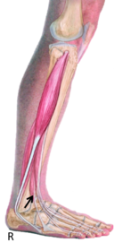
- 6- Middle anterior surface of fibula, interosseus membrane -> dorsal aspect of distal phalanx of Hallux - extends Hallux; dorsiflexes ankleExtensor Hallucis Longus
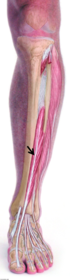
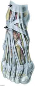
- 7SUP: Anterior border if tibia-> lower end of fibula INF: Stem attached to calcaneous laterally, Upper limb attached to medial malleolus, Lower limb passes around medial border of foot to blend w/ dense fascia over Add. HallucisExtensor Retinacula
- 8Superior surface of calcaneous, inferior extensor retinaculum -> Base of proximal phalanx of Hallux - Deep Peroneal N (L5, S1)Extensor Hallucis Brevis
(black)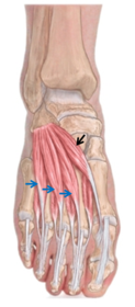
- 9Superior surface of calcaneous inferior extensor retinaculum -> Long extensor tendon of toes 2-4 - Deep Peroneal (L5, S1)Extensor Digitorum Brevis
(blue)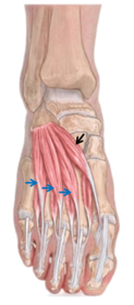
- 10- Branch of Common Peroneal between Peroneus Longus and neck of fibula - Lateral compartment of leg supplying muscles and continuous as Cutaneous N - Cutaneous innervation to distal anterior surface of leg and most of dorsum of footSuperficial Peroneal N
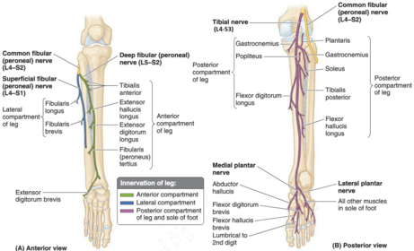
- 11- Branch of Common Peroneal between Peroneus Longus & neck of fibula - Passes through Extensor Digitorum Longus (EDL) and travels w/ Anterior Tibial A supplying all anterior compartment muscles - Crosses ankle jt to suppply EDB & EHB - Cutaneous innervation to skin between digits 1 and 2Deep Peroneal N
- 12Arteries of the Leg: 1) Through interosseus membrane and descends on this 2) gives fibular artery that provides perforating branches to the lateral compartmentPopliteal Arteries:
1) Anterior Tibial A
2) Posterior Tibial A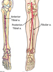
- 131) Popliteal surface of femur superior to medial condyle -> posterior surface of calcaneous via Tendocalcaneous 2) Lateral aspect of lateral condyle of femur -> posterior surface of calcaneous via Tendocalcaenous - Tibial N (S1, S2) - plantar flexes ankle; flexes kneeGastrocnemius
1) Medial
2) Lateral - 14Lateral supracondylar line of femur (proximal to lateral head of Gastrocnemius) -> posterior surface of calcaneous via Tendocalcaneous - Tibial N (S1, S2) - Assists in plantarflexion of anklePlantaris
- most of length is tendinous
- absent in about 10% of population - 15Soleal line of tibia, upper 1/3rd posterior fibula, tendinous arch between bony attachments -> posterior surface of calcaneous via Tendocalcaneous - Tibial N (S1, S2) - Planarflexes ankle jtSoleus
- 16Lateral condyle of femur (pit for politeus) and lateral meniscus -> popliteal area of tibia (above soleal line) - Tibial N (L4, L5, S1) - Unlocks knee jt by laterally rotating femur on fixed tibia - Creates stable jt, important in bipedalismPopliteus
- 17Posterior surface of tibia -> Base of distal phalanx of digits 2-4 - Tibial N (L5, S1, S2) - Flexes lateral 4 digits - Weak plantar flexion of ankleFlexor Digitorum Longus (FDL)
- 18Posterior surface of fibula -> Base of distal phalanx of Hallux - Tibial N (L5, S1, S2) - Flexes Hallux - Weak plantar flex of ankleFlexor Hallucis Longus
- 19Posterior surface of tibia and fibula, interosseous membrane -> tuberosity of navicular, cuneiforms, cuboid, sustentaculum tail of calcaneous, base of 2nd, 3rd and 4th metatarsals - Tibial N (L4, L5) - Plantarflexes ankle; inverts footTibialis Posterior
- 20Medial malleolus -> Calcaneous - Posterior tibial vessels and tibial nerve found between FDL and FHL tendons - Tom, Dick, And, Very, Naughty, HarryFlexor Retinaculum
- 21- Terminal branches of Popliteal A - Between heads of Gastrocnemius and deep to soleus to run on surface of TP and FDL - Gives Fibular A as a branch - Terminates as medial and lateral plantar As in footPosterior Tibial A
- 22Runs medial to fibula, usually within FHLFibular A
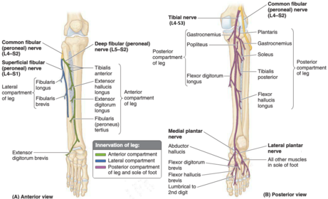
- 23- Passes between heads of Gastrocnemius and deep to tendinous arch of Soleus w/ posterior Tibial A - Between FHL and FDL at the ankle jt - Gives terminal branches that supply the sole of the foot: Medial and Lateral Plantar NsTibial N
- 24- Interosseous tibiofibular lig continous w/ interosseous membrane - Anterior and Posterior ligaments - Slight movement to accomodate wedging of trochlea of talus during dorsiflexionSyndemosis - Inferior Tibiofibular Jt
- 25- Attaches around the articular margins - Anteriorly extends onto the neck of the Talus - Weak anteriorly and posteriorly - Supports lateral and medially by strong collateral ligsAnkle Capsule
- 26- Anterior Talofibular -> neck of talus - Posterior Talofibular -> lateral tubercle of talus - Calcaneofibular -> lateral surface of calcaneousLateral Collateral Ligaments
- 27Fans out from medial malleolus and has 4 parts: - Anterior tibiotalar - Tibionavicular - Tibiocalcaneal - Posterior TibiotalarMedial Collateral ligament (deltoid)
- 281) Medial tubercle calcaneous, flexor retinaculum -> medial base of proximal phalanx 2) Medial tubercle calcaneous -> middle phalanges of lateral 4 toes 3) Medial and lateral tubercle calcaneous -> lateral base proximal phalanxFoot - Layer 1
1) Abd Hallucis
2) FDB
3) Abd Digiti Minimi - 291) Long flexor tendons -> toes 2) Tendon of FDL -> medial side of dorsal tendon expansion 3) Medial and lateral surfaces of calcaneous -> lateral side of FDL tendonFoot - Layer 2
1) FDL and FHL
2) Lumbricles
3) Quadratus Plantae (QP) - 301) Cuboid and lateral cuneiform -> both sides base proximal phalanx of Hallux 2) Transverse Head - Plantar lig of MTP jt Oblique Head - Base of metatarsals 2-4 -> lateral side base of proximal phalanx of Hallux 3) Base 5th metatarsal -> base prox phalanx of 5th digitFoot - Layer 3
1) Flexor Hallucis Brevis (FHB)
2) Add Hallucis
3) Flexor Digiti Minimi Brevis - 311) Bases and medial side of metatarsals 3-5 -> medial side of proximal phalanx of 3-5th digit - Adduct 2) Adjacent sides of metatarsals 1-5 -> 1st on medial side proximal phalanx 2nd digit -> 2nd-4th lateral side proximal phalanx 2nd -4th digitsFoot - Layer 4
1) 3x Plantar interossi PAD
2) 4x Dorsal interossi DAB - 32- Terminal branches of Tibial N deep to flexor retinaculum - Enters foot deep to Abductor Hallucis 1) Between abductor Hallucis and FDB 2) Between layers 1 and 2, deep and superficial branches. Deep branch between layers 3 and 4Plantar Ns
1) Medial Plantar N
2) Lateral Plantar N - 33Innervation of muscles of foot (S2, S3) 1) To: Abd Hallucis, FDB, 1st lumbricle, FHB 2) All the other muscles1) Medial Plantar N
2) Lateral Plantar N - 34Supported by: 1) Tendon of Tibialis Post 2) Tendon of Peroneus Longus 3) Adductor Hallucis 4) Deep transverse metatarsal ligTransverse Arch
1) & 2) Tarsal arch
3) Metatarsal arch
4) Anterior arch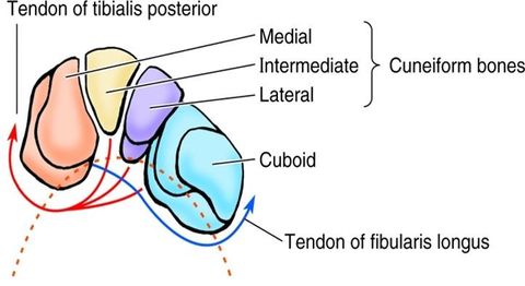
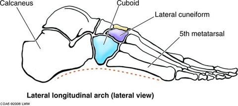
- 35Calcaneous, Cuboid, Metatarsals 4 and 5Bones of lateral arch
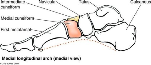
- 36Calcaneous, Talus, 3 Cunieforms, metatarsals 1-3Bones of medial arch
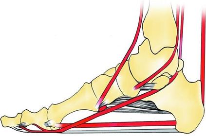
- 37Intrinsic muscles of the foot, FHL, FDLDynamic Support
- 381) Plantar calcaenonavicular (spring) lig 2) Short plantar lig (calcaneocuboid) 3) Long plantar lig 4) Plantar aponeurosisPassive support
- 39Talonavicular and Calcaneocuboid jts - Talcocalcaneal part is - Talonavicular part is Calcaneocuboid jt isTransverse (mid) tarsal jt
- plane synovial
- ball and socket
- plane synovial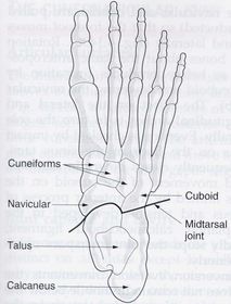
- 40Ligaments of Midtarsal JT 1) Calcaenous-> Navicular and cuboid 2) Calcaneus -> Cuboid 3) Calcaneus-> Cuboid and Metatarsals 4) Calcaneus -> Cuboid 5) Calcaneus-> NavicularLigaments of Midtarsal jt
1) Bifurcate lig
2) Dorsal Calcaneocuboid lig
3) Long Plantar lig
4) Short plantar lig
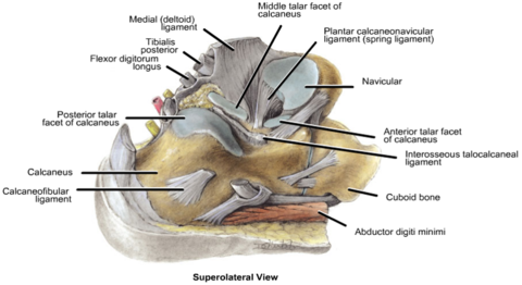
5) Spring lig (Calcaneonavicular) - 41What is thisJoint of inversion/eversion
- Subtalar jt
- Talocalcaneal part of Talocalcanonavicular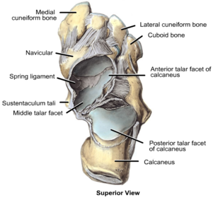
- 42Movements of jts of inversion/eversion: 1) Inversion/eversion. Forefoot, midfoot and calcaneus rotate under talus 2) Augments inversion/eversion, forefoot and midfoot rotate on hindfoot1) Subtalar jt
2) Midtarsal jt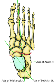
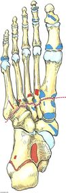
- 43—Tarsometatarsal jts
(Lisfranc's jt)
- Ant tarsal bones (cuboid and cuneiforms) artic w/ base of metatarsals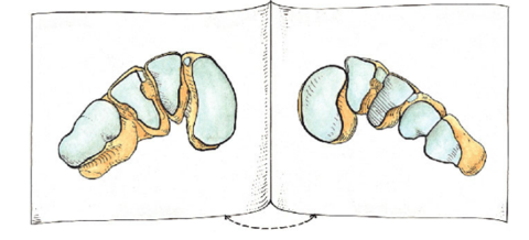
- 44—Tarsometatarsal ligs
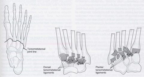
- 451) synovial condyloid jt, - flexion, extension, abduction and adduction 2) Synovial hinge jt - flexion and extension1) Metatarsophalangeal (MTP) jts
2) Interphalangeal (IP) jts - 46Weight distributed almost equally between 1) and 2)1) Calcaneus
2) Heads of metatarsals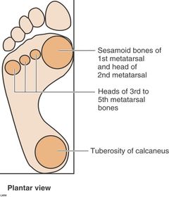
- 47- Talonavicular (ant part talocalcaneonavicular) - Subtalar - Calcaneocuboid - Cuneonavicular - Tarsometatarsal - Metatarsophalangeal - Intermetatarsal - InterphalangealJoints of the foot
{Talonavicular + Calcaneocuboi= Transv (mid) tarsal jt}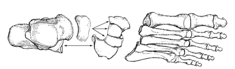
- 48Plane synovial jt 1) Slightly concave posterior calcaneal artic surface 2) Convex posterior articular facet1) Talus
2) Calcaneus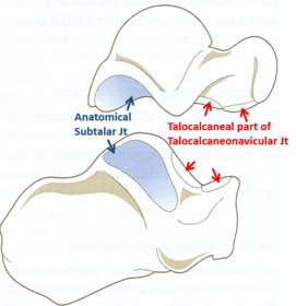
- 49What joint is this?Subtalar jt
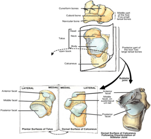
- 50Weak capsule supported by Medial, Lateral, Interosseous and Posterior Talocalcaneal ligsLigaments of subtalar jt
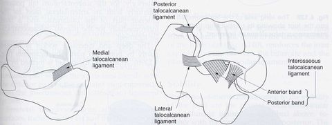
- 51—Transverse (mid)tarsal jt
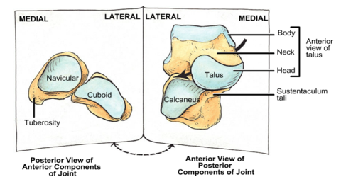
- 52—
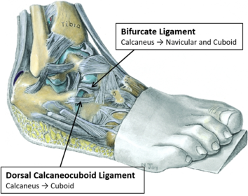
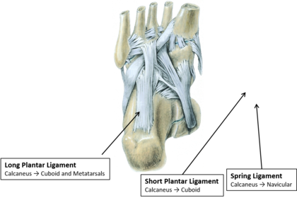
- 531) Dorsalis Pedis A is a cont of ? 2) Branches: ?, ?, ? 3) Runs across metatarsals to anastamose with lateral tarsal A Metatarsal and digital AArteries of dorsum of foot:
1) Ant tibial A
2) Lateral tarsal A, Deep plantar A (sole of foot),
3) Arcuate
Information
- Description
- Anatomy Flashcards on Lower Limb Flashcards, created by Emma MacRae on 15/02/2017.
- Created by
- Emma MacRae
- Views
- 1
- Rating