
Flashcards
Gastrointestinal
Cards in this deck (122)
- 1Normal gastrointestinal embryology (which gut forms what structure? midgut development?)Foregut:
Pharynx to duodenum
Midgut:
Duodenum to proximal 2/3 of transverse colon
Hindgut:
distal 1/3 of transverse colon to anal canal above pectinate line
Midgut development:
6th week --> physiologic midgut herniates through umbilical ring
10th week --> midgut returns to abdominal cavity and rotates around superior mesenteric artery (SMA) 270 counterclockwise - 2Ventral wall defects (rostral/lateral/caudal fold closure failure)Developmental defects due to failure of:
1. Rostral fold closure:
a) sternal defects
2. Lateral fold closure
a) gastroschisis - extrusion of abdominal contents through abdominal folds, not covered by peritoneum
b) omphalocele - persistence of herniation of abdominal contents into umbilical cord, sealed by peritoneum AND amnion
3. Caudal fold closure
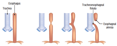
a) bladder exstrophy - 3—(tracheoesophageal anomalies)
1. normal anatomy
2. Pure EA - CXR shows gasless abdomen
3. Pure TEF (H-type)
a) TEF allows air to enter stomach - seen on CXR
b) Cyanosis 2* to laryngospasm (to prevent reflux aspiration)
c) fistula resembles the letter H
4. EA with distal TEF (most common)
a) polyhydramnios in utero (since the fetus isn't swallowing amniotic fluid)
b) first feed --> neonate chokes + vomit
c) TEF allows air to enter stomach
d) cyanosis 2* to laryngospasm
3 + 4 --> clinical test: failure to pass nasogastric tube into stomach - 4(presentations? types? and their presentations)"intestinal atresia"
Presentation:
1. Bilious vomiting
2. Abdominal distention within 1st 1-2 days of life
Type 1: Duodenal atresia
- failure to recanalize --> dilation of stomach and proximal duodenum -->
1. CXR --> double bubble
2. Assoc. w. Down Syndrome
3. Polyhydramnios (just like TEF)
4. Bilious vomiting (not like TEF)
Type 2: Jejunal and illeal atresia
- disruption of mesenteric vessels --> ischemic necrosis --> segmental resorption
1. Bowel discontinuity or "apple peel"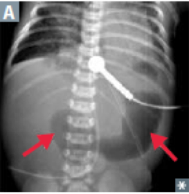
- 5Hypertrophic pyloric stenosis (epidemiology, findings, complications, treatment)Congenital hypertrophy of pyloric smooth muscle
Epidemiology
1. Most common cause of gastric outlet obstruction in infants
2. First born males (common)
3. Macrolide exposure
Findings:
1. palpable "olive" mass in epigastric region
2. Nonbilious projectile vomiting at 2-6 weeks old (not at birth)
3. Visible peristalsis
Complications
1. Hypokalemic, hypochloremic metabolic alkalosis (2* to gastric acid depletion + volume contraction)
Treatment
surgical incision (pyloromyotomy) - 6Pancreas and Spleen embryologyPancreas
- from foregut
- ventral pancreatic buds contribute to uncinate process + main pancreatic duct
- dorsal pancreatic bud alone becomes body, tail, isthmus, and accessory pancreatic duct
- both contribute to pancreatic head
Problem:
a) Annular pancreas
- ventral pancreatic bud abnormally encircles 2nd part of duodenum --> forms a ring around duodenum --> narrowing:
1. Nonbilious vomiting
b) Pancreas divisum
- ventral and dorsal parts fail to fuse at 8 weeks
- common anomaly --> mostly asymptomatic, but:
1. maybe chronic abdominal pain
2. maybe pancreatitis
Spleen - arises in mesentery of stomach (mesodermal) but has foregut supply (celiac trunk --> splenic artery)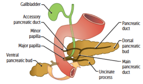
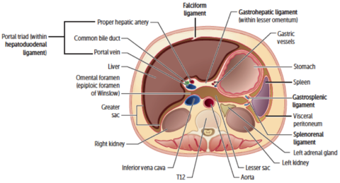
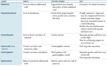
- 7Retroperitoneal structures (what structures? injury?)- GI structures w/o mesentery
- non GI structures
- injury --> blood/gas accumulation
SAD PUCKER
Suprarenal (adrenal) glands
Aorta and IVC
Duodenum (2nd, 3rd, 4th parts)
Pancreas (not tail)
Ureters
Colon (descending + ascending)
Kidneys
Esophagus (thoracic portion)
Rectum (partially)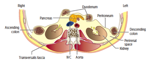
- 8—
- 9—Layers of gut wall - MSMS
Mucosa:
1. Epithelium -->
2. Lamina propria -->
3. Muscularis mucosa
Submucosa:
1. submucosal glands (fluid secretion) +
2. Meissner plexus
Muscularis externa:
1. Inner circular layer -->
2. Auerbach (myenteric) plexus -->
3. Outer longitudinal layer
^motility
Serosa (if intraperitoneal)
Adventitia (if retroperitoneal)
Ulcers - extend into as far as outer muscular layer
Erosions - in mucosa only
Basal electric rhythm:
Stomach - 3 waves/min
Duodenum - 12 waves/min
Ileum - 8-9 waves/min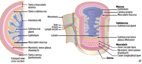
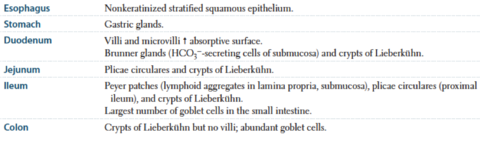
- 10Digestive tract histology (esophagus, stomach, duodenum, jejunum, ileum, colon)
- 11Abdominal aorta and branches (levels and how they branch: Celiac, superior mesenteric, inferior mesenteric, renal, gonadal , first lumbar, middle suprarenal, second lumbar, third lumbar, four lumbar, aortic bifurcation, median sacral artery, etc)arteries supplying GI structures - branch anteriorly
arteries supplying non-GI structures - branch laterally + posteriorly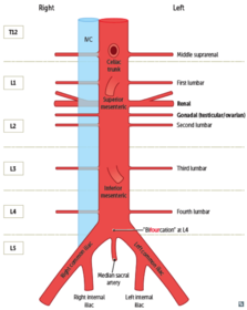
- 12Superior mesenteric artery syndrome (predisposition, pathogenesis, symptoms)Predisposition
1. Diminished mesenteric fat (low body weight/malnutrition)
Pathogenesis
Transverse duodenum trapped between SMA and aorta
Symptoms
1. Postprandial pain - 13Gastrointestinal blood supply and innervation (foregut, midgut, hindgut)
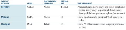
- 14—Branches of celiac trunk:
1. Left gastric
2. Splenic artery + 3. Common hepatic artery
Strong anastomoses
- left and right gastroepiploic arteries
- left and right gastric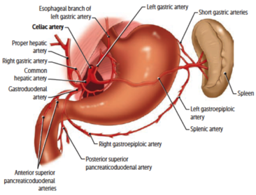
- 15In portal hypertension: (sites of anastomoses? clinical sign? portal vein <--> systemic vein? treatment?)Treat with TIPS (trans jugular intrahepatic portosystemic shunt) between portal vein and hepatic vein
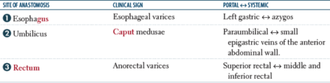
- 16Above pectinate line (what's pectinate line? presentations? arterial supply? venous drainage? lymphatic drainage? internal hemorrhoids?)Pectinate line - formed where endoderm (handgun) meets ectoderm
Presentations
1. Internal hemorrhoids
2. Adenocarcinomas
Arterial supply
1. Superior rectal artery (IMA)
Venous drainage:
superior rectal vein --> inferior mesenteric vein --> portal system
Lymphatic drainage
- internal iliac lymph nodes
Internal hemorrhoids
- visceral innervation
- not painful - 17Below pectinate line (pectinate line? presentations? arterial supply? venous drainage? hemorrhoid characteristics? anal fissure?)Pectinate line - where endoderm (hindgut) meets ectoderm
Presentations:
1. External hemorrhoids
2. Anal fissures
3. Squamous cell carcinoma
Arterial supply
1. Inferior rectal artery (internal pudendal artery <-- internal iliac artery)
Venous drainage:
inferior rectal vein --> internal pudendal vein --> internal iliac vein --> common iliac vein --> IVC
External hemorrhoids:
1. somatic innervation (inferior rectal branch of pudendal nerve)
2. Painful if thrombosed
Anal fissure
- Pain while Pooping
- Blood on toilet Paper
- located Posteriorly b/c it's Poorly Perfused - 18Liver tissue architectures (hepatocyte layout, kupffer cell, ito cells, zones 1/2/3)Hepatocytes
1. Apical surface --> face bile canaliculi
2. Basolateral surface --> face sinusoids
Kupffer cells
- speciacialized macrophages
- form lining of sinusoids
Hepatic stellate (Ito) cells
- stay in space of Disse
- store Vit A (when quiescent)
- produce extracellular matrix (active)
Zone 1 - periportal zone
- affected 1st by viral hepatitis
- ingested toxins
Zone 2 - intermediate zone
- yellow fever
Zone 3 - pericentral vein zone
- affected 1st by ischemia
- contains cytochrome P450
- most sensitive to metabolic toxins
- site of alcoholic hepatitis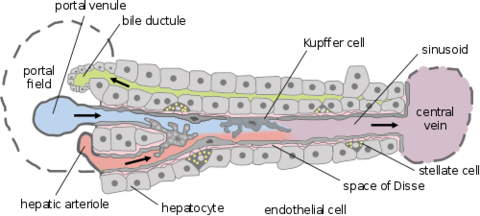
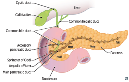
- 19Biliary structures (cystic duct? common hepatic duct? main pancreatic duct? accessory pancreatic duct? ampulla of vater?)
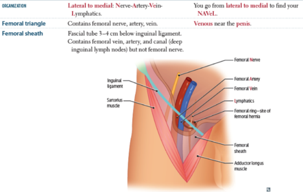
- 20Femoral region (what forms the femoral triangle? contents of the femoral triangle? femoral sheath?)
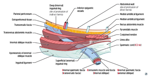
- 21Inguinal canal (picture all the contents of the region)
- 22—"diaphragmatic hernia"
About:
abdominal structures enter the thorax
Cause:
1. Congenital defect of pleuroperitoneal membrane
2. Trauma
Location:
1. Commonly on the left side (since right side has liver)
Types:
1. Sliding hiatal hernia - GE junction is displaced upwards --> "hourglass stomach"
2. Paraesophageal hernia - GE junction is usually normal --> funds protrudes into thorax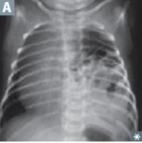
- 23—Indirect inguinal hernia
Risk
1. failure of processus vaginalis to close
Epidemiology
1. Infants
2. Males
Process:
content goes through internal (deep) inguinal ring (lateral to inferior epigastric vessels) --> external (superficial) inguinal ring --> into scrotum
*follows path of testis descent
*covered by all 3 layers of spermatic fascia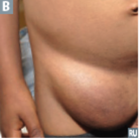
- 24Direct inguinal hernia (pathway, covered by? epidemiology)Abdominal content --> bulges through Hesselbach triangle (medial to inferior epigastric vessel, above inguinal ligament, lateral to rectus abdominus muscle) --> goes through external (superficial) inguinal ring only --> covered by external spermatic fascia
Epidemiology
1. Usually in older men - 25Femoral hernia (pathway? epidemiology?)- protrudes below inguinal ligament through femoral canal
- below + lateral pubic tubercle
Epidemiology
- females
- tend to have incarceration (can't put back into abdomen)
- strangulation (ischemia --> necrosis)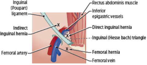
- 26Gastrin (source, action, regulation, pathologic conditions?)Source:
1. G cells (antrum of stomach, duodenum)
Action:
1. (+) gastric H secretion
2. (+) growth of gastric mucosa
3 (+) gastric motility
Regulation
1. (+) by stomach distention/alkalinization
2. (+) by amino acids (Phe) + peptides
3. (+) by vagal stimulation via GRP (gastrin-releasing peptide)
4. (-) by pH < 1.5
Pathologic:
1. high in chronic PPI use
2. high in chronic atrophic gastritis (H. pylori)
3. high in ZES (zollinger-ellison, gastronome) - 27Somatostatin (source? regulation? action? notes?)Source:
1. D cells in GI mucosa
2. D cells in pancreatic islets
Regulation
1. (+) by acid
2. (-) by vagal stimulation
Actions
1. (-) gastric acid and pepsinogen secretion
2. (-) pancreatic and SI fluid secretion
3. (-) gallbladder contraction
4. (-) insulin and glucagon release
Notes
1. Inhibits secretion of various hormones
2. Octreotide, an analog, used to treat acromegaly, carcinoid syndrome, and variceal bleeding - 28Cholecystokinin (source? regulation? action? how does it act?)Source
1. I cells in duodenum + jejunum
Regulation
1. (+) by fatty acids
2. (+) by amino acids
Action
1. (+) pancreatic secretion
2. (+) gallbladder contraction
3. (-) gastric emptying
4. (+) sphincter of Oddi relaxation
Notes:
1. Action on neural muscarininc pathways to cause pancreatic secretion - 29Secretin (source? regulation? action? notes?)Source:
1. S cells (duodenum)
Regulation
1. (+) by acid
2. (+) by fatty acids in duodenal lumen
Action:
1. (+) pancreatic HCO3 secretion
2. (-) gastric acid secretion
3. (+) bile secretion
Notes:
HCO3 neutralizes gastric acid in duodenum --> allows pancreatic enzymes to function - 30Glucose-dependent insulinotropic peptide (source? regulation? actions?)AKA GIP (gastric inhibitory peptide)
Source:
1. K cells (duodenum, jejunum)
Regulation
1. (+) by fatty acids
2 (+) by amino acids
3. (+) by oral glucose
Actions:
1. (-) gastric H secretion
2. (+) insulin release - 31Motilin (source? regulation? action? therapeutic potential?)Source:
1. Small intestine
Regulation:
1. (+) in fasting state
Action:
1. Produces MMCs (migrating motor complexes)
Motilin receptor agonists (erythromycin) are used to stimulate intestinal peristalsis - 32Vasoactive intestinal polypeptide (VIP) (source, regulation, action, VIPoma?)Source:
1. Parasympathetic ganglia in:
a) sphincters
b) gallbladder
c) small intestine
Regulation:
1. (+) by distention
2. (+) by vagal stimulation
3. (-) by adrenergic input
Actions:
1. (+) intestinal water and electrolyte secretion
2. (+) relaxation of intestinal smooth muscle and sphincters
VIPoma
- non-a, non-B islet cell pancreatic tumor that secretes VIP
- Watery Diarrhea
- Hypokalemia
- Achlorhydria - 33Nitric oxide (action? loss of NO?)Action:
1. (+) smooth muscle relaxation (i.e. lower esophageal sphincter LES)
Loss of NO secretion --> (+) LES tone of achalasia - 34Ghrelin (source? regulation? action? pathologic states?)Source:
1. Stomach
Regulation:
1. (+) in fasting state
2. (-) by food
Action:
1. (+) appetite
Pathologic states:
1. (+) in Prader-Willi syndrome
2. (-) after gastric bypass surgery - 35Intrinsic factor (source? action? pathologic states?)Source
1. Parietal cells (stomach)
Action
1. Vit B12 binding protein (required for B12 uptake in terminal ileum)
Pathologic state
autoimmune destruction of parietal cells --> chronic gastritis + pernicious anemia - 36Gastric acid (source? action? regulation?)Source:
1 Parietal cells (stomach)
Action:
1. Acidify the stomach
Regulation
1. (+) by histamine (ECL cells)
2. (+) by ACh
3. (+) by gastrin
4. (-) by somatostatin
5. (-) by GIP
6. (-) by prostaglandin
7. (-) by secretin - 37Pepsin (source? regulation? action?)Source:
1. Chief cells (stomach)
Regulation
1. (+) by vagal stimulation
2. (+) by local acid
Action:
1. Protein digestion
Pepsinogen (inactive) --> [H] --> pepsin (active) - 38Bicarbonate (source? regulation? action?)Source:
1. Mucosal cells of
a) salivary gland
b) stomach
c) duodenum (Brunner glands)
d) pancreas
Regulation
1. (+) by pancreatic secretion w/ secretin
2. (+) by biliary secretion w/ secretin
Action:
1. Neutralizes acid
Trapped in mucus that covers the gastric epithelium - 39Locations of GI secretory cells (parietal cells? chief cells? G cells? mucous cells? ECL cells? D cells? I cells? S cells? K cells?)
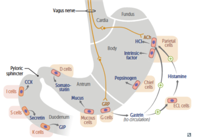
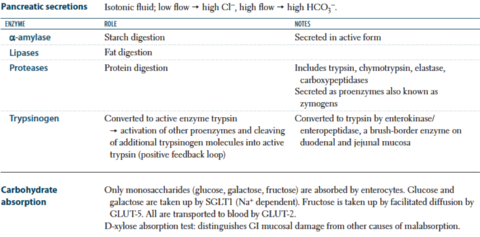
- 40Pancreatic secretions (secretes what? what do they do? low flow has what? high flow has what? carbohydrate absorption?)
- 41Vitamin/mineral absorption (location: Fe, Folate, B12)

- 42Peyer Patches (what is it? where is it? function/action?)
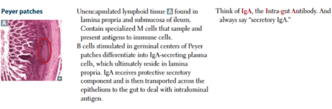
- 43Bile (composition, enzymes, functions)Composition
1. Bile salts (bile acid + glycine, taurine)
2. Phospholipids
3. Cholesterol
4. Bilirubin
5. Water
6. Ions
Enzymes
1. Cholesterol 7a-hydroxylase catalyzes rate-limiting step of bile acid synthesis
Functions
1. Digestion and absorption of lipids and fat-soluble vitamins
2. Cholesterol excretion (body's only means of eliminating cholesterol)
3. Antimicrobial activity (via membrane disruption) - 44Bilirubin (where it's from? enzymes involved? how excreted?)[macrophages]
RBCs --> Heme --> (heme oxygenase) --> biliverdin --> *reduced* --> unconjugated bilirubin (indirect bilirubin - water insoluble)
+ albumin
[bloodstream]
unconjugated bilirubin-albumin complex
- albumin
[liver]
UDP glucuronosyl-transferase --> conjugated bilirubin (bilirubin-glucuronate) --> bile
+ gut bacteria
[gut]
1. --> urobilinogen --> excreted in feces
2. - urobilinogen reabsorbed --> excreted in kidney as urobilin - 45Salivary gland tumors (characteristics? presentation? types?Characteristics
1. most commonly benign and in parotid gland
2. tumors in smaller glands --> more likely malignant
Presentation
1. Painless mass/swelling
2. Facial pain/paralysis --> malignant involvement of CN VII
Types:
1. Pleomorphic adenoma (benign mixed tumor)
- most common salivary gland tumor
- composed of chondromyxoid stroma (cartilage) and epithelium
- arises in parotid --> mobile, painless (no facial invasion), circumscribed mass at angle of the jaw
- recurs if incompletely excised (due to irregular margin)/ruptured intraoperatively
2. Mucoepidermoid carcinoma
- most common malignant tumor
- has mucinous and squamous components
- arises in the parotid --> commonly involves the facial nerve (since VII passes through the parotid)
3. Warthin tumor
(papillary cyst adenoma lymphomatosum)
- benign cyst tumor w/ germinal centers containing abundant lymphocytes
- almost always arises in the parotid (since it's the last gland to separate from stromal tissue --> often has lymph tissues in it) - 46What? Cause? Findings? Lab? Complications?Achalasia
What
1. Failure of relaxation of LES
2. Disordered esophageal motility
Cause:
1. loss of myenteric auerbach plexus (which regulates bowel motility and LES relaxation)
2. 2* achalasia from Chagas (T. cruzi)
3. 2* achalasia from extra esophageal malignancies
Findings:
1. High LES resting pressure and uncoordinate/absent peristalsis --> progressive dysphagia to solids and liquids
2. Putrid breath (when food rots)
Lab
1. Barium swallow --> dilated esophagus w/ area of distal stenosis
2. Esophageal manometry - high LES pressure
Complications
1. inc. risk of esophageal cancer (squamous cell carcinoma)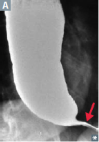
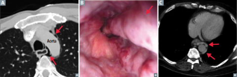
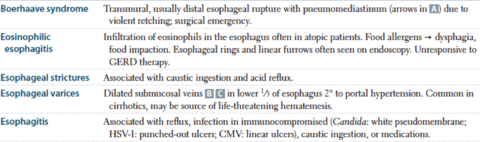
- 47eosinophilic esophagitis? esophageal stricture (what? assoc?) esophagitis (assoc.? causative agents? signs?)
- 48Sclerodermal esophageal dysmotility? (pathogenesis? findings? part of what?)esophageal smooth muscle atrophy --> dec. LES pressure + esophageal dysmotility --> dysphagia + acid reflux -->
1. Stricture
2. Barrett esophagus
3. Aspiration
Part of CREST syndrome - 49—Barrett Esophagus
What:
1. specialized intestinal metaplasia --> replacement of nonkeratinized stratified squamous epithelium w/ intestinal epithelium (conciliated columnar w/ goblet cells)
Location
1. Distal esophagus
Cause:
1. Chronic GERD
Complication
1. Risk of esophageal adenocarcinoma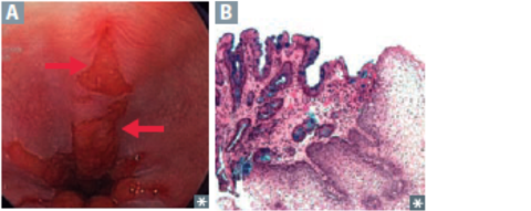
- 50Esophageal cancer (presentation? types? their location/risk factors? metastasis?)Presentations:
1. Progressive dysphagia (first solids, then liquids)
2. Weight loss
3. Hematemesis
4. Poor prognosis
Type:
1. Squamous cell carcinoma
- upper 2/3 of esophagus
- risk factors
a) alcohol + tobacco
b) hot liquids
c) caustic injury
d) achalasia/esophageal web (plummer-vinson)
--> more common worldwide
--> poor prognosis since it presents late
--> additional presentation
a) hoarse voice (recurrent laryngeal nerve involvement)
b) cough (tracheal involvement)
2. Adenocarcinoma
- lower 1/3 of esophagus
- risk factors
1. Chronic GERD
2. Barrett esophagus
3. Obesity
4. Smoking
5. Achalasia
- more common in the U.S.
3. Metastasis to lymph nodes
1. Upper 1/3 --> cervical nodes
2. Middle 1/3 --> mediastinal/tracheobronchial nodes
3. Lower 1/3 --> celiac and gastric nodes - 51Acute Gastritis (types? causes? epidemiology?)- Acute gastritis -
*acid damage to the stomach mucosa*
Causes --> imbalance between mucosal defense and acidic environment:
1. NSAIDs --> dec. PGE2 --> dec gastric mucosa protection
2. Burns (Curling ulcer) --> hypovolemia (systemically) --> mucosal ischemia --> less blood to pick up excess acid
3. Brain injury (Cushing ulcer) --> (+) vagal stimulation --> (+) ACh --> (+) H production
4. Heavy alcohol consumption
5. Chemotherapy (destroys stem cells)
Epidemiology:
1. Alcoholics
2. Daily NSAIDs takers (rheumatoid arthritis patients) - 52—What:
gastric hyperplasia of mucosa --> hypertrophied rug (looks like brain gyri)
Pathogenesis:
1. Excess mucus production -->
a) protein loss
b) parietal cell atrophy
c)^ --> (-) acid production
Complications
- precancerous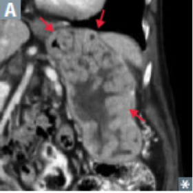
- 53Gastric cancer (types? characteristics? presentation? location? nodes?)Types:
1. Gastric adenocarcinoma (most common)
2. Lymphoma
3. GI stromal tumor
4. Carcinoid (rare)
Characteristics
1. Early aggressive local spread w/ node/liver metastasis
2. Often presents late
Presentation
1. Weight loss
2. Abdominal pain/ early satiety
3. Anemia
4. Acanthosis nigricans
5. Leser-trelat sign
Location:
A) intestinal --> assoc. w/:
1. H. pylori
2. Dietary nitrosamine (smoked food)
3. Tobacco/Smoking
4. Achlorhydria
5. Chronic gastritis
6. Blood type A
- commonly on lesser curvature --> looks like ulcer w/ raised margins
B) diffuse - not assoc. w/ H pylori, intestinal metaplasia, or nitrosamines
- signet ring cells (mucin-filled cells w/ peripheral nuclei)
- Desmoplasia --> linitis plastica (stomach wall grossly thickened and leathery)
Nodes:
1. Virchow node - involvement of left supraclavicular node by metastasis from stomach
2. Krukenberg tumor - bilateral metastasis to ovaries; abundant mucin-secreting, signet ring cells
(diffuse type)
3. Sister Mary Joseph nodule - subcutaneous periumbilical metastasis (intestinal type) - 54Peptic Ulcer disease (gastric ulcer vs duodenal ulcer)- duodenal ulcer pain improves with meals because duodenum activates protective mechanisms during meals
- complications of duodenal ulcer:
1. if arise in posterior duodenum (usually anterior) --> rupture --> gastroduodenal artery bleed --> acute pancreatitis
- Gastric ulcer usually at lesser curvature
- rupture --> bleed from left gastric artery
- can be due to malignancy (but never duodenal ulcer)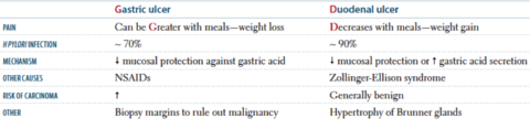
- 55Ulcer complications (which ones? signs?)Hemorrhage
- gastric, duodenal (posterior > anterior)
- most common complication
- ruptured gastric ulcer on lesser curvature of stomach --> bleed from left gastric artery
- ulcer on posterior wall of duodenum --> bleed from gastroduodenal artery
Obstruction
- pyloric channel
- duodenal
Perforation
- duodenal (anterior > posterior)
- may see free air under diaphragm w/ referred pain to the shoulder via phrenic nerve - 56Celiac sprue (about? pathogenesis? genetics? presentation? lab findings? pathology? tests? complications?)About:
Autoimmune-mediated intolerance of gliadin (in gluten) --> damage of small bowel villi
Pathogenesis
Gliadin absorbed --> deaminated by tissue transglutaminase (tTG) --> deaminated gliadin presented via MHC-class II --> helper T cells mediate tissue damage
Genetics
1. HLA-DQ2, HLA-DQ8
2. Northern european descent
Presentation
1. Diarrhea --> steatorrhea
2. Weight loss/weakness
3. Vitamin and mineral deficiencies (due to malabsorption at distal duodenum - jejunum/ileum less involved)
4. Dermatitis herpetiformis
5. Dec. bone density
Lab Findings
1. IgA anti-tissue transglutaminase
2. IgA Anti-endomysial antibodies
3. IgA Anti-deamidated gliadin peptide antibodies
Pathology
1. Villous atrophy (flattened)
2. crypt hyperplasia
3. Intraepithelial lymphocytosis
Tests
1. D-xylose test (passively absorbed in proximal small intestine --> blood/urine level drops w/ mucosa defects/bacterial overgrowth)
2. Sudan stain (stains for fecal fat)
Complications (occurs despite gluten-free diet)
1. T-cell lymphoma risk
2. Small bowel carcinoma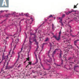
- 57Lactose intolerance (due to? findings? pathology? lab findings?)Due to:
1. Lactase deficiency (AR)
2. This^ could be 2* to viral enteritis (this would be temporary)
3. Acquired deficiency (occurs in late childhood)
Findings
1. Osmotic diarrhea
Pathology
1. Normal-appearing villi
2. Unless 2* to viral enteritis (villi tip injury)
Lab-findings
1. Dec. stool pH
2. Lactose hydrogen breath test (+ for lactose intolerance if post lactose breath hydrogen value rises > 20 ppm compared w/ baseline) - 58Pancreatic insufficiency (causes? complications? lab findings?)Causes:
1. Chronic pancreatitis
2. Cystic fibrosis
3. Obstructing cancer
Complications
1. malabsorption of fat --> steatorrhea
2. Malabsorption of fat-soluble vitamins
A-D-E-K-VitB12
Lab findings
1. Dec. duodenal pH (bicarbonate)
2. Dec. fecal elastase - 59Tropical sprue (cause? pathogenesis? findings? treatment?)Cause:
unknown, but it's assoc. w/ residents of OR recent visitors to the tropics
Pathogenesis
- most prominent in jejunum and ileum --> Vit B12 + folate deficiency
- duodenum less involved
Findings:
1. Malabsorption --> steatorrhea
2. Megaloblastic anemia (due to folate deficiency, and later B12 deficiency)
Treatment
It does respond to antibiotics - 60Whipple disease (cause? epidemiology? pathology? pathogenesis? findings?)Cause:
1. Infection with Tropheryma whipplei (intracellular gram +)
Epidemiology
1. Older men
Pathology
1. PAS(+) foamy macrophages in intestinal lamina propria (partially destroyed infectious organisms)
2. Mesenteric nodes
Pathogenesis
Foamy macrophages compress lacteals --> chylomicrons cannot be transferred from enterocytes to lymphatics -->
Findings:
1. Fat malabsorption --> steattorhea
2. cardiac symptoms
3. arthralgia
4. neurologic symptoms
*Foamy Whipped cream in a CAN*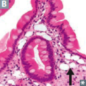
- 61Location? Gross morphology? Micro morphology? Presentation? Complications? Extraintestinal manifestations? Treatment?Crohn disease (IBD)
Location
- any portion of the GI tract (but rectal sparing)
- usually the terminal ileum and colon
- skip lesions
Gross morphology
- Transmural inflammation (wall thickening)
- cobblestone mucosa
- creeping fat (myofibroblasts)
- strictures (string sign - myofibroblasts)
- linear ulcers + fissures
Microscopic morphology
- noncaseating granulomas
- lymphoid aggregates
Presentation
1. Diarrhea that may or may not be bloody
2. RLQ pain
Complications
1. malabsorption/malnutrition
2. colorectal cancer
3. enterovesical fistula --> recurrent UTI
4. strictures --> obstruction
5. perianal disease**
Extraintestinal manifestations (common)
1. Rash (erythema nodosum --> pyoderma gangrenosum)
2. Eye - (episcleritis, uveitis)
3. Oral ulcerations (aphthous stomatitis)
4. Arthritis
Extraintestinal manifestations (only to CD)
1. Ca-oxalate nephrolithiasis
2. Gallstones
Treatment
1. Corticosteroids
2. Azathioprine
3. Antibiotics (cipro, metronidazole)
4. Infliximab
5. Adalimumab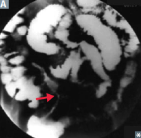
- 62location? gross morphology? micro. morphology? presentation? extraintestinal manifestations? complications? Treatment?Ulcerative colitis (IBD)
Location
Rectal involvement --> continuous lesion
Gross morphology
- mucosal and submucosal inflammation ONLY
- mucosal pseudopolyps (polyps are the normal epithelium)
- loss of haustra --> "lead pipe" appearance on imaging
Microscopic morphology
- crypt abscesses + ulcers
- bleeding
- no granulomas
Presentation
1. Recurrent bouts of bloody diarrhea
2. Abdominal pain
Extraintestinal manifestations (common)
1. Rash (erythema nodosum --> pyoderma gangrenosum)
2. Eye inflammation (episcleritis, uveitis)
3. Oral ulcerations (more common in CD)
4. Arthritis (peripheral, spondylitis)
Extraintestinal manifestations
1. 1* sclerosis cholangitis (assoc. w/ p-ANCA)
Complications
1. Malabsorption/malnutrition
2. CRC (inc. risk w/ pancolitis, > 10 yrs of disease)
3. Fulminant colitis
4. Toxic megacolon
5. Perforation
Treatment
1. 5-aminosalicylic preparations (mesalamine)
2. 6-mercaptopurine
3. Infliximab (anti-TNF-a)
4. Colectomy
*smoking protects against UC*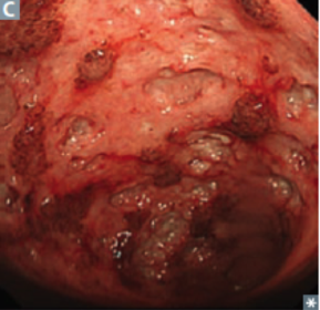
- 63Irritable bowel syndrome (criteria? epidemiology? chronic symptoms?)Recurrent abdominal pain assoc. w/ 2 of the following:
1. Pain improves w/ defecation
2. Changes in stool frequency
3. Changes in stool appearance
No structural abnormalities
Epidemiology
- most common in middle-aged women
chronic symptoms
1. either - diarrhea-predominant
2. or - constipation-predominant - 64etiology? presentation? complications? differentials? treatment?Appendicitis
- acute inflammation of the appendix
Etiology
- obstruction by fecalith (adults)
- lymphoid hyperplasia (children)
Presentation
1. initial diffuse periumbilical pain migrates to McBurney point (1/3 the distance from right anterior superior iliac spine to umbilicus)
2. Nausea
3. Fever
4. Psoas/obturator signs
5. Guarding and rebound tenderness
Complications
1. Rupture --> peritonitis
Differential
1. Diverticulitis (elderly)
2. Ectopic pregnancy (use B-hCG) to rule out
Treatment
1. Appendectomy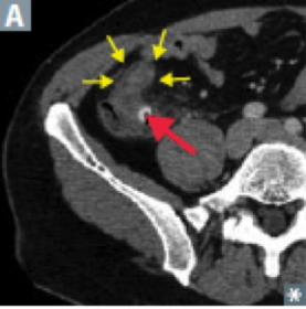
- 65Diverticula of the GI tract (diverticulum? types? diverticulosis? diverticulitis? presentation? complications of? treatment?)Diverticulum
- blind pouch that protrudes from the alimentary tract that communicates w/ the lumen of the gut
- most are acquired; most often in sigmoid colon
True diverticulum
- all 3 gut wall layers outpouch (meckel)
False diverticulum
- only mucosa and submucosa outpouch
- occur esp. where vasa recta perforate muscularis externa
Diverticulosis
- many false diverticula of the colon (sigmoid)
- common (esp. as ppl age)
- caused by inc. intraluminal pressure and focal weakness in colonic wall
- assoc. w/ low-fiber diets
- usually asymptomatic/assoc. w/ vague discomfort
- complications:
1. diverticular bleeding (painless hematochezia)
2. Diverticulitis
Diverticulitis
- diverticulosis w/ inflamed microperforations
- presentations:
1. LLQ pain
2. Fever
3. Leukocytosis
^treatment - antibiotics
- complications
1. Abscess
2. Fistula (colovesicular fistula --> pneumaturia)
3. Obstruction (inflammatory stenosis)
4. Perforation (peritonitis)
Treatment
- percutaneous drainage/surgery - 66Zenker diverticulum (what? etiology? presentation? epidemiology?)What:
1. Pharyngoesophageal false diverticulum
Etiology
esophageal dysmotility --> herniation of mucosal tissue at Killian triangle between thyropharyngeal and cricopharyngeal parts of the inferior pharyngeal constrictor (where the esophagus joins pharynx)
Presentation
1. Dysphagia
2. Obstruction
3. Aspiration
4. Halitosis
5. Gurgling
6. Neck mass
Most commonly in elderly males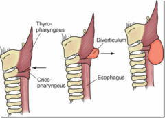
- 67Etiology? Presentation? Diagnosis?Meckel diverticulum - True Diverticulum
Etiology
- Persistence of vitelline duct (it doesn't involute)
- may contain ectopic acid-secreting gastric mucosa and/or pancreatic tissue
- most common congenital anomaly of GI tract
Presentation
1. Melena
2. RLQ pain
3. Intussusception
4. Obstruction near terminal ileum
Diagnosis
1. Pertechnetate study (uptake by the ectopic gastric mucosa)
The six 2's
2 x higher in males
2 inches long
2 ft from ileocecal valve
2% of population
presents before 2
2 types of epithelia (gastric/pancreatic)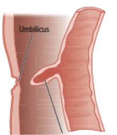
- 68Hirschsprung disease (what? pathophysiology? presentation? epidemiology? diagnosis? treatment?)What
- congenital megacolon characterized by lack of ganglion cells/enteric nervous plexuses (Auerbach AND meissner)
Pathophysiology
- RET mutation --> neural crest cell migration problem --> lack of Auerbach/Meissner plexuses in distal colon
Presentation
1. Bilious emesis
2. Abdominal distention --> chronic constipation
3. Failure to pass meconium within 48 hrs
4. Dilated proximal colon (transition zone)
Epidemiology
1. Inc. risk in Down syndrome patients
Diagnosis
1. Rectal suction biopsy (normal biopsy isn't good enough)
Treatment
1. Resection of the involved bowel - 69MalrotationAnomaly of midgut rotation during fetal development -->
1. Improper positioning of bowel
2. formation of fibrous bands (Ladd bands)
3. Complications:
a) volvulus
b) duodenal obstruction - 70Volvulus (what? epidemiology?)Twisting of portion of bowel around its mesentery --> obstruction and infarction
Midgut volvulus - more common in infants and children
Sigmoid volvulus - more common in elderly - 71Intussuception (findings? children causes? adult causes?)Telescoping of proximal bowel segment into distal segment, commonly at ileocecal junction
Findings:
compromised blood supply -->
1) intermittent abdominal pain
2) Currant jelly stools
Usually in children
1. Idiopathic
2. Lymphoid hyperplasia (rotavirus)
- arises in terminal ileum (payer's patch) --> intussusception into ceum
In adult
- tumor most likely cause - 72Acute mesenteric ischemia Chronic mesenteric ischemia Colonic ischemia AngiodysplasiaA) Acute mesenteric ischemia:
- Critical blockage of intestinal blood flow (embolic occlusion of SMA) --> small bowel necrosis --> abdominal pain out of proportion to physical findings
- "currant jelly" stools
B) Chronic mesenteric ischemia
- "intestinal angina"
- atherosclerosis of celiac/SMA/IMA --> intestinal hypoperfusion --> postprandial epigastric pain
- food aversion and weight loss
C) Colonic ischemia
- Reduction in intestinal blood flow --> ischemia (worsens w/ meals)
- Crampy abdominal pain followed by hematochezia
- occurs commonly at watershed areas:
1. Splenic flexure
2. Distal colon
- affects elderly typically
D) Angiodysplasia
- Tortuous dilation of mucosal and submucosal vessels --> hematochezia
- often found in:
terminal ileum - cecum - ascending colon
- more common in elderly patients
- confirmed by angiography - 73Adhesion Ileus Meconium ileus Necrotizing enterocolitisAdhesion
- most common cause of small bowel obstruction
- surgery --> formation of fibrous band of scar tissue
- well-demarcated necrotic zones
Ileus
- intestinal hypomotility w/o obstruction
- presentation:
1. constipation
2. distended/tympanic abdomen w/o bowel sounds
- assoc. w/:
1. Abdominal surgeries
2. Opiates
3. Hypokalemia
4. Sepsis
- treatment
1. Bowel rest
2. Electrolyte correction
3. Cholinergic drugs (to stimulate intestinal motility)
Meconium ileus
- cystic fibrosis --> meconium plug obstructs intestine --> prevents stol passage at birth
Necrotizing enterocolitis
- seen in premature, formula-fed infants w/ immature immune system
- necrosis of intestinal mucosa (primarily colonic) w/ possible perforation -->
1. pneumatosis intestinalis (free air in abdomen)
2. portal venous gas - 74—
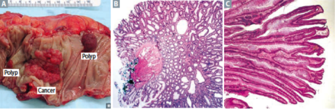
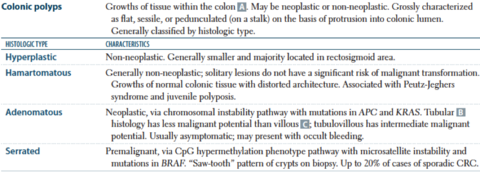
- 75Polyposis Syndromes (1, part a and b, 2, and 3)A) Familial adenomatous polyposis
- AD mutation of APC (tumor suppressor) on 5q
- 2-hit hypothesis
- thousands of polyps arise starting after puberty (pancolonic + involves rectum)
- prophylactic colectomy or else 100% CRC
B) Gardner Syndrome
- FAP +
1. osteomas and fibrzomatosis - osseous and soft tissue tumors
2. Congenital hypertrophy of retinal pigment epithelium (CHRPE)
3. Supernumerary teeth
C) Turcot syndrome
-if FAP(+): medulloblastoma
-if MHL1, PSM2 (+): glialblastoma
D) Peutz-Jeghers syndrome (STK11)
- AD syndrome w/ numerous hamatomas throughout GI tract +
1. Hyperpigmented mouth/lips/hands/genitals
4+2+1
1. Colorectal carcinoma
2. Gastric carcinoma
3. Pancreatic carcinoma
4. Hepatocellular carcinoma
5. Breast cancer
6. Ovarian cancer
7. Lung cancer
E) Juvenile polyposis syndrome
- AD syndrome in children (< 5 yrs)
- numerous hamartomatous polyps in colon - small bowel - stomach
- assoc. w/ inc. risk of CRC - 76Lynch Syndrome (genetic mutation? complications? other cancers?)AKA HNPCC (hereditary nonpolyposis colorectal cancer)
- AD mutation of DNA mismatch repair genes (MLH1, MSH2, MSH6, EPCAM, PMS2) --> subsequent microsatellite instability
- 80% progression to CRC
- proximal colon is always involved
- assoc. w/:
1. ENDOMETRIAL cancer
2. Ovarian cancer
3. Skin cancer - 77Molecular pathogenesis of colorectal cancer (APC? K-RAS? p53? DCC?)
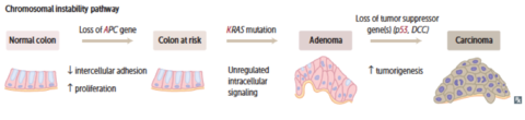
- 78Cirrhosis (pathology? complications? etiologies? findings?)Cirrhosis
- diffuse bridging fibrosis (via TGF-B from stellate cells) and regenerative nodules
- ^ disrupt normal architecture of liver
- complication --> inc. risk of HCC
- Etiologies:
1. Alcohol
2. non-alcoholic steatohepatitis
3. chronic viral hepatitis
4. autoimmune hepatitis
5. biliary disease
6. genetic metabolic disorders
Findings:
1. Jaundice (UC)
2. Spider angiomas (hyperestrinism)
3. gynecomastia
4. testicular atrophy
5. Peripheral edema (hypoalbuminemia)
6. Cardiomyopathy (from ^)
7. Thrombocytopenia - anemia (portal hypertension --> hypersplenism)
8. Coagulation disorders (dec. coag protein synthesis --> inc. PT)
9. Hepatic encephalopathy (from high [ammonia])
10. Asterixis (flapping tremor)
11. Anorexia
12. Nausea, vomiting - 79Portal hypertension (what? etiologies? findings?)- inc. pressure in portal venous system
- etiologies:
1. cirrhosis
2. Portal vein thrombosis (vascular obstruction)
3. Budd-Chiari syndrome (vascular obstruction)
4. Schistosomiasis
Findings:
1. Esophageal varices (esophageal vain --> left gastric vein --> portal vein --> painless hematemesis --> most common cause of death in cirrhosis)
2. Caput medusa
3. Internal hemorrhoids
4. Ascites
5. Splenomegaly
6. Gastric varices --> melena - 80Serum markers of liver pathology (AST, ALT, AST:ALT, Alk Phos, g-glutamyl transpeptidase, bilirubin, albumin, prothrombin, platelets)AST, ALK:
- ALT > AST --> liver disease
- AST > ALT --> alcoholic liver disease
- AST > ALT in nonalcoholic liver disease --> progression to adv. fibrosis/cirrhosis
Alk Phos
- if inc:
1. cholestasis (biliary obstruction)
2. infiltrative disorders
3. bone disease
g-glutamyl transpeptidase
- if inc:
1. cholestasis (just like alk phos)
2. various liver disease
3. alcohol use
- NOT IN BONE DISEASE -
Bilirubin
- inc. in:
1. biliary obstruction
2. alcoholic/viral hepatitis
3. cirrhosis
4. hemolysis
Albumin
- dec. in adv. liver disease
Prothrombin
- inc. in adv. liver disease (dec. production of clotting factors)
Platelets
- dec. in adv. liver disease (dec. thrombopoietin via liver sequestration)
- dec. in portal hypertension (splenomegaly/splenic sequestration) - 81Reye syndrome (what? etiology? mechanism? finding? kawasaki?)What
- rare, often fatal childhood hepatic encephalopathy
Etiology
- Viral infection (VZV, influenza B) that's been treated with aspirin
Mechanism:
- aspirin metabolites dec. B-oxidation by reversible inhibition of mitochondrial enzymes
Findings:
1. Hypoglycemia
2. Fatty liver (microvesicular fatty change)
3. Vomiting
4. Hepatomegaly --> elevated liver enzymes
5. Coma
Avoid aspirin in children, except in those w/ Kawasaki disease - 82What is this (left? middle? right?)Alcoholic liver disease
Hepatic steatosis
- macrovesicular fatty change that may be reversible w/ alcohol cessation
Alcoholic hepatitis
- sustained, long-term consumption of alcohol
- swollen and necrotic hepatocytes w/ neutrophilic infiltration
- mallory bodies (intracytoplasmic eosinophilic inclusions of damaged keratin filaments)
- AST > ALT ( > 2:1)
- painful hepatomegaly
Alcoholic cirrhosis
- final and irreversible form
- micro-nodular, irregularly shrunken liver with "hobnail" appearance
- sclerosis around central vein (zone III)
- Manifestations of chronic liver disease:
1. Jaundice
2. hypoalbuminemia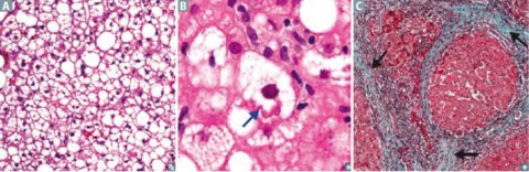
- 83What is this? Mechanism? complications?Non-alcoholic fatty liver disease
Mechanism
- metabolic syndrome (insulin resistance)
- obesity --> fatty infiltration of hepatocytes --> cellular "ballooning" --> eventual necrosis --> cirrhosis
Complications
- cirrhosis
- HCC
ALT > AST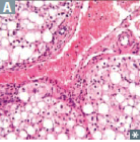
- 84Hepatic encephalopathy (pathophysiology? findings? triggers? treatment?)Pathophysiology
Cirrhosis --> portosystemic shunts --> dec. NH3 metabolism --> neuropsychiatric dysfunction
Spectrum of findings:
1. Disorientation/asterixis (mild)
2. Difficult arousal or coma (severe)
Triggers:
a) Inc. NH3 production and absorption
1. Dietary protein
2. GI bleed
3. Constipation
4. Infection
b) Dec. NH3 removal
1. Renal failure
2. Diuretics
3. Bypassed hepatic blood flow (post TIPS)
Treatment
1. Lactulose (inc. NH4 generation)
2. Rifaximin or neomycin (dec. NH4 producing gut bacteria) - 85Epidemiology? Assoc? Complications? Findings? how does it spread? Diagnosis?Hepatocellular carcinoma/hepatoma
Epidemiology
1. Most common 1* malignant tumor of liver in adults
Assoc:
1. HBV (can occur w/o cirrhosis)
2. HCV (after cirrhosis)
and all causes of cirrhosis:
3. Alcoholic fatty liver disease
4. Non-alcoholic fatty liver disease
5. Autoimmune disease (primary biliary cirrhosis)
6. Hemochromatosis
7. a1-antitrypsin deficiency
8. Aflatoxin (from aspergillus)
Complications:
1. Budd-Chiari syndrome (liver infarction due to hepatic vein obstruction --> painful hepatomegaly and ascites)
Findings:
1. jaundice
2. tender hepatomegaly
3. ascites (when there's portal HTN) -->
4. polycythemia**
5. anorexia
Spread hematogenously (unlike other carcinomas)
Diagnosis
1. inc. a-fetoprotein (aFP)
2. ultrasound or contrast CT/MRI
3. biopsy (for background cirrhosis)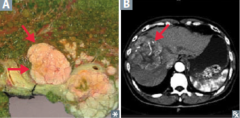
- 86Other liver tumors (cavernous hemangioma, hepatic adenoma, angiosarcoma, metastasis)A) Cavernous hemangioma
- common benign liver tumor
- ages 30-50
- biopsy contraindicated because of hemorrhage
B) Hepatic adenoma
- Rare benign liver tumor
- related to:
1. oral contraceptives
2. anabolic steroid use
- can regress
- if rupture --> abdominal pain/shock
C) Angiosarcoma
- malignant tumor of endothelial origin
- assoc. w/ exposure to:
1. arsenic
2. vinyl chloride
Metastases
- most common overall
- from:
1. GI malignancies
2. Breast cancer
3. Lung cancer - 87Budd-Chiari syndrome (pathophysiology? risk factors? findings?)Pathophysiology
Thrombosis/compression of hepatic veins --> centrilobular congestion and necrosis --> congestive liver disease
Risk factors
1. Hypercoagulable states
2. Polycythemia vera
3. Postpartum state
4. HCC
Findings:
a) due to congestive liver disease:
1. Hepatomegaly
2. Ascites
3. Varices
4. Abdominal pain
5. Liver failure
6. Absence of JVD
7. Maybe: nutmeg liver - 88a1-antitrypsin deficiency (genetics? pathogenesis? findings?)Genetics
- codominant trait
Pathogenesis
- misfiled gene product protein --> aggregates in hepatocellular ER --> cirrhosis w/ PAS (+) globules in liver
- dec. a1-antitrypsin --> uninhibited elastase in alveoli --> dec. elastic tissue --> panacinar emphysema
Findings
1. Cirrhosis
2. Panacinar emphysema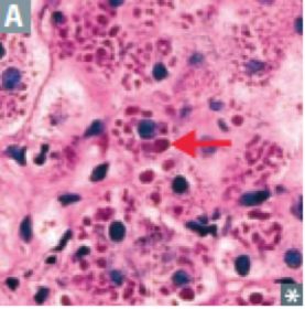
- 89Unconjugated vs conjugated vs mixed hyperbilirubinemia (causes for each)Unconjugated (indirect) hyperbilirubinemia
1. Hemolytic anemia
2. Physiologic (newborns)
3. Crigler-Najjar
4. Gilbert syndrome
Conjugated (direct) hyperbilirubinemia
A) Biliary track obstruction
1. Gallstones
2. Cholangiocarcinoma
3. Pancreatic cancer
4. Liver cancer
5. Liver fluke (chlonorchis sinensis)
B) Biliary tract disease
6. 1* sclerosis cholangitis
7. 1* biliary cirrhosis
C) Excretion defect
8. Dubin-Johnson syndrome
9. Rotor syndrome
Mixed (direct and indirect) hyperbilirubinemia
1. Hepatitis
2. Cirrhosis - 90Physiologic neonatal jaundice (pathophysiology, timeline, treatment)Pathophysiology
At birth: immature UDP-glucuronosyltransferase --> unconjugated hyperbilirubinemia --> jaundice + kernicterus (bilirubin deposition in brain - esp. Basal Ganglia)
Timeline:
Happens at 24 hrs of life --> resolves by 1-2 weeks
Treatment:
1. Phototherapy (non-UV) --> isomerize unconjugated bilirubin to water-soluble form - 91Hereditary hyperbilirubinemias*all AR*
A) Gilbert syndrome
- mildly dec. UDP-glucuronosyltransferase conjugation + impaired bilirubin uptake
- asymptomatic OR mild jaundice
- high indirect bilirubin w/o hemolysis
- stress/fasting --> (+) bilirubin
B) Crigler-Najjar syndrome type I
- absent UDP-glucuronosyltransferase
- presents early in life --> patients die within a few years
- jaundice + kernicterus
- high indirect bilirubin
- treat via plasmapheresis + phototherapy
C) Crigler-Najjar Type II
- less severe as type I
- responds to phenobarbital --> inc. liver enzyme synthesis
D) Dubin-Johnson syndrome
- defective liver excretion --> conjugated hyperbilirubinemia
- grossly black liver
- benign
E) Rotor syndrome
- similar as Dubin-Johnson syndrome
- due to impaired hepatic uptake + excretion
- milder w/o black liver - 92Wilson disease (hepatolenticular degeneration) (genetics? pathophysiology? presentation? lab? treatment? complication?)Genetics:
- AR in ATP7B, chromosome 13 (for copper-transport)
Pathophysiology
- Inadequate copper excretion into bile and blood
- Inadequate copper incorporation into ceruloplasmin
- Copper accumulates in liver, brain, cornea, kidneys, joints
Presentation
- before 40:
A) liver disease
1. hepatitis
2. acute liver failure
3. cirrhosis
B) neurologic disease (copper deposits in basal ganglia)
4. Dementia
5. Dystonia
6. Tremor
7. Parkinsonism
8. psychiatric disease
C) eye problems
9. Kayser-Fleischer rings (Descemet membrane of cornea)
D) blood problems
10. Hemolytic anemia
E) Kidney problems
11. Fanconi syndrome --> renal failure
Lab:
1. (+) urinary copper
2. (-) serum ceruloplasmin
3. (+) copper on liver biopsy
Treatment
1. Chelation w/ penicillamine or tridentine
2. Oral zinc
Complications
1. Hepatocellular carcinoma - 93Hemochromatosis (genetics, other causes, pathophysiology, presentation, complications, diagnosis, treatment)Genetics
- AR mutation in HFE gene (C282Y > H63D, chromosome 6) --> abnormal iron sensing + inc. intestinal absorption
- Inc. Ferritin, Inc. Fe, Dec. TIBC --> Inc. transferrin saturation
Other causes:
- 2* to chronic transfusion therapy (B-thalassemia major)
Pathophysiology
- iron accumulates in liver, pancreas, skin, heart, pituitary, joints
Presentation
- after 40 (when today body Fe > 20g)
- iron loss via menstruation slows progression in women
-triad:
1. Cirrhosis
2. Diabetes mellitus
3. Skin pigmentation
4. Dilated cardiomyopathy (reversible)
5. Hypogonadism
6. Arthropathy (Ca pyrophosphate in MCP)
Complication:
1. HCC --> death
Diagnosis
1. Liver MRI --> identify hemosiderin
2. Biopsy + Prussian blue stain
Treatment
1. Repeated phlebotomy
2. Chelation w/ deferasirox, deferoxamine, oral deferiprone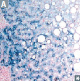
- 94Primary sclerosis cholangitis (presentations? lab? pathology? diagnosis via? epidemiology? assoc. w/? serum markers? complications?)- a Biliary tract disease -
Presentations
1. Pruritus
2. Jaundice
3. Dark urine + light stool
4. Hepatosplenomegaly
Lab presentations:
1. Cholestatic pattern of LFTs (inc. conj. bilirubin, inc. cholesterol, inc. Alk Phos)
- Primary Sclerosing Cholangitis -
pathology
- unknown cause
- concentric "onion skin" periductal fibrosis --> "beading" of intra-/extrahepatic bile ducts
Diagnosis:
- ERCP + MRCP (magnetic resonance cholangiopancreatography)
Epidemiology:
Middle-aged men
Markers
1. p-ANCA +
2. (+) IgM
Assoc. w/ ulcerative colitis
Complications:
1. 2* biliary cirrhosis (a late presentation)
2. Cholangiocarcinoma
3. Gallbladder cancer - 95Primary Biliary Cirrhosis (Presentations? lab? pathology? epidemiology? markers? complications?)- a Biliary tract disease -
Presentations
1. Pruritus
2. Jaundice
3. Dark urine + light stool
4. Hepatosplenomegaly
Lab presentations:
1. Cholestatic pattern of LFTs (inc. conj. bilirubin, inc. cholesterol, inc. Alk Phos)
- Primary Biliary Cirrhosis -
Pathology
Autoimmune reaction --> lymphocytic infiltrate + granulomas --> destruction of interlobular bile ducts
Epidemiology
- middle-aged women
Markers
- IgM anti-mitochondrial antibody +
Complications - other autoimmune diseases:
1. Sjogren syndrome
2. Hashimoto thyroiditis
3. CREST
4. Rheumatoid arthritis
5. Celiac disease - 96Secondary biliary cirrhosis (presentations? lab? pathology? epidemiology? complications?)- a Biliary tract disease -
Presentations
1. Pruritus
2. Jaundice
3. Dark urine + light stool
4. Hepatosplenomegaly
Lab presentations:
1. Cholestatic pattern of LFTs (inc. conj. bilirubin, inc. cholesterol, inc. Alk Phos)
- secondary biliary cirrhosis -
Pathology
Extrahepatic biliary obstruction --> pressure in intrahepatic ducts --> injury/fibrosis + bile stasis
epidemiology
Patients w/ known obstructive lesions:
1. gallstones
2. biliary strictures
3. pancreatic carcinoma
Complications:
1. Ascending cholangitis - 97Causes? Types and their assoc.? Presentations? Diagnosis? Complications?Gallstones - cholelithiasis
causes:
1. Inc. cholesterol/bilirubin
2. Dec. bile salts/phospholipids
3. Gallbladder stasis
Types:
A) Cholesterol stones
- radiolucent, 80% of stones
- Associations:
1. Obesity
2. Crohn disease
3. Adv. age
4. Estrogen therapy
5. Multiparity
6. Rapid weight loss
7. Native american origin
B) Pigment stones
- Black: radiopaque, Ca-bilirubinate, hemolysis
- Brown: radiolucent, infection
Assoc. w/:
1. crohn disease
2. chronic hemolysis
3. alcoholic cirrhosis
4. advanced age
5. biliary infections
6. total parenteral nutrition
Presentations:
A) uncomplicated --> biliary colic
(neurohormonal activation via CCK after fatty meal --> contraction of gallbladder --> stone passes into cystic duct)
Diagnosis: ultrasound
Treatment: cholecystectomy (symptomatic)
Complications:
1. Cholecystitis (most common)
2. Acute pancreatitis
3. Ascending cholangitis
4. Gallstone ileus (fistula between gallbladder and GI tract --> gallstones pass into GI tract)
Charcot triad of cholangitis:
1. Jaundice
2. Fever
3. RUQ pain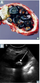
- 98What is this? causes? Presentation? Complications? Diagnosis? Treatment?Cholecystitis
Acute/chronic inflammation of gallbladder, usually from cholelithiasis (stone at neck of gallbladder) w/ gallbladder wall thickening
Causes:
1. Gallstones commonly block the cystic duct --> 2* infection
2. Rarely acalculus:
2a - ischemia and stasis (impacted gallstone --> dilatation --> pressure ischemia)
2b - CMV
Presentation:
1. Murphy sign (+) - inspiratory arrest on RUQ palpation due to pain
2. If bile duct becomes involved (ascending cholangitis) --> (+) Alk Phos
Complications:
1. If chronic - herniation of gallbladder mucosa into muscular wall (Rokitansky-Aschoff sinus)
2. If chronic - porcelain gallbladder --> inc. risk for carcinoma
Diagnosis
1. Ultrasound
2. Cholescintigraphy (HIDA - hepatobiliary iminodiacetic acid scan)
Cholecystectomy is the definitive treatment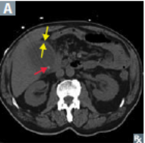
- 99—Porcelain gallbladder
- calcified gallbladder due to chronic cholecystitis
- usually found incidentally on imaging
Complications
- adenocarcinomas of gallbladder
Treatment
- prophylactic cholecystectomy (cuz of cancer risk)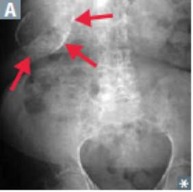
- 100What is it? Causes? Diagnosis? Findings? Complications?Acute Pancreatitis
Autodigestion of pancreas by pancreatic enzymes
Causes: (I GET SMASHED)
1. Idiopathic
2. Gallstones
3. Ethanol
4. Trauma
5. Steroids
6. Mumps
7. Autoimmune disease
8. Scorpion sting
9. Hypercalcemia/Hypertriglyceridemia (> 1000 mg/dL)
10. ERCP
11. Drugs (sulfa drugs, NRTIs, protease inhibitors)
Diagnosis (2 out of 3 criteria):
1. Acute epigastric pain (radiating to back)
2. Inc. serum amylase/lipase to 3x normal upper limit
3. Characteristic imaging findings
Findings:
1. Liquefactive hemorrhagic necrosis of pancreas
2. Fat necrosis of peri-pancreatic fat
3. Nausea and vomiting
Complications:
1. Pseudocysts (lined by granulation tissue, not epithelium)
2. Necrosis
3. Hemorrhage
4. Infection
5. Shock --> organ failure (renal failure, ARDS)
6. Hypocalcemia (precipitation of Ca soaps)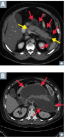
- 101What is this? causes? Clinical features? Complications? Long term complication?Chronic pancreatitis
1. Chronic inflammation --> atrophy + calcification of pancreas
Causes:
1. Alcohol abuse (ppl can't get over habit)
2. Idiopathic
3. CFTR mutation --> cystic fibrosis
Clinical features
1. Epigastric pain that radiates to the back
2. Pancreatic insufficiency (see below)
3. Dystrophic calcification of pancreatic parenchyma on imaging
Chronic pancreatic insufficiency
1. Steatorrhea
2. Fat-soluble vitamin deficiency
3. 2* diabetes mellitus
4. Amylase/lipase may/may not be elevated (but ALWAYS elevated in acute pancreatitis)
Complications
1. Inc. risk for pancreatic carcinoma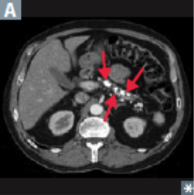
- 102What is this? Prognosis? Markers? Risk factors? Presentations? Treatment?Pancreatic adenocarcinoma
- very aggressive tumor arising from pancreatic ducts
- tumors more common in pancreatic head
Prognosis
- often metastatic at presentation
- avg survival ~1 yr after diagnosis
Tumor markers:
- CA 19-9
- CEA (but less specific)
Risk factors:
1. Tobacco use
2. Chronic pancreatitis (esp. > 20 yrs)
3. Diabetes
4. Age > 50 yrs
5. Jewish + African-American males
Presentations
1. Abdominal pain that radiates to back
2. Weight loss (malabsorption + anorexia)
3. Migratory thrombophlebitis (Trousseau syndrome) - redness and tenderness on palpation of extremities
4. Obstructive jaundice w/ palpable, contender gallbladder (Courvoisier sign) - since tumor is most likely to be in the head
Treatment
- Whipple procedure
- chemotherapy
- radiation therapy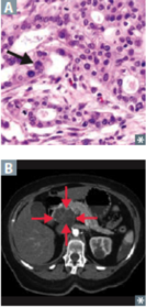
- 103Cleft Lip and Palate (what? etiology? timeline?)Full thickness defect of lip/palate, although both usually occur together
Etiology
- failure of facial prominences to fuse
Timeline
- early pregnancy --> facial prominences (superior, two lateral, two inferior) grow --> fuse together to form face - 104Behcet syndrome (what is it? presentation? pathogenesis?)What is it?
- recurrent triad of:
1. Aphthous ulcers (superficial ulceration of the oral mucosa due to stress - resolves automatically but often recurs)
2. genital ulcers
3. uveitis
Pathogenesis
- immune complex vasculitis involving small vessels
- seen after viral infection, but etiology is unknown - 105Squamous cell carcinoma of mouth (what? risk factors? precursor lesions?)What?
- malignant neoplasm of squamous cells lining the oral mucosa --> most likely at the floor of mouth
Risk factors
- tobacco
- alcohol
Precursor lesions:
1. Leukoplakia
- squamous cell dysplasia
- white plaque that can't be scraped away
- no oral candidiasis OR hairy leukoplakia (which is on lateral tongue - EBV induced)
2. Erythroplakia
- red plaque
- represents vascularized leukoplakia --> suggestive of squamous cell dysplasia
- biopsied to r/o carcinoma - 106Sialadenitis (what? etiology? complication)What?
- inflammation of the salivary gland
Etiology
- due to an obstructing stone (sialolithiasis)
Complication
- S. aureus infection --> usually unilateral - 107Esophageal web (what? where? presentation? complications? could be part of?)What
- thin protrusion of esophageal MUCOSA
- often in the upper esophagus
Presentation
- dysphagia (for poorly chewed food)
Complications
- esophageal squamous cell carcinoma
Could be part of: Plummer-Vinson syndrome
1. SEVERE iron deficiency anemia
2. esophageal web
3. beefy-red tongue (due to atrophic glossitis)
4. risk of esophageal squamous cell carcinoma - 108Mallory-Weiss Syndrome? (what? etiology? presentation? complications) GERD? (what? risk factors? clinical features? complications?)Mallory-Weiss Syndrome
- Longitudinal laceration of mucosa at the GE junction
Etiology
- severe vomiting due to:
1. Alcoholism
2. Bulimia
Presentation:
1. Painful hematemesis
complication:
1. Boerhaave syndrome
rupture of esophagus --> air in mediastinum --> subcutaneous emphysema (air bubbles beneath the skin)
GERD
Reflux of acid from stomach due to reduced LES tone
Risk factors:
1. Alcohol
2. Tobacco
3. Obesity
4. Fat-rich diet
5. Caffeine
6. Hiatal hernia
Clinical features
1. Heartburn (mimics cardiac chest pain)
2. Asthma (adult onset) and cough
3. Damage to enamel of teeth
Complications
1. Ulceration w/ strictures (mucosa + submucosa both knocked out --> no stem cell --> fibrosis --> stricture)
2. Barrett Esophagus (metaplasia --> columnar epithelium + goblet cells) - 109Chronic gastritis (about? causes? presentation? complications? treatment?)Chronic gastritis
chronic stomach mucosal inflammation
Causes:
1. H. pylori
- most common
- H pylori secretes urease and proteases --> inflammation --> weaken mucosal defenses
- affects antrum first --> spreads to body
- Presentation:
1. Epigastric abdominal pain
Increased risk of:
1. peptic ulcer disease
2. MALT lymphoma (inflammation produces germinal centers, which has marginal zones)
3. Gastric adenocarcinoma
Treatment
- triple therapy (clarithromycin, PPI, amoxicillin)
- confirm via lack of stool antigen
2. Autoimmune
- autoantibodies to parietal cells + intrinsic factor (body/fundus of stomach)
- pathogenesis mediated by T cells (Type IV hypersensitivity)
Complications:
1. inc. risk of pernicious anemia
2. Achlohydria (which (+) gastrin levels --> antral G-cell hyperplasia)
3. Gastric adenocarcinoma (atrophy of mucosa --> intestinal metaplasia) - 110Small bowel infarction (findings? types and causes?)Since small bowel does a lot of digestion - it's highly susceptible to ischemic injury
Findings:
1. Abdominal pain
2. Bloody diarrhea
3. Decreased bowel sounds
Transmural infarction
- occurs w/ thrombosis/embolism of SMA OR thrombosis of mesenteric vein
Causes
1. atrial fibrillation
2. vasculitis (polyarderitis nodosa)
3. polycythemia vera
4. anti-phospholipid syndrome
Mucosal infarction
- occurs w/ marked hypotension (since mucosa is the farthest away from blood supply) - 111Abetalipoproteinemia (what? consequences?)Autosomal recessive deficiency of apolipoprotein B-48 and B100
No B-48:
- malabsorption of fat due to defective chylomicron formation
No B-100:
- absent plasma VLDl and LDL - 112Carcinoid tumor (what? location? pathology? if metastasis?)Malignant proliferation of neuroendocrine cells --> low grade malignancy
Location - small bowel most common site
Pathology
1. Chromogranin (+) - it contains neurosecretory granules:
a) serotonin --> portal circulation --> liver MAO metabolize it to 5-HIAA --> excreted in urine
2. Grows as submucosal polyp-like nodule
If metastasis to liver:
serotonin released into hepatic vein --> systemic circulation -->
A) carcinoid syndrome
1. bronchospasms
2. diarrhea
3. flushing of skin
- serotonin release stimulated by alcohol -
B) carcinoid heart syndrome
- right-sided valvular fibrosis -->
1. tricuspid regurgitation
2. pulmonary valve stenosis
- MAO in lung --> left side valvular lesions not seen - 113Colonic polyps (what? types? progression pathway? screening? prognostic indicators?)- raised protrusions of colonic mucosa
- most common: hyper plastic and adenomatous polyps
A) Hyperplastic polyps
- due to hyperplasia of glands
- most common type of polyp
- arises in left colon
- benign AND no malignant potential
B) Adenomatous polyps
- due to NEOplastic proliferation of glands
- 2nd most common type of polyps
- benign but premalignant --> may progress to adenocarcinoma via:
adenoma-carcinoma sequence:
1. APC --> increase risk for polyps
2. K-ras --> formation of polyps
3. P53 and (+) COX --> progression to carcinoma
(aspirin may help)
Screening via
1. colonoscopy (gold standard)
2. test for fecal occult blood
Greatest risk for cancer:
1. Size > 2 cm
2. Sessile growth
3. Villous histology - 114Colorectal carcinoma (epidemiology? risk factors? pathogenesis? location and presentation? assoc. w/? staging? assessment of treatment response/recurrence?)Carcinoma arising from colonic or rectal mucosa
Epidemiology
- peak: age 60-70
Risk factor
1. IBD
2. Smoking
3. Diet high in meat and fat, low in fiber
4. FAP
5. Adenomatous/serrated polyps
Pathogenesis
1. Adenoma-carcinoma sequence
2. Microsatellite instability (MSI) - Lynch Syndrome
a) AD mutation of: MLH1, MSH2, MSH6, EPCAM, PMS2 - all DNA mismatch repair enzymes
b) increased risk of:
1. CRC (arises de novo)
2. Ovarian carcinoma
3. Endometrial carcinoma
Presentation:
1. If left sided -
a) napkin-ring lesion (squeezed)
b) decreased stool caliber
c) LLQ pain
d) blood-streaked stool
2. If right sided -
a) iron-deficiency anemia (Old ppl w/ anemia are presumed to have CRC)
b) vague pain
Assoc. w/:
1. Strep bovis endocarditis
Staging:
TNM (metastasis to liver)
CEA - useful for assessing treatment response and detecting recurrence (NOT for screening) - 115Pancreatic carcinoma (what is it? epidemiology? risk factors? clinical features? tumor marker? treatment? prognosis?)Adenocarcinoma arising from pancreatic ducts
Epidemiology
- seen in the elderly - avg age 70
Risk factors
1. Smoking
2. chronic pancreatitis
Clinical features (late in disease)
1. Epigastric abdominal pain
2. Weight loss
3. Obstructive jaundice (pale stool, palpable gallbladder) - if tumor is in head
4. 2* diabetes mellitus - if tumor is in tail
5. Pancreatitis (if tumor blocks ducts)
6. Migratory thrombophlebitis (Trousseau syndrome)
Tumor marker: CA 19-9
Surgical resection - en block removal of duodenum, head and neck of pancreas, gallbladder
Prognosis: 1 yr survival < 10% - 116Biliary atresia (pathogenesis? result? presentation?)Pathogenesis:
1. failure to form OR
2. early destruction OF
- extra hepatic biliary tree
- biliary obstruction within first 2 months of life
Presentation:
1. Jaundice (conj. bilirubin)
2. Cirrhosis - 117Ascending Cholangitis (What? due to? presentation? risk factor?)Bacterial infection of the bile ducts
Due to:
1. Ascending infection w/ enteric gram-neg bacteria
Presentation:
1. Sepsis (high fever + chills)
2. Jaundice
3. Abdominal pain
Risk factor:
Choledocholithiasis (stone in biliary duct blocking the natural expulsion of bile) - 118Gallbladder carcinoma (what? risk factor? presentation? prognosis?)Adenocarcinoma arising from glandular epithelium that lines the gallbladder
Risk factor
1. Gallstones
2. Porcelain gallbladder complication
Presentation:
1. Cholecystitis in an elderly woman
Poor prognosis - 119Hepatitis A Hepatitis E (transmission, disease characteristics, markers, complications)Transmission
- fecal oral
- HAV - acquired by travelers
- HEV - contaminated water/undercooked seafood
Disease characteristics
- acute hepatitis - no chronic states
1. Jaundice (mixed CB and UCB)
2. Dark urine (due to CB)
3. Fever/malaise/nausea
4. Elevated Liver enzymes (ALT>AST)
Pathology
1. Inflammation involving the lobules of liver and portal tracts AND
2. Apoptosis of hepatocytes
Markers
- Active infection: anti-virus IgM
- Protective: anti-virus IgG
Complications
- HEV in pregnant woman --> fulminant hepatitis (liver failure w/ massive liver necrosis) - 120Hepatitis B (HBV) (transmission, disease characteristics, pathology, markers)Transmission:
- Parenteral (childbirth, sex, IVD)
Disease characteristics:
1. Acute hepatitis
2. Chronicity occurs in 20% of cases
Findings:
1. Jaundice (mixed CB and UCB)
2. Dark urine (due to CB)
3. Fever/malaise/nausea
4. Elevated Liver enzymes (ALT>AST)
Pathology
1. Inflammation involving the lobules of liver and portal tracts AND
2. Apoptosis of hepatocytes
3. If chronic - inflammation of JUST portal tracts --> cirrhosis
Markers:
1. HBsAG - marker of infection
(+) = acute infection
(+) after 6 months = chronicity
2. HBcAB - marker of battle
IgM = acute or window period
IgG = resolved or chronic
3. HBsAB - marker of victory
IgG = resolved or immunized
4. HBeAG and HBV DNA - infectivity
(+) = acute
- or + in chronic states - 121Hepatitis C (HCV) (transmission, disease characteristics, findings, pathology, markers)Transmission
- Parenteral transmission (IVD, sex, no childbirth)
Disease characteristics
1. Acute hepatitis --> 80% chronicity
Findings:
1. Jaundice (CB and UCB)
2. Dark urine (CB)
3. Fever/nausea/malaise
4. elevated liver enzymes (ALT>AST)
Pathology
1. Inflammation of lobules and portal tracts
2. Apoptosis of hepatocytes
3. If chronic - portal tract inflammation only
Markers
1. HCV-RNA = infection
2. Decreased RNA = recovery
3. Persistence = chronicity - 122Hepatitis D (HDV)Dependent on HBV for infection
Superinfection upon existing HBV more severe than coinfection
Information
- Description
- Organ modules Flashcards on Gastrointestinal, created by Eric Zhang on 07/03/2017.
- Created by
- Eric Zhang
- Views
- 2
- Rating