
Mind map
Chapter 43: The Immune System
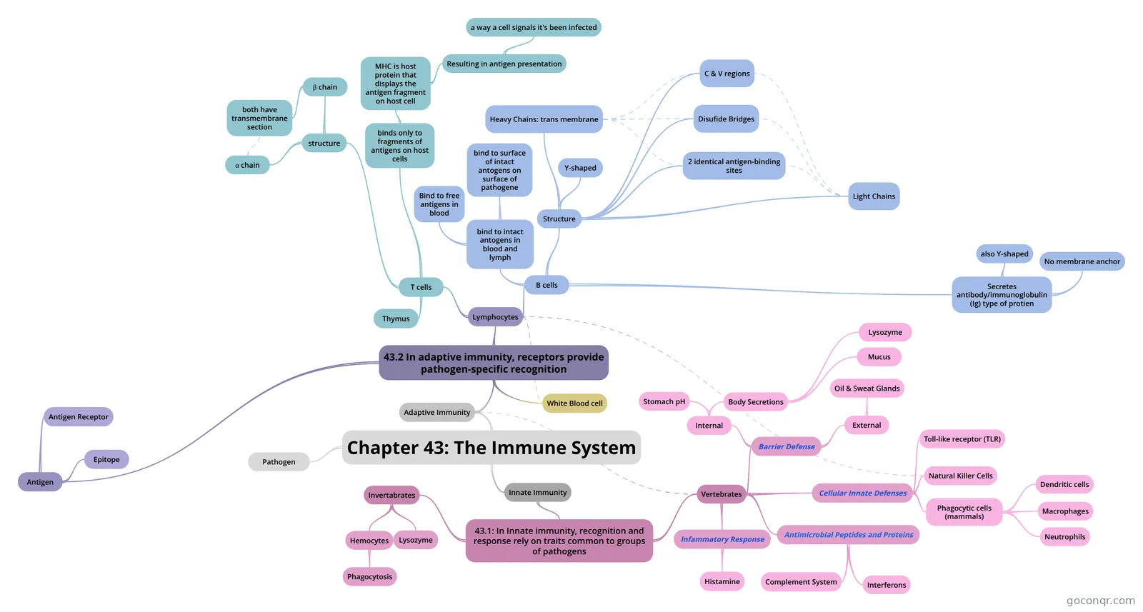
Mind map: The immune system
Map content
Chapter 43: The Immune System
- the bodily system that protects the body from foreign substances, cells, and tissues by producing the immune response and that includes especially the thymus, spleen, lymph nodes, special deposits of lymphoid tissue (as in the gastrointestinal tract and bone marrow), macrophages, lymphocytes including the B cells and T cells, and antibodies
Pathogen
- bacterium, fungus, virus or other disease-causing agent
Innate Immunity
- immunity possessed by a group (as a species or race) that is present in an individual at birth prior to exposure to a pathogen or antigen and that includes components (as intact skin, salivary enzymes, neutrophils, natural killer cells, and complement) which provide an initial response against infection — called also natural immunity; compare acquired immunity, active immunity, passive immunity
- 43.1: In Innate immunity, recognition and response rely on traits common to groups of pathogens
- Invertabrates
- Lysozyme
- an enzyme that breaks down bacterial cell walls, further protects the insect digestive system.
- Hemocytes
- immune cells of insects
- Phagocytosis
- some hemocytes ingest and break down microooganisms : process by which certain living cells called phagocytes ingest or engulf other cells or particles. The phagocyte may be a free-living one-celled organism, such as an amoeba, or one of the body cells, such as a white blood cell. In some forms of animal life, such as amoebas and sponges, phagocytosis is a means of feeding. In higher animals phagocytosis is chiefly a defensive reaction against infection and invasion of the body by foreign substances (antigens). The process by which cells engulf solid matter is called phagocytosis. There are four essential …Encyclopædia Britannica, Inc. Early observations The presence of foreign particles within cells was first described in the 1860s by pathologist Kranid Slavjansky. In the 1880s Russian-born zoologist and microbiologist Élie Metchnikoff introduced the term phagocyte in reference to immune cells that engulf and destroy foreign bodies such as bacteria. Metchnikoff also recognized that phagocytes play a major role in the immune response, a discovery that earned him a share of the 1908 Nobel Prize for Physiology or Medicine. Types of phagocytes The particles commonly phagocytosed by white blood cells include bacteria, dead tissue cells, protozoa, various dust particles, pigments, and other minute foreign bodies. In humans, and in vertebrates generally, the most-effective phagocytic cells are two kinds of white blood cells: the macrophages (large phagocytic cells) and the neutrophils (a type of granulocyte). The macrophages occur especially in the lungs, liver, spleen, and lymph nodes, where their function is to free the airways, blood, and lymph of bacteria and other particles. Macrophages also are found in all tissues as wandering amoeboid cells, and the monocyte, a precursor of the macrophage, is found in the blood. The smaller phagocytes are chiefly neutrophils that are carried along by the circulating blood until they reach an area of infected tissue, where they pass through the blood vessel wall and lodge in that tissue. Both macrophages and neutrophils are drawn toward an area of infection or inflammation by means of substances given off by the bacteria and the infected tissue or by a chemical interaction between the bacteria and the complement system of blood serum proteins. Neutrophils may also engulf particles after colliding with them accidentally. Time-lapse photography of a macrophage (the light-coloured, globular structure) consuming bacteria.Encyclopædia Britannica, Inc. Particle adherence Similar Topics erythroblast myeloblast normoblast lymphoblast blood cell formation Before phagocytosis is accomplished, the phagocyte and the particle must adhere to each other, the possibility of which depends largely on the chemical nature of the surface of the particle. In the case of bacteria, if the phagocyte cannot adhere directly, protein components of the blood known as opsonins (e.g., complement and antibodies) form a surface film on bacteria—a process known as opsonization. Phagocytes adhere to the opsonins, and phagocytosis follows. Encapsulated bacteria are ingested with more difficulty. In the absence of specific antibodies that recognize the bacteria, opsonization cannot occur, and the bacteria repel phagocytes. The surfaces of such bacteria are coated with special antibodies only after the body has mounted an immune response to the presence of that particular kind of bacterium. Such antibodies are of great importance in establishing immunity to diseases. Special staining and imaging techniques were used to create these photographs of bacteria (yellow) …Encyclopædia Britannica, Inc. Particle engulfment and digestion The speed with which a phagocytic cell ingests a particle varies somewhat with the size of the particle. Small particles, such as bacteria or minute grains of charcoal, are ingested almost instantaneously. Larger objects, such as clumps of bacteria or tissue cells, are phagocytosed over the course of a more-prolonged response. The cell flows around the object until it has been completely engulfed. The engulfed object is thus enclosed within a membrane-bound vacuole called a phagosome. The phagocyte digests the ingested particle with hydrolytic enzymes, which are contained within membrane-enclosed sacs called lysosomes found within the cell. Phagocytic enzymes are secreted into the vacuole in which digestion takes place. Small organic components of the particle are used to build larger molecules needed by the cell. Learn More in these related articles: human disease: Phagocytic cells of the bodyPhagocytosis is the process by which certain cells ingest particulate material. When a phagocytic cell comes in contact with some particle such as a bacterium or even inert material such as dust, the ... Read This Article immune system: Immune capacity among invertebrates...from their own. When tissue grafts are made in animals higher up the evolutionary tree—between individual annelid worms or starfish, for example—the foreign tissue is commonly invaded by phagocytic... Read This Article circulatory system: Blood...Many different types of hemocytes have been described in different species, but they have been studied most extensively in insects, in which four major types and functions have been suggested: (1) ... Read This Article in Karl Albert Ludwig Aschoff German pathologist who recognized the phagocytic (capable of engulfing bacteria and other substances) activity of certain cells found in diverse tissues and named them the reticuloendothelial...Read This Article in bone Rigid body tissue consisting of cells embedded in an abundant, hard intercellular material. The two principal components of this material, collagen and calcium phosphate, distinguish...Read This Article in bone marrow Soft, gelatinous tissue that fills the cavities of the bones. Bone marrow is either red or yellow, depending upon the preponderance of hematopoietic (red) or fatty (yellow) tissue....Read This Article in cell Cell, in biology, the basic membrane-bound unit that contains the fundamental molecules of life and of which all living things are composed.Read This Article in Kupffer cell Any of the stellate (star-shaped) cells in the linings of the liver sinusoids. The sinusoids are microscopic blood channels. The Kupffer cells are phagocytic, i.e., capable of...Read This Article in white blood cell White blood cell, cellular component of blood that helps defend the body against infection.Read This Article Additional Media More about phagocytosis 17 References found in Britannica ArticlesAssorted Referencescomplement system (in complement (immune system component)) disease prevention (in human disease: Phagocytic cells of the body) effect on pneumococcus (in pneumococcus) practice of Protozoa (in protozoan: Mechanisms of food ingestion) reticuloendothelial system (in mononuclear phagocyte system) occurrence inchronic granulomatous disease (in chronic granulomatous disease) connective tissue (in connective tissue: Migrating cells) human blood (in blood (biochemistry): Blood cells) (in blood (biochemistry): Destruction of red blood cells) (in circulatory system: Blood) External Links Rader’s Biology4Kids - Phagocytosis Article History Feedback Corrections? Updates? Help us improve this article! Contact our editors with your feedback. × LEARN MORE IntroductionEarly observationsTypes of phagocytesParticle adherenceParticle engulfment and digestion
- Lysozyme
- Vertebrates
- Barrier Defense
- The blocking of many pathogens by the mucous membranes and skin
- Internal
- Body Secretions
- Lysozyme
- Present in tears, saliva and mucous secretions destroys the cell walls of susceptible bacteria
- Mucus
- Lysozyme
- Stomach pH
- Body Secretions
- External
- Oil & Sweat Glands
- Secreation from these glands give human skin a pH ranging from 3 to 5, acidic enough to prevent the growth of many bacteria
- Oil & Sweat Glands
- Cellular Innate Defenses
- Toll-like receptor (TLR)
- Toll-like receptors (TLRs) were the first pattern recognition receptors (PRRs) identified in mammals and to date are the best characterized. They initiate key inflammatory responses and also shape adaptative immunity. All TLRs (10 in humans and 11 in mice) are type I transmembrane proteins characterized by an extracellular leucine-rich domain and a cytoplasmic tail. They recognize diverse pathogen-associated molecular patterns (PAMPs) from bacteria, fungi, parasites and viruses, including lipid-based bacterial cell wall components such as lipopolysaccharide (LPS) and lipopeptides, microbial protein components such as flagellin, and nucleic acids such as single-stranded or double-stranded RNA and CpG DNA. They also react to certain damage-associated molecular patterns (DAMPs) from endogenous cells and the environment. InvivoGen offers numerous tools to study TLR signaling pathways: TLR-expressing cell lines; a comprehensive choice of TLR ligands (agonists and antagonists); TLR antibodies for detection or neutralization; TLR inhibitors such as signal transduction inhibitors and shRNAs; fully-sequenced TLR and related genes; and TLR-detection products such as PRR signaling reporter plasmids
- Phagocytic cells (mammals)
- Neutrophils
- Neutrophils (engulf and destroy the infecting pathogens. smaller than macrophages) are a type of white blood cell. In fact, most of the white blood cells that lead the immune system’s response are neutrophils. There are four other types of white blood cells. Neutrophils are the most plentiful type, making up 55 to 70 percent of your white blood cells. White blood cells, also called leukocytes, are a key part of your immune system.
- Macrophages
- A macrophage is a large white blood cell that is an important part of our immune system. The word 'macrophage' literally means 'big eater.' It's an amoeba-like organism, and its job is to clean our body of microscopic debris and invaders. A macrophage has the ability to locate and 'eat' particles, such as bacteria, viruses, fungi, and parasites. Macrophages are born from white blood cells called monocytes, which are produced by stem cells in our bone marrow. Monocytes move through the bloodstream and when they leave the blood, they mature into macrophages. They live for months, patrolling our cells and organs and keeping them clean.
- Dendritic cells
- Dendritic cells (DCs) get their name from their surface projections (that resemble the dendrites of neurons). They are found in most tissues of the body and are particularly abundant in those that are interfaces between the external and internal environments (e.g., skin, lungs, and the lining of the gastrointestinal tract) where they are ideally placed to encounter extrinsic antigens, including those expressed by invading pathogens. Two dendritic cells (arrows) from the spleen of a mouse. Compare their smooth surface with that of the two macrophages visible at the upper left. (Courtesy of Ralph Steinman, from R. M. Steinman et al., J. Exp. Med. 149:1, 1979.) Although there are several distinct subtypes of DCs, they all share these features: They are actively motile. They continuously sample their surroundings — ingesting antigens by endocytosis (using phagocytosis, receptor-mediated endocytosis, and pinocytosis). Many of these antigens are "self" antigens, e.g., dead cells, proteins in the extracellular fluid. But the antigens can also be foreign antigens, for example, bacteria that are resident in the body (e.g., in the colon) or that invade the body. In either case, the ingested antigens are degraded in lysosomes into peptide fragments that are then displayed at the cell-surface nestled in class II MHC molecules [Link]. Having ingested antigen in the tissue, they migrate to lymph nodes and spleen where they can meet up with T cells bearing the appropriate T-cell receptor for antigen (TCR). What happens next depends on the nature of the antigen. Self antigens are presented to T cells without any costimulatory molecules. This interaction causes the T cells to divide for a brief time, but then they commit suicide by apoptosis and so cannot attack tissues of the body. The animal becomes tolerant to that antigen. Foreign antigens produce a different outcome. The dendritic cells becomes "activated' and begin to display not only the MHC-peptide complex for the TCR of the T cells [Link] but also costimulatory molecules, e.g. B7 which binds to CD28 on the T cell. [Link] The importance of dendritic cells in developing immunity to pathogens is dramatically shown in those rare infants who lack a functioning gene needed for the formation of dendritic cells. They are so severely immunodeficient that they are at risk of life-threatening infections. What accounts for the activation of dendritic cells by foreign antigens but not by self antigens?
- Neutrophils
- Natural Killer Cells
- Natural killer cells (also known as NK cells, K cells, and killer cells) are a type of lymphocyte (a white blood cell) and a component of innate immune system. NK cells play a major role in the host-rejection of both tumours and virally infected cells. NK cells are cytotoxic; small granules in their cytoplasm contain special proteins such as perforin and proteases known as granzymes. Upon release in close proximity to a cell slated for killing, perforin forms pores in the cell membrane of the target cell through which the granzymes and associated molecules can enter, inducing apoptosis. The distinction between apoptosis and cell lysis is important in immunology - lysing a virus-infected cell would only release the virions, whereas apoptosis leads to destruction of the virus inside. NK cells are activated in response to interferons or macrophage-derived cytokines. They serve to contain viral infections while the adaptive immune response is generating antigen-specific cytotoxic T cells that can clear the infection. Patients deficient in NK cells prove to be highly susceptible to early phases of herpes virus infection.
- These lymphocytes stay in the b
- Toll-like receptor (TLR)
- Antimicrobial Peptides and Proteins
- Interferons
- a group of signaling proteins[1] made and released by host cells in response to the presence of several pathogens, such as viruses, bacteria, parasites, and also tumor cells. In a typical scenario, a virus-infected cell will release interferons causing nearby cells to heighten their anti-viral defenses.
- Complement System
- The complement system is a part of the immune system that enhances (complements) the ability of antibodies and phagocytic cells to clear microbes and damaged cells from an organism, promotes inflammation, and attacks the pathogen's plasma membrane.
- They are activated by substances on the surface of many pathogens
- Interferons
- Infammatory Response
- The inflammatory response (inflammation) occurs when tissues are injured by bacteria, trauma, toxins, heat, or any other cause. The damaged cells release chemicals including histamine, bradykinin, and prostaglandins. These chemicals cause blood vessels to leak fluid into the tissues, causing swelling.
- Histamine
- an organic nitrogenous compound involved in local immune responses as well as regulating physiological function in the gut and acting as a neurotransmitter for the brain, spinal cord and uterus.[3][4] Histamine is involved in the inflammatory response and has a central role as a mediator of itching.[5] As part of an immune response to foreign pathogens, histamine is produced by basophils and by mast cells found in nearby connective tissues. Histamine increases the permeability of the capillaries to white blood cells and some proteins, to allow them to engage pathogens in the infected tissues.[6]
- Barrier Defense
- Invertabrates
Adaptive Immunity
- The adaptive immune system, also known as the acquired immune system or, more rarely, as the specific immune system, is a subsystem of the overall immune system that is composed of highly specialized, systemic cells and processes that eliminate pathogens or prevent their growth.
- 43.2 In adaptive immunity, receptors provide pathogen-specific recognition
- Lymphocytes
- A lymphocyte is one of the subtypes of white blood cell in a vertebrate's immune system. Lymphocytes include natural killer cells (NK cells) (which function in cell-mediated, cytotoxic innate immunity), T cells (for cell-mediated, cytotoxic adaptive immunity), and B cells (for humoral, antibody-driven adaptive immunity). They are the main type of cell found in lymph, which prompted the name "lymphocyte".
- Originate from stem cells in the bone marrow
- T cells
- migrate to thymus from bone marrow
- Thymus
- structure
- α chain
- β chain
- both have transmembrane section
- binds only to fragments of antigens on host cells
- MHC is host protein that displays the antigen fragment on host cell
- major histocompatibility complex molecule
- Resulting in antigen presentation
- a way a cell signals it's been infected
- MHC is host protein that displays the antigen fragment on host cell
- B cells
- Develop and stay in bone marrow
- Structure
- Heavy Chains: trans membrane
- Light Chains
- Disufide Bridges
- C & V regions
- 2 identical antigen-binding sites
- Y-shaped
- Secretes antibody/immunoglobulin (Ig) type of protien
- also Y-shaped
- No membrane anchor
- bind to intact antogens in blood and lymph
- bind to surface of intact antogens on surface of pathogene
- Bind to free antigens in blood
- White Blood cell
- Antigen
- Any substance that elicits a B or T cell response
- Antigen Receptor
- Both B cells and T cells have surface receptors for antigen. Each cell has thousands of receptors of a single specificity; that is, with a binding site for a particular epitope. T-cell receptors (TCRs) enable the cell to bind to and, if additional signals are present, to be activated by and respond to an epitope presented by another cell called the antigen-presenting cell or APC. B-cell receptors (BCRs) enable the cell to bind to and, if additional signals are present, to be activated by and respond to an epitope on molecules of a soluble antigen. The response ends with descendants of the B cell secreting vast numbers of a soluble form of its receptors. These are antibodies.
- Epitope
- the small, accessible portion of an antigen that binds to an antigen receptor
- Each B or Tcell thus displays specificity for a particular epitope, enabling it to respons to any pathogen tat produces molecules containg that epitope
- Lymphocytes
Information
- Description
- I Biology Mind Map on Chapter 43: The Immune System, created by Christina . on 25/10/2017.
- Created by
- Christina .
- Views
- 4
- Rating