
Mind map
Cardiogenic Shock
Map content
Cardiogenic Shock
Main Causes (Mebazaa, 2018)
- Shock
- A syndrome characterized by decreased tissue perfusion and impaired cellular metabolism.
- Results in an imbalance between the supply and demand of oxygen and nutrients.
- This exchange is essential for life. When a cell is in a state of hypoperfusion the demand for oxygen and nutrients exceeds the supply.
- Three Stages of Shock (EMS1, 2016).
- Stage I
- Compensated/ Non-Progressive (Lewis, 2014).
- During the compensatory stage, the body activates neural, hormonal and biochemical compensatory mechanisms to try and compensate for anaerobic metabolism and to help maintain homeostasis.
- Compensated/ Non-Progressive (Lewis, 2014).
- Stage II
- Decompensated / Progressive (Lewis, 2014).
- Begins when compensatory mechanisms fail.
- In this stage of shock, aggressive interventions are necessary to prevent further development. Systemic interstitial edema increases due to the decrease in circulating volume.
- Begins when compensatory mechanisms fail.
- Decompensated / Progressive (Lewis, 2014).
- Stage III
- Irreversible (Lewis, 2014).
- Final stage of shock.
- Anaerobic metabolism is exacerbated due to decreased cardiac output and decreased perfusion from peripheral vasoconstriction. Lactic acid builds up and contributes to increased capillary permeability and dilation.
- Because of the increased permeability, fluid and plasma proteins leave the vascular space and move to the interstitial space. This loss of fluids worsens hypotension and tachycardia; decreasing coronary blood flow. This is the start of mulitsystem organ failure and recovery at this point is highly unlikely.
- Anaerobic metabolism is exacerbated due to decreased cardiac output and decreased perfusion from peripheral vasoconstriction. Lactic acid builds up and contributes to increased capillary permeability and dilation.
- Final stage of shock.
- Irreversible (Lewis, 2014).
- Stage I
- Three Stages of Shock (EMS1, 2016).
- This exchange is essential for life. When a cell is in a state of hypoperfusion the demand for oxygen and nutrients exceeds the supply.
- Results in an imbalance between the supply and demand of oxygen and nutrients.
- Cardiogenic Shock.
- A condition caused by the inability of the heart to pump blood sufficiently to meet metabolic demands. This condition is due to impaired contractility of the heart (Martin, 2017).
- Four Stages of Cardiogenic Shock (Urden, Stacy, & Lough (2014).
- Initial Stage.
- Diminished cardiac output without clinical symptoms.
- Compensatory.
- Baroreceptors respond to decreased cardiac output by stimulating SNS.
- SNS releases catecholamines to improve myocardial contractility and vasoconstriction.
- Leads to increased venous return and arterial blood pressure.
- SNS releases catecholamines to improve myocardial contractility and vasoconstriction.
- Baroreceptors respond to decreased cardiac output by stimulating SNS.
- Progressive.
- As tissue perfusion remains inadequate, cells begin anaerobic metabolism.
- Leads to metabolic acidosis and fluid leakage from capillaries into interstitial space.
- Decrease in circulating volume and increase in blood viscosity may cause clotting in capillaries and tissue death.
- Leads to metabolic acidosis and fluid leakage from capillaries into interstitial space.
- As tissue perfusion remains inadequate, cells begin anaerobic metabolism.
- Refractory.
- Shock becomes unresponsive to therapy.
- Considered irreversible.
- Shock becomes unresponsive to therapy.
- Initial Stage.
- A syndrome characterized by decreased tissue perfusion and impaired cellular metabolism.
- Either systolic or diastolic dysfunction of the pumping act of the heart results in compromised cardiac output.
- Systolic
- Dysfunction
- Inability of the heart to pump blood forward.
- Precipitating Causes
- Include myocardial infarction, cardiomyopathy, blunt cardiac injury, sever or systemic pulmonary hypertension and myocardial depression.
- Affects the right chambers of the heart.
- blood flow through the pulmonary circulation is compromised.
- Dysfunction
- Diastolic
- Effects the ability to fill the ventricle.
- Decreased filling of the ventricle results in decreased stroke volume.
- Diastolic dysfunction includes cardiac tamponade and cardiomyopathy.
- Decreased filling of the ventricle results in decreased stroke volume.
- Effects the ability to fill the ventricle.
- Systolic
- Heart Failure.
- Causes of Heart Failure.
- Valvular Problems.
- Ex: Aortic outflow tract is damaged or narrowed. This makes it hard for blood to be pushed out.
- Stiffness of ventricular wall.
- Decreased myocardial contractility.
- Myocardial infarctions are the main cause of decreased contractility.
- Decreased myocardial contractility.
- Fluid overload.
- Valvular Problems.
- Causes of Heart Failure.
- Shock
Management and Treatment
- Surgical Interventions (Mayo Clinic, 2017; El Sibai, Bachir, & El Sayed, 2018).
- Cardiac repair.
- To repair tears in heart chambers or valves.
- Ventricular Assist Device.
- Mechanical device that is impacted in patient's abdomen and attaches to the heart in order to help it pump.
- Helps extend and improve the lives of people with end-stage heart failure awaiting transplant or that are unable to undergo heart transplant.
- Mechanical device that is impacted in patient's abdomen and attaches to the heart in order to help it pump.
- Coronary Artery Bipass Surgery (CABG)
- A CABG is a procedure in which doctors use a vein from your leg or your inner chest wall and use it to bypass the area of the heart that has been affected. This helps restore blood flow .
- Significantly improves chances of survival (Lewis, 2014).
- Cardiac repair.
- Medications (Fox, 2016).
- Vasodilators
- Nitroglycerin
- Causes relaxation of vascular smooth muscle resulting in decrease in preload and blood pressure.
- Sodium Nitroprusside
- Increases cardiac output by decreasing after load and produces peripheral and systemic vasodilation by direct action to the smooth muscles of the blood vessels.
- Nitroglycerin
- Vasopressors
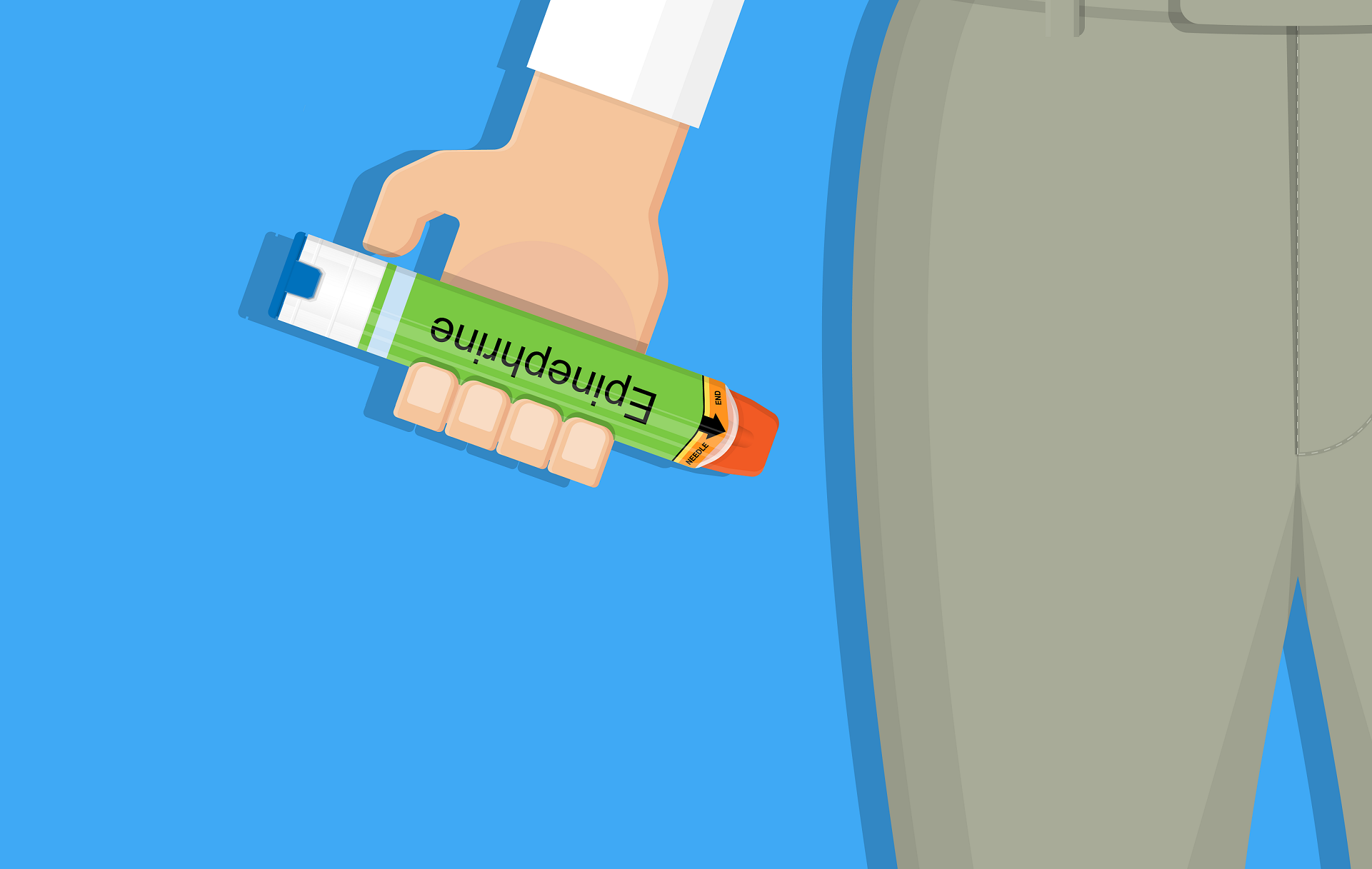
- Epinephrine
- Norepinephrine
- Vasodilators
- Surgical Interventions (Mayo Clinic, 2017; El Sibai, Bachir, & El Sayed, 2018).
Nursing Considerations (Martin, 2017).
- Ineffective Tissue Perfusion: Decreased oxygen leads to poor oxygenation at capillary level.
- Assess capillary refill.
- Capillary refill should be less than two second.
- Restrict activity level. Bedrest may be ordered.
- Restricting activity level or maintaining bedrest decreases oxygen demand.
- Obtain vital signs.
- Vital signs should include heart rate, pulse oximetry, temperature, blood pressure, pulse pressure and pain level.
- Assess for changes in level of consciousness.
- Changes in level of consciousness could be a sign of cerebral hypoxia
- Assess capillary refill.
- Anxiety
- Encourage the patient to verbalize their feelings or worries.
- Talking about their anxieties validates their feelings and communicates acceptance.
- Explain procedures using appropriate terminology.
- Information helps reduce anxiety. Patient's that are anxious tend to retain less information.
- Assess patient's level of anxiety.
- Maintain confident composure.
- Assess coping mechanisms.
- Reduce stressful stimuli.
- By maintaining an environment free from excessive conversation, noise and equipment, anxiety my start to fade.
- Encourage the patient to verbalize their feelings or worries.
- Excess Fluid Volume: Increased isotonic fluid retention.
- Assess for edema.
- Edema generally starts in the feet and ankles.
- Leads to gradual weight gain.
- Assess fluid balance and weight gain.
- Compromised regulatory mechanisms lead to fluid and sodium retention thus increasing body weight.
- Edema generally starts in the feet and ankles.
- Monitor intake and output
- Check for jugular vein distention.
- May indicate fluid excess.
- Auscultate lungs.
- Listen for presence of adventitious breath sounds.
- Monitor for presence of cough, dyspnea or orthopnea.
- Listen for crackles or wheezes.
- Listen for presence of adventitious breath sounds.
- Monitor electrolyte level.
- Some diuretics promote renal potassium secretion. Monitor for hypokalemia.
- Monitor urine output.
- Concentrated in colour.
- Scant amounts.
- Monitor for decreased renal perfusion.
- Assess for edema.
- Impaired Gas Exchange.
- Auscultate lungs for decreased ventilation and adventitious breath sounds.
- Listening for the presence of most crackles can help establish presence of increased pulmonary capillary permeablilty and increased intra-alveolar edema.
- Check for cyanosis or pallor.
- Assess patient's skin, nail beds and mucous membranes.
- O2 sats.
- Oxygen should be maintained at 90% or higher.
- Oxygen administration.
- As needed.
- Supplemental oxygen may be required to help maintain an appropriate oxygen level.
- If no standing order is in place, an order from the physician may be required.
- As needed.
- Monitor changes in level of conciousness.
- Early signs of hypoxia may include headache, restlessness, confusion, etc.
- Encourage coughing and/ or suctioning when needed.
- Suctioning out excess secretions will aid the patient when they are unable to effectively clear their airway.
- Ineffective oxygen therapy may lead to mechanical ventilation. Preparation is needed.
- Mechanical ventilation provides supportive care to maintain adequate oxygenation and ventilation.
- Early intubation and mechanical ventilation is recommended in order to prevent rapid decompensation.
- Assess respiratory rate, rhythm and depth.
- As shock progresses, hypoventilation will be present. This may lead to respiratory failure.
- Auscultate lungs for decreased ventilation and adventitious breath sounds.
- Decreased Cardiac Output: Inadequate blood pumped by the heart to meet metabolic demands of the body.
- Assess blood work.
- Check potassium and magnesium for hypomagnesemia and hypokalemia.
- Can lead to the development of arrhythmias.
- May further reduce cardiac output.
- Can lead to the development of arrhythmias.
- Check potassium and magnesium for hypomagnesemia and hypokalemia.
- Replace electrolytes as prescribed.
- Electrolyte imbalance may lead to dysrhythmias.
- Prepare for Surgery.
- Surgical treatment may be required for septal defects or acute valvular problems.
- Assess for any changes in level of consciousness.
- Obtain vital signs.
- Assess the patient's electrocardiogram as well as their cardiac rate and rhythm.
- Assess heart sounds.
- Listening for extra heart sounds such as S3, S4 or the presence of a murmur.
- Check central and peripheral pulses.
- Assess fluid balance and weight gain.
- Monitor urine output.
- Assess blood work.
- Ineffective Tissue Perfusion: Decreased oxygen leads to poor oxygenation at capillary level.
Manifestations (Lewis, Dirksen, Heitkemper, Bucher, & Camera, 2014)
- Signs
- Laboured or tachypnic breathing.
- Cyanosis.
- Cool, clammy or mottled skin.
- Decreased time for capillary refill.
- Anxiety.
- Confusion.
- Agitation.
- Symptoms
- Pulmonary congestion or crackles.
- Increased jugular vein distention.
- Angina
- Organ failure or disruption.
- Decreased urine output
- Tachycardia
- Hypotension
- Weak or narrowed pulse pressure.
- Low cardiac output.
- Weak peripheral pulses.
- Signs
Diagnostic Testing (Kosaraju and Hai, 2018).
- Electrocardiogram.
- Cardiac enzyme test.
- Brain natriuretic peptide.
- Complete Blood Panel.
- May include a metabolic panel, magnesium, phosphorus, coagulation profile and thyroid-stimulating hormone.
- Echocardiogram (BMJ, 2003).
- Two-dimensional echocardiography.
- Should be performed early in order to triage the patient correctly. This will help determine if there are any valvular abnormalities between the left or ventricle.
- Valvular abnormalities may include severe aortic valve stenosis; severe mitral or aortic valve regurgitation; acute endocarditis.
- Chest x-ray.
- With left-sided heart failure, an x-ray is an important diagnostic tool to help establish if there is presence of pulmonary edema; pulmonary vascular redistribution; enlarged hila; kerley B lines; and bilateral plural effusion (Gibson, 2014).
- Arterial blood bass draw (Mohammed & Abdelatief, 2015).
- May be taken from your femoral, brachial or radial artery. An arterial blood gas draw provides the most efficient assessment of pO2 and pCO2 and gives valuable insight into ventilation.
- Ultrasonography.
- To guide fluid management.
- Coronary angiography (Mayo Clinic, 2018).
- Also known as a cardiac catheterization. An angiography uses a type of dye that can be read with an x-ray machine. After the dye is injected, images are rapidly taken in order to view the blood vessels of a heart.
- Lactate.
Cardiac Risk Factors (Kosaraju and Hai, 2018).
- Men: 45+ years old Women: 55+ years old.
- Because heart disease becomes more common as you age, it is important to have regular checkups with your family physician.
- Hyperlipidemia.
- Too high of a cholesterol level can lead to a build up of plaque inside your arteries. This narrows the diameter of the arteries and makes it harder for blood to pass through. This process can trigger heart disease.
- Hypertension.
- Uncontrolled high blood pressure can negatively affect your heart in a number of ways. Ex: Coronary heart disease.
- Family history of premature coronary artery disease.
- Certain genes can increase your risk of cardiac-related health issues. Some of these issues may include high blood pressure and/ or cholesterol levels.
- Diabetes Mellitus.
- The longer you have diabetes the more chances of negative effects can occur. Over time, high blood glucose levels can damage both your vessels and your nerves that control your heart.
- Poor diet.
- A poor diet can play a major role to increase your chances of heart disease. Unhealthy food choices can lead to a multitude of problems including high blood pressure, diabetes, obesity, high cholestoeral and several others that affect your heart.
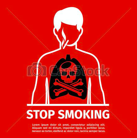
- Smoking.
- Long-term smokers damage the lining of their arteries. This damage causes a build up of fatty material which causes a narrowing to occur. It's never too late to quit smoking. After smoking cessation, physiological benefits start as early as 12 hours after your last cigarette.
- Physical inactivity.
- Living a sedentary lifestyle negatively affects your heart. Physical activity lowers your blood pressure and halves your chances of coronary heart disease, angina and/ or heart attack.
- Men: 45+ years old Women: 55+ years old.
Images and files in the map


Information
- Description
- Nursing Mind Map on Cardiogenic Shock, created by Christina West on 05/11/2018.
- Created by
- Christina West
- Views
- 8
- Rating