
Mind map
Digestive System
Map content
Digestive System
mouth cavity
- vestibule of the mouth
- slit like space
- bound externally by the lips and cheeks
- mouth cavity proper
- bounded by
- ant+later
- gums
- teeth
- post
- communicates with the pharynx through the ismuth of fauces
- ismuth is bounded by the palato-glossal arch on each side
- communicates with the pharynx through the ismuth of fauces
- roof
- hard and soft palate
- floor
- anterior 2/3 of the tongue
- ant+later
- bounded by
- vestibule of the mouth
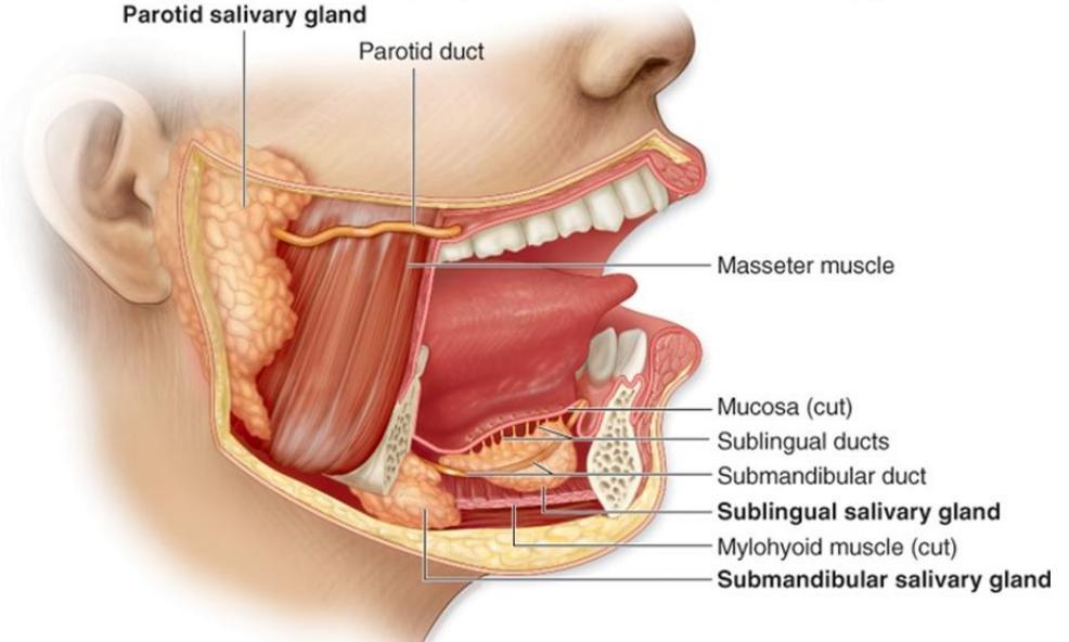
salivary glands
- paired parotid
- sublingual
- paired submandibular
- numerous small glands scattered throughout the mouth cavity
pharynx
- musculomembranous tube
- post
- mouth
- nose
- larynx
- extends from the base of the skull
- to the C6
- WHERE it joins the osephagus
- to the C6
osephagus
- long muscular tube(10 inch)
- extends through
- neck
- thorax
- abdomen
- begins as a lower continuation of the pharynx
- ends at the caridac end of the stomach
- parts
- cervical
- opposite the lower border of the circoid cartilage
- C6
- thoracic
- longest and the main part
- it lies in the super. then post. mediastina
- abdominal
- shortest
- enter abdomen through an opening in the right crus of the diaphragm
- cervical
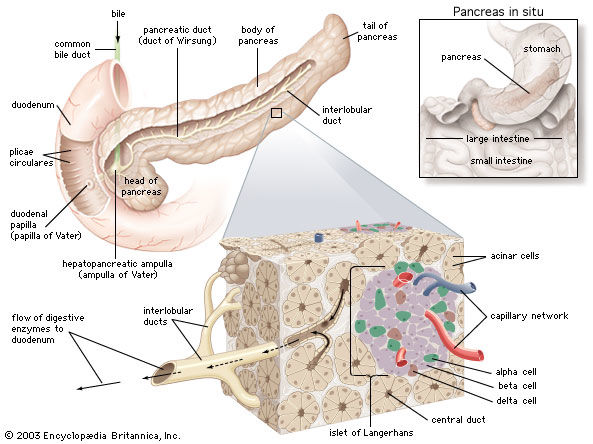
Pancreas
- It`s a combined exocrine&endocrine glan
- lies transversly across the posterior abdominal wall
- extends from the concavity of the duodenum on the R.side
- to the spleen on the L.side
- parts
- head
- neck
- body
- tail
- lower part forms a projection
- uncinate process
- ducts
- main
- unites with the comm.bile.duct
- to form ampulla of veter
- open in 2nd duodenum
- in A.v
- open in 2nd duodenum
- to form ampulla of veter
- unites with the comm.bile.duct
- accessory
- opens in the 2nd part of the duodenum
- above ampulla of vater
- opens in the 2nd part of the duodenum
- main
- It`s a combined exocrine&endocrine glan
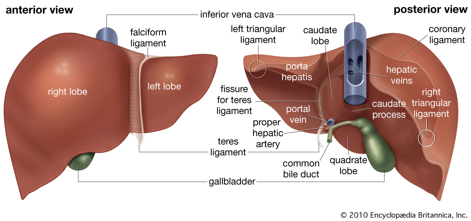
liver
- shape
- wedge
- covered by peritoneum except small area on the posterior surface
- bare area
- lobes
- R.
- LARGER
- L.
- QUADRATE
- CAUDATE
- 2 LOBES SEPARATED BY A PERITONEUM FOLD
- falciform ligament
- R.
- blood supply
- portal vein
- 75%
- carries nutrients and substances absorbed from GIT
- heaptic art.
- 25%
- portal vein
- PORTA HEPATIS(HILUM OF LIVER)
- PORTAL.V.
- HEPATIC.ART.
- COMM. HEP. BILE DUCT
- shape
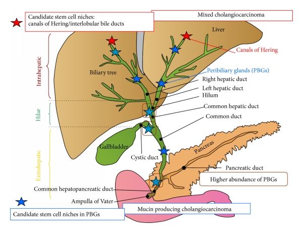
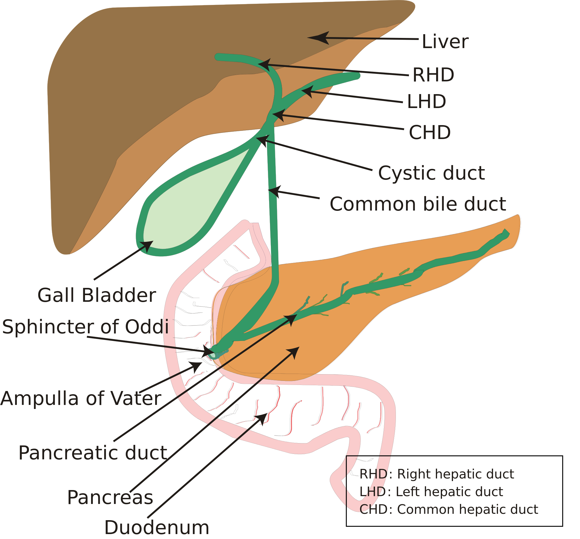
Biliary sys
- gall bladder+biliary duct
- GALL BLADDER
- pear-shaped
- situated on the inf.sur.of liver
- 3 parts
- fundus
- R.9TH C.C.
- BODY
- NECK
- fundus
- BILIARY DUCTS
- hepatic duct
- R.+L.
- ONE FROM each lobe
- in front of the 2 branches of portal v.+hep.art.
- unit in the right part of porta hepatis
- comm.hep.duct
- R.+L.
- Comm.hep.duct
- 1inch long
- descends in front of portal v.+R. HEP.ART
- + CYSTIC= BILE DUCT
- CYSTIC DUCT
- 1.5 inch long
- s-shaped
- joins com.hep.duct at an acute angle just below the porta hepatis
- bile duct
- 3 inch
- joins pan.duct. to form ampulla of vater
- which open in post.wall.of 2nd duod.below its middle
- the opening is guarded by the sphincter of oddi
- which open in post.wall.of 2nd duod.below its middle
- hepatic duct
Images and files in the map
Information
- Description
- Mind Map on Digestive System, created by ahmed elsayed on 02/05/2016.
- Created by
- ahmed elsayed
- Views
- 9
- Rating