
Mind map
Comparing Photosynthetic RC
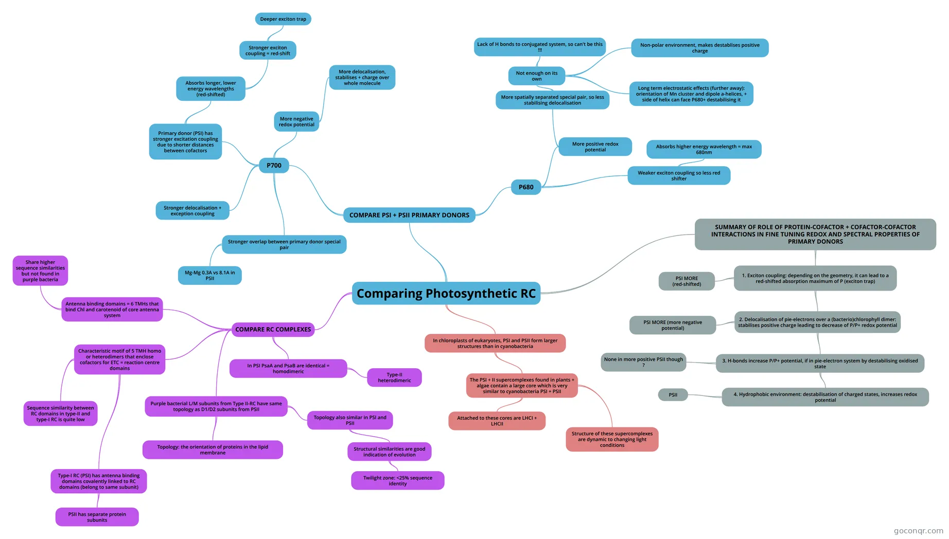
Mind map: Comparing photosynthetic reaction centres
Map content
Comparing Photosynthetic RC
COMPARE PSI + PSII PRIMARY DONORS
- P700
- Primary donor (PSI) has stronger excitation coupling due to shorter distances between cofactors
- Absorbs longer, lower energy wavelengths (red-shifted)
- Stronger exciton coupling = red-shift
- Deeper exciton trap
- Stronger exciton coupling = red-shift
- Absorbs longer, lower energy wavelengths (red-shifted)
- More negative redox potential
- More delocalisation, stabilises + charge over whole molecule
- Stronger delocalisation + exception coupling
- Stronger overlap between primary donor special pair
- Mg-Mg 0.3A vs 8.1A in PSII
- Primary donor (PSI) has stronger excitation coupling due to shorter distances between cofactors
- P680
- More positive redox potential
- More spatially separated special pair, so less stabilising delocalisation
- Not enough on its own
- Lack of H bonds to conjugated system, so can't be this !!!
- Non-polar environment, makes destablises positive charge
- Long term electrostatic effects (further away): orientation of Mn cluster and dipole a-helices, + side of helix can face P680+ destabilising it
- Not enough on its own
- More spatially separated special pair, so less stabilising delocalisation
- Weaker exciton coupling so less red shifter
- Absorbs higher energy wavelength = max 680nm
- More positive redox potential
- P700
SUMMARY OF ROLE OF PROTEIN-COFACTOR + COFACTOR-COFACTOR INTERACTIONS IN FINE TUNING REDOX AND SPECTRAL PROPERTIES OF PRIMARY DONORS
- 1. Exciton coupling: depending on the geometry, it can lead to a red-shifted absorption maximum of P (exciton trap)
- PSI MORE (red-shifted)
- 2. Delocalisation of pie-electrons over a (bacterio)chlorophyll dimer: stabilises positive charge leading to decrease of P/P+ redox potential
- PSI MORE (more negative potential)
- 3. H-bonds increase P/P+ potential, if in pie-electron system by destabilising oxidised state
- None in more positive PSII though ?
- 4. Hydrophobic environment: destabilisation of charged states, increases redox potential
- PSII
- 1. Exciton coupling: depending on the geometry, it can lead to a red-shifted absorption maximum of P (exciton trap)
COMPARE RC COMPLEXES
- Purple bacterial L/M subunits from Type II-RC have same topology as D1/D2 subunits from PSII
- Topology: the orientation of proteins in the lipid membrane
- Topology also similar in PSI and PSII
- Structural similarities are good indication of evolution
- Twilight zone: <25% sequence identity
- Structural similarities are good indication of evolution
- Characteristic motif of 5 TMH homo or heterodimers that enclose cofactors for ETC = reaction centre domains
- Type-I RC (PSI) has antenna binding domains covalently linked to RC domains (belong to same subunit)
- PSII has separate protein subunits
- Sequence similarity between RC domains in type-II and type-I RC is quite low
- Type-I RC (PSI) has antenna binding domains covalently linked to RC domains (belong to same subunit)
- Antenna binding domains = 6 TMHs that bind Chl and carotenoid of core antenna system
- Share higher sequence similarities but not found in purple bacteria
- In PSI PsaA and PsaB are identical = homodimeric
- Type-II heterodimeric
- Purple bacterial L/M subunits from Type II-RC have same topology as D1/D2 subunits from PSII
In chloroplasts of eukaryotes, PSI and PSII form larger structures than in cyanobacteria
- The PSI + II supercomplexes found in plants + algae contain a large core which is very similar to cyanobacteria PSI + PSII
- Attached to these cores are LHCI + LHCII
- Structure of these supercomplexes are dynamic to changing light conditions
- The PSI + II supercomplexes found in plants + algae contain a large core which is very similar to cyanobacteria PSI + PSII
Information
- Description
- Universität Membrane Proteins Mind Map on Comparing Photosynthetic RC, created by George West on 18/05/2016.
- Created by
- George West
- Views
- 0
- Rating