
Notes
Spinal Cord - created from Mind Map
Content of the notes (1)
Page 1
White Matter: myleinated axons
Grey matter: unmyelinated cell bodies/soma
Roots: Connect spinal nerves to horns
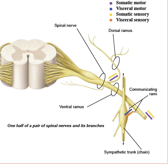
Rami: Travel from the body to the spinal nerve
Pathways
Plexuses: Groups of nerves from the ventral rami
Reflexes
Anatomy
Meninges
Vertebrae
Dorsal column
Ventral Column
Lateral column
posterior
Anterior
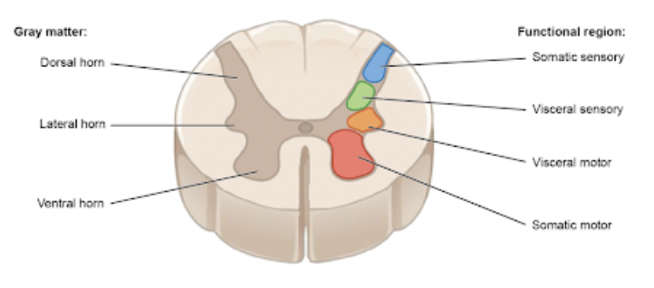
Horns
Dorsal Horn
Lateral Horn
Ventral Horn
distinguished by type of information they carry
Sensory (visceral and somatic)
Visceral motor
Somatic motor
Dorsal Root
Ventral Root
Distinguished by type of information (sensory or motor)
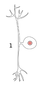
Dorsal root ganglion
Contains cell bodies
Psuedounipolar neurons
Contains cell bodies
Classified by location of destination or origin
Ventral Ramus
Dorsal Ramus
Communicating Rami
Front sides of body
Back side of the body
Smooth/cardiac muscle, glands
Cell bodies located in sympathetic ganglion
Visceral motor and sensory
Sympathetic chain
Runs parallel and anterior to spinal cord
Carried to sympathetic trunk
Ascending: Send sensory information to the brain
Descending: Send instructions to muscles for motor function
Neurons
2 synapses
Types
1st order neuron
2nd order neuron
3rd order neuron
Cell bodies
Peripheral sensor neuron
Body to dorsal horn
Interneuron
Travels from dorsal horn up lateral column to thalamus
Interneuron
Travels from thalamus to cerebral cortex
Decussates at pyramids of medulla
Located in dorsal horn and thalamus
Dorsal column-medial leminscus
Spinothalamic
Spinocerebellar
Neurons
1 synapse
Upper motor neuron
Lower motor neuron
Cell Bodies
Interneuron
Travels from primary motor cortex to ventral horn through the ventral column
Motor neuron
Travels from the ventral horn to the muscle
Located in ventral horn
Intercostal nerves
Cervical plexus
Brachial Plexus
Lumbosacral plexus
Runs along ribs
Innervates muscles and skin of abdominal wall, bones
Innvervates neck
Innervates arm
C5-T1
Innervates legs
L2-S4
Polysynaptic Reflex Arc
Monosynaptic
Sensory Neuron (travels along rami to spinal nerve)
Dorsal Horn
Interneuron (gray matter)
Where sensory neuron synapses with interneuron
Ventral horn
Can also go outside of spinal column
Ventral Root
Where interneuron synapses
Muscle
Sensory neuron (travels along ramus)
Ventral horn
Muscle
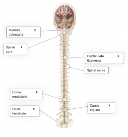
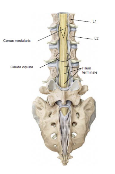
Conus Medullaris
Cauda Equina
Filum Terminale
Denticulate ligaments
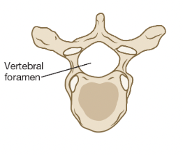
Vertebral foramen
Vertebral canal
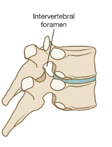
Intervertebral foramen
Termination of spinal cord at L1/L2
Bundle of spinal nerve roots that lumbar, sacral, and coccygeal nerves pass through
Holds spinal cord down to sacrum and coccyx
Continuation of pia mater
Extention of pia mater that stabilize spinal cord in vertebral canal
Where spinal nerves emerge through foramina in vertebrae, forms vertebral canal
Location of spinal cord
Where the spinal cord runs through
Spaces between vertebrae where spinal nerves exit
Between vertebrae
Dura Mater
Arachnoid Mater
Subarachnoid Space
Pia Mater
Central Canal
Anterior median fissure
Epidural Space
Location of CSF
Fluid-filled
Continuity with ventricles of brain
Filled with fat and blood vessels
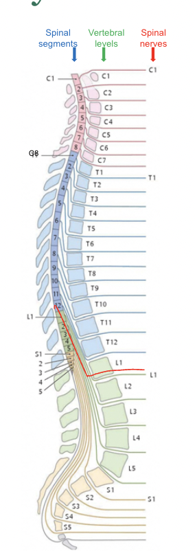
Cervical
Thoracic
Lumbar
Sacral
Coccygeal
8
12
5
5
1
Information
- Description
- Anatomy Note on Spinal Cord - created from Mind Map, created by Julia Morneau on 14/09/2022.
- Created by
- Julia Morneau
- Views
- 0
- History
-
- Created by Julia Morneau · Sep 14, 2022
- Copied to Note by Julia Morneau · Sep 14, 2022
- Rating