
Fluxograma
Cell Cycle
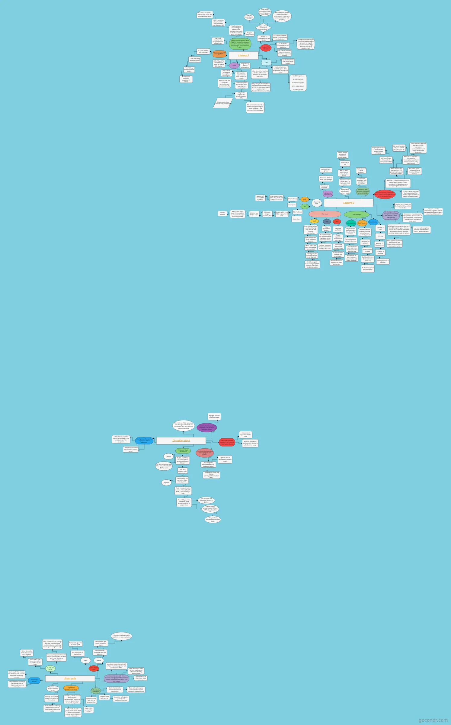
Flowchart: Cell cycle
Conteúdo do fluxograma
Lecture 1
Stages of the cell cycle
Mitosis + cytokinesis- the cell divides
G1- Cellular contents (except chromosomes) are duplicated
S- All the 46 chromosomes are duplicated
G2- The cell ensures the chromosomes have been replicated properly
During mitosis the double chromosomes are split to produce 92 single chromosomes, half of which go into each daughter cell.
3 types of non-kinetochore microtubules
Overlap- Assemble the spindle
Polar- Move centrosomes apart by pushing against each other
Astral- Mediate spindle displacement and chromosome movement; extend toward the cell periphery
Cdks
Are inactive unless bound to cyclin. Drive progression through cell cycle.
Cyclins help direct cdks to the target protein.
Active kinase has its active site (for ATP) located between its small and large lobe.
They catalyze the transfer of the gamma phosphate (from ATP) to the hydroxyl group of ser, thr or tyr. Phosphorylation is reversible.
Cyclins
G2: Cdk1-Cyclin A.
M: Cdk1-Cyclin B
G1: Cdk4/6- Cyclin D
G1/S: Cdk 2-Cyclin E
S: Cdk2- Cyclin A
Degraded by the 26S proteasome.
E2F regulates expression of genes needed for S phase.
Unphosphorylated Rb can bind to E2F and stop the transcription
Cell growth triggers the phosphorylation of Rb
Active G1-Cdk and S-Cdk can accumulate and phosphorylate Rb.
Free E2F can increase the transcription of its own gene.
P27, P16 and P21 block the G1/S phase by making Cdk inactive
Controlling the M phase
1. Cyclin binding (cdk1-cyclin B).
2. Cdk-activating phosphorylation
3. Inhibitory phosphorylation (Wee1)
4. Removal of inhibitory phosphate (P-Cdc25).
Mitogens stimulate proliferation by inhibiting Rb
Platelet-derived growth factor (PDGF) is released by platelets, smooth muscle cells, activated macrophages and endothelial cells.
The PDGF receptor is a receptor tyrosine kinase (RTK).
PDGFR activation causes a signal transduction pathway that results in the transcription of cell cycle genes
PDGFR activation phosphorylates and inhibits Rb, thus liberating E2F.
Recombinant PDGF is used to treat ulcers and stimulate bone repair.
Triggers dimerization
RB1 (on chromosome 13) is a tumour suppressor gene. When mutated, it is a common childhood cancer
Lecture 2
P53 is produced at low levels when there is no damage. Active in its tetrameric form.
When P53 is not needed it produces MDM2 which inhibits P53 by stimulating its degradation and transporting it to the cytosol.
When there is DNA damage then P53 gets phosphorylated and inhibits MDM2.
When P53 is activated it increases the transcription of p21 and therefore blocks entry into S phase.
ATM and ATR are kinases that phosphorylate P53.
Puma activates BAX and BAK which cause membrane permeablization and inhibit Bcl-2 (stops apoptosis).
P21 binds to cyclin-cdk so it cannot phosphorylate Rb.
P53 active form- 2 antiparallel beta sheets and 4 alpha helices.
TP53 is a cancer mutation that impacts the DNA binding region of P53 so it cannot form a tetramer.
Are phosphoinositide 3 kinase-related protein kinases (PI3KKs).
M checkpoint: DNA damage activates ATM, ATR and Chk1 which inactivate the phosphatase cdc25.
Inactive cdc25 blocks the dephosphorylation/activation of cdk1.
Kinetochore microtubules (K fibre) bind to chromosomes at the centromere. Kinetochores have an inner, central and outer layer.
Cohesins hold the centromere together and can be cleaved by separase which is inactivated by securin.
Anaphase promoting complex (APC) is an E3 ubiquitin ligase that uses securin as a substrate to degrade anaphase so mitosis does not continue. Needs cdc20 for activity.
An issue with anaphase (APC off) activates MAD1, MAD2, BubR1 and Bub3
If the spindle checkpoint is off then the APC (cdc20 is recruited) and separase are active.
DNA damage
Deamination
Cytosine -> uracil
CG -> UA
Adenine -> hypoxanthine
Guanine -> xanthine
5-methylcytosine -> thymine
Bulky adducts
Thymine/cytosine residues in a DNA strand become covalently linked.
Caused by UV radiation
Photoactivates the base
Blocks transcription and replication
Cyclobutylthymine (linked thymine residues)
DNA alkylation
Ethylmethane sulfonate (EMS) transfers ethyl to bases.
O6-ethylguanine pairs with thymine.
Environmental carcinogens are electrophiles that alkylate DNA.
Alkylation of N7 guanine causes depurination and strand breakage.
DNA repair
BER
5-enzyme pathway
Treats cytosine deamination (C:G ->A:T)
Uracil DNA glycosylase removes uracil.
AP endonuclease removes the uracil sugar.
Phosphodiesterase removes the uracil phosphate.
NER
Nuclease cleaves the phosphodiester backbone of the abnormal strand.
Fixes pyrimidine dimers.
Helicase separates the abnormal strand from the DNA helix.
MMR
DNA mismatches are produced during DNA rep, HR and cytidine deamination.
MSH2-MSH6 bind to the mismatch location.
MLH1, PMS2 and EX01 exonuclease are recruited.
EX01 digests the lesion and is filled in by DNA pol.
Proliferating cell nuclear antigen (PCNA) links the DNA pol to the DNA template.
Repairing DSBs
HR
G2 of cell cycle
RAD50, MRE11 and NBS1 expose the 3' ends.
RPA coats single-stranded DNA.
BRCA1,2 and RAD51 enter.
RAD51 uses intact sister chromatid DNA as a template to repair the damaged DNA.
Strand invasion
Error-free.
NHEJ
G1 of cell cycle
Error-prone
DNA-protein kinase (DNA-PKc), Ku70 and Ku80 are recruited.
Ligation of broken DNA by XRCC4.
Xeroderma pigmentosum
Autosomal recessive
Decreased ability to repair DNA damage.
Defects in NER genes.
Breast cancer susceptibility genes 1 and 2.
The expression of BRCA1 and 2 is highest in the S phase.
BRCA1 and 2 expression is shared with RAD51.
Participate in HR.
Defect causes autosomal dominant breast cancer.
Hereditary non-polyposis colorectal cancer (HNPCC).
Autosomal dominant. Also called lynch syndrome.
Inactivation of MMR proteins.
Circadian clock
Environmental cues, called zeitgebers, can reset our biological clocks.
Eg, light, exercise and food intake.
The suprachiasmatic nucleus (SCN) is located in the hypothalamus.
It is a central regulator/ master clock.
However, peripheral oscillators are located in all cells in the body.
Intrinsically photosensitive retinal ganglion cells (ipRGCs).
Detect light for environmental cues. Located in retina.
Send signals to the SCN via the retinohypothalamic tract (RHT).
Internal time-of day signals are sent throughout the body via hormones, SNS, PNS and core body temperature.
Light can also be detected by rods and cones.
Molecular clock mechanism
CLOCK and NPAS2 are transcription factors that bind to BMAL1.
This then binds to DNA.
This enhances the expression of per and cry genes.
Negative
Positive.
These translocate to the nucleus and stop CLOCK-BMAL1 from binding to DNA.
Per and Cry are then degraded using phosphorylation or ubiquination.
This increases the expression of CLOCK-BMAL1.
Casein kinase phosphorylate PER and AMPK phosphorylates CRY.
They are then polyubiquinated by E3 ligase.
The Wee1 promoter has 3 E-box sites, where CLOCK-BMAL1 bind.
Clock genes influence the expression of Cdk inhibitors.
In tumour cells, there are mutations in clock genes.
A defect in the circadian rhythm will increase C-myc and decrease P53-apoptosis.
Stem cells
Types of stem cells
Early
Are embryonic or blastocystic.
Pluripotent - give rise to all cell types.
Mature
Obtained from embiblical cord and placenta.
Multipotent - give rise to limited cell types.
Ectoderm, mesoderm and endoderm are also multipotent.
Hematopoietic stem cells (in bone marrow) undergo transdifferentiation (turn to progenitor cells) which turn into organs.
Lymphoid progenitor cells (B/T cells) or myeloid progenitor cells (neutrophils, RBCs).
Long term HSCs- Rare. Limited proliferation. Maintain a healthy blood system throughout life.
Short term HSCs- Short-lived. More common.
Differentiation pathways
Bone marrow cells (mesenchymal stem cells) turn to :
Bone cells (osteocytes), connective tissue cells (tendons) and adipocytes.
Epithelial stem cells turn to:
Absorptive cells, goblet, paneth and enterendocrine cells.
Epidermal stem cells turn to keratinocytes.
Therapeutic approaches to cancer.
Leukemia patients have a defect in their hematopoietic system so they are given a bone marrow transplant.
Patients that are too young or old would need a stem cell transplant instead, where they turn into erythrocytes.
Issue: HSCs are hard to isolate.
Somatic cell nuclear transfer (SCNT).
Isolating an ovulated oocyte and replacing the nucleus.
The membrane of the egg and donor nucleus are fused using an electrical pulse.
Parkinson's disease
Very common. Caused by the degeneration of dopamine-producing neurons.
Nurr1 is a TF that ensures the midbrain cells become dopamine-producing neurons.
ES cells and OCT4
OCT4 is a TF that keeps stem cells in an undifferentiated state.
When stem cells divide they become OCT4 negative.
Induced pluripotent stem cells (iPSCs) use OCT3.4, SOX2, Klf4 and c-myc to reverse differentiation.
They avoid blastocyst damage and help in personalized medicine, disease modelling and drug screnning/toxicology.
Informações
- Descrição
- 2nd Year College Biochemistry Fluxograma sobre Cell Cycle, criado por Emma N em 03-04-2024.
- Criado por
- Emma N
- Visualizações
- 1
- Avaliação
Mais sobre Cell Cycle and Division