
Mapa mental
Brain Structure-Function, Blood Supply, Formation
Conteúdo do mapa
Brain Structure-Function, Blood Supply, Formation
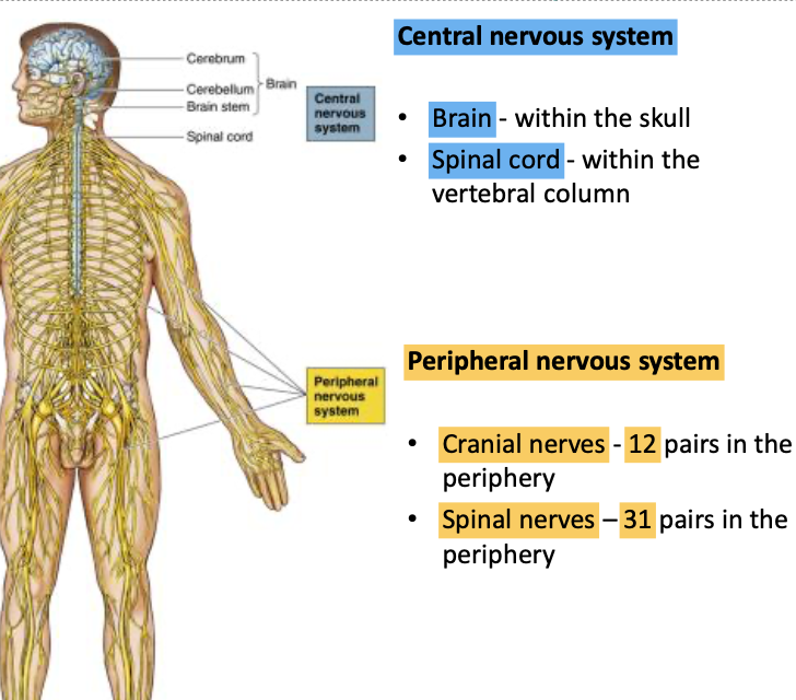
Functional divisions of the brain - CNS
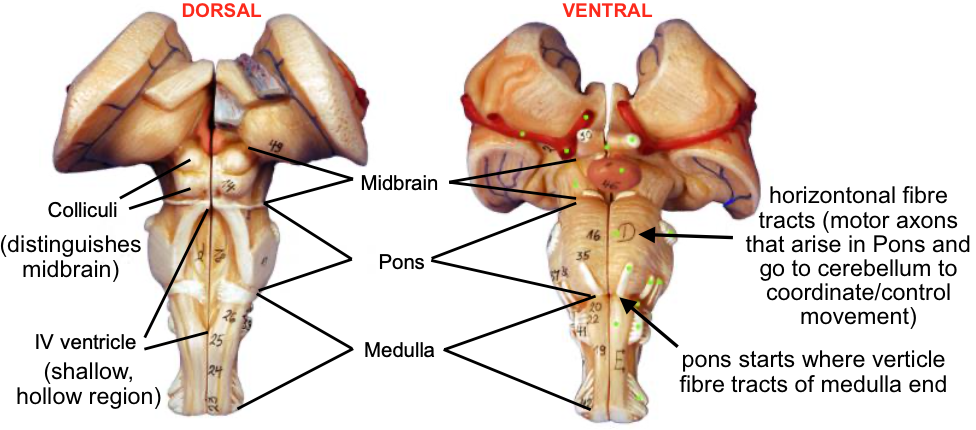
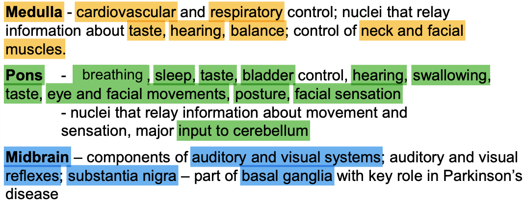
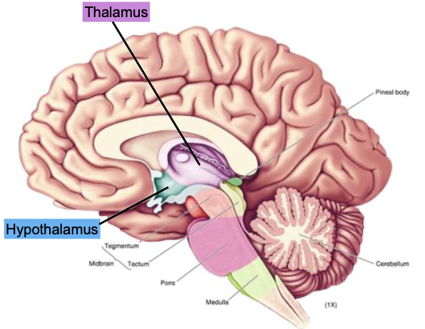
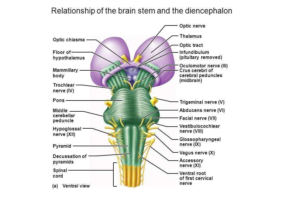
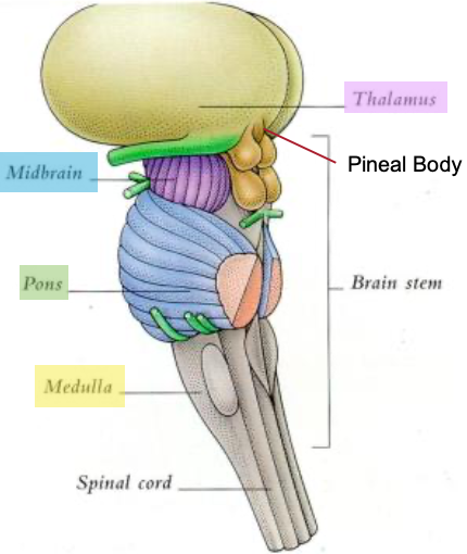
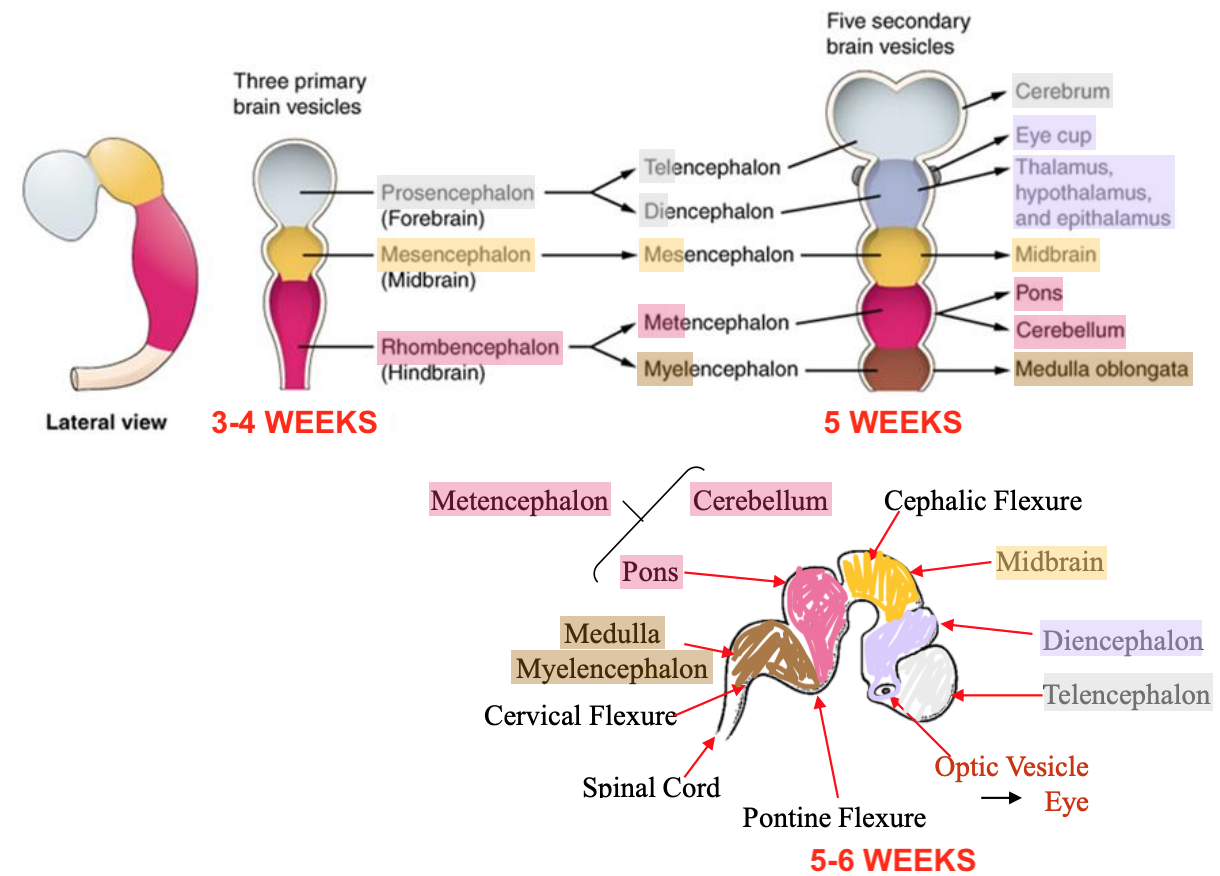
- Brainstem
- Medulla
- Pons
- Midbrain
- Superior (visual) & inferior (auditory) COLLICULI
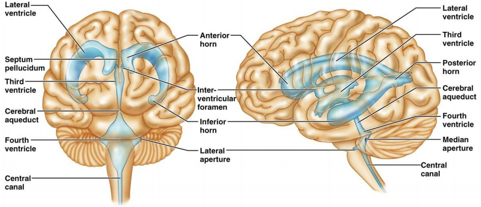
- PINEAL BODY: regulates circadian rhythms - synthesise melatonin. Pineal tumours can compress midbrain and block cerebral aqueduct - vCSF from 3rd to 4th ventricle
- SENSORY and MOTOR functions via CN's and BODY VIA SPINAL CORD
- Cerebellum
- Maintaining POSTURE. Coordinate HEAD & EYE mvmts. FINE-TUNING mvmts. MOTOR LEARNING.
- Diencephalon
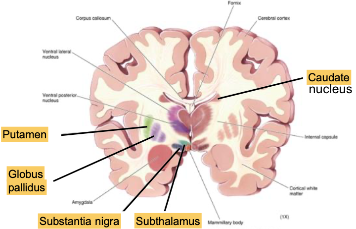
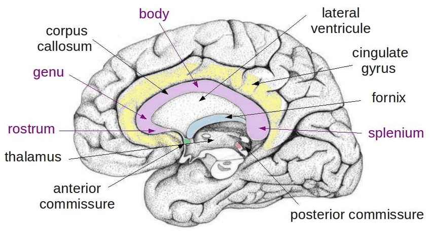
- Thalamus
- Essential for transferring ALL SENSORY INFO (minus olfaction). Relay sensory info to CORTEX.
- GATES & MODULATES sensory info
- Integrates MOTOR control
- Influences ATTENTION & CONSCIOUSNESS
- Essential for transferring ALL SENSORY INFO (minus olfaction). Relay sensory info to CORTEX.
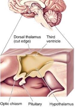
- Hypothalamus
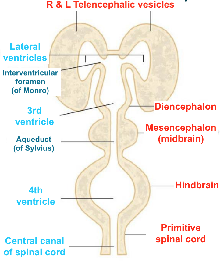
- Associated with 3rd ventricle & cerebral aqueduct
- Regulates HOMEOSTASIS & SEXUAL REPRODUCTION BEHAVIOURS
- eg. drinking, growth, eating, maternal behaviour, circadian rhythms
- Connected to Pituitary (HPA Axis) - hormonal secretions
- Extensive connections to rest of CNS
- Thalamus
- Cerebrum
- Cortex
- 'HIGHER FUNCTIONS': PERCEPTION. MOTOR PLANNING. COGNITION. EMOTION. MEMORY
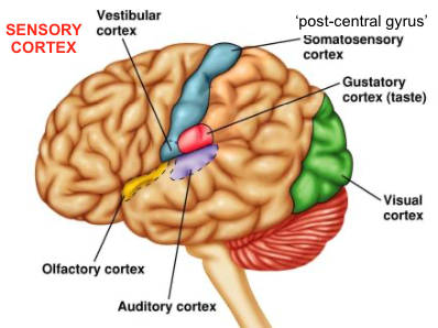
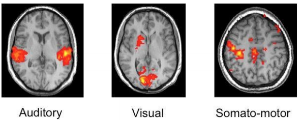
- SENSORY CORTEX
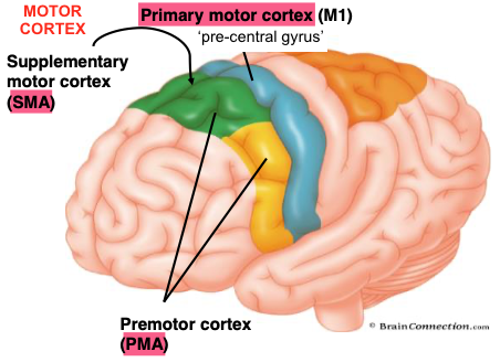
- MOTOR CORTEX
- SMA + PMA provide info INTO Primary motor cortex (M1). First step for voluntary mvmt. Then from M1, motor neurones in cerebral cortex > spinal cord > skeletal muscle
- > PNS
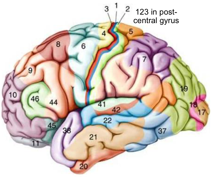
- Brodmann's Areas
- Anatomical differences/organisation across brain mean cortex areas have different function. Structure of axons relates to function.
- SMA + PMA provide info INTO Primary motor cortex (M1). First step for voluntary mvmt. Then from M1, motor neurones in cerebral cortex > spinal cord > skeletal muscle
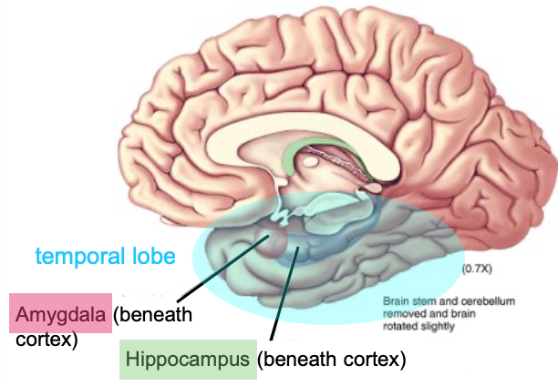
- Discovered via Temporal Lobectomy 1953 - Hippocampus removed > cured epilepsy but induced anterograde amnesia
- 'HIGHER FUNCTIONS': PERCEPTION. MOTOR PLANNING. COGNITION. EMOTION. MEMORY
- Amygdala
- Social BEHAVIOUR & EMOTION
- Hippocampus
- MEMORY
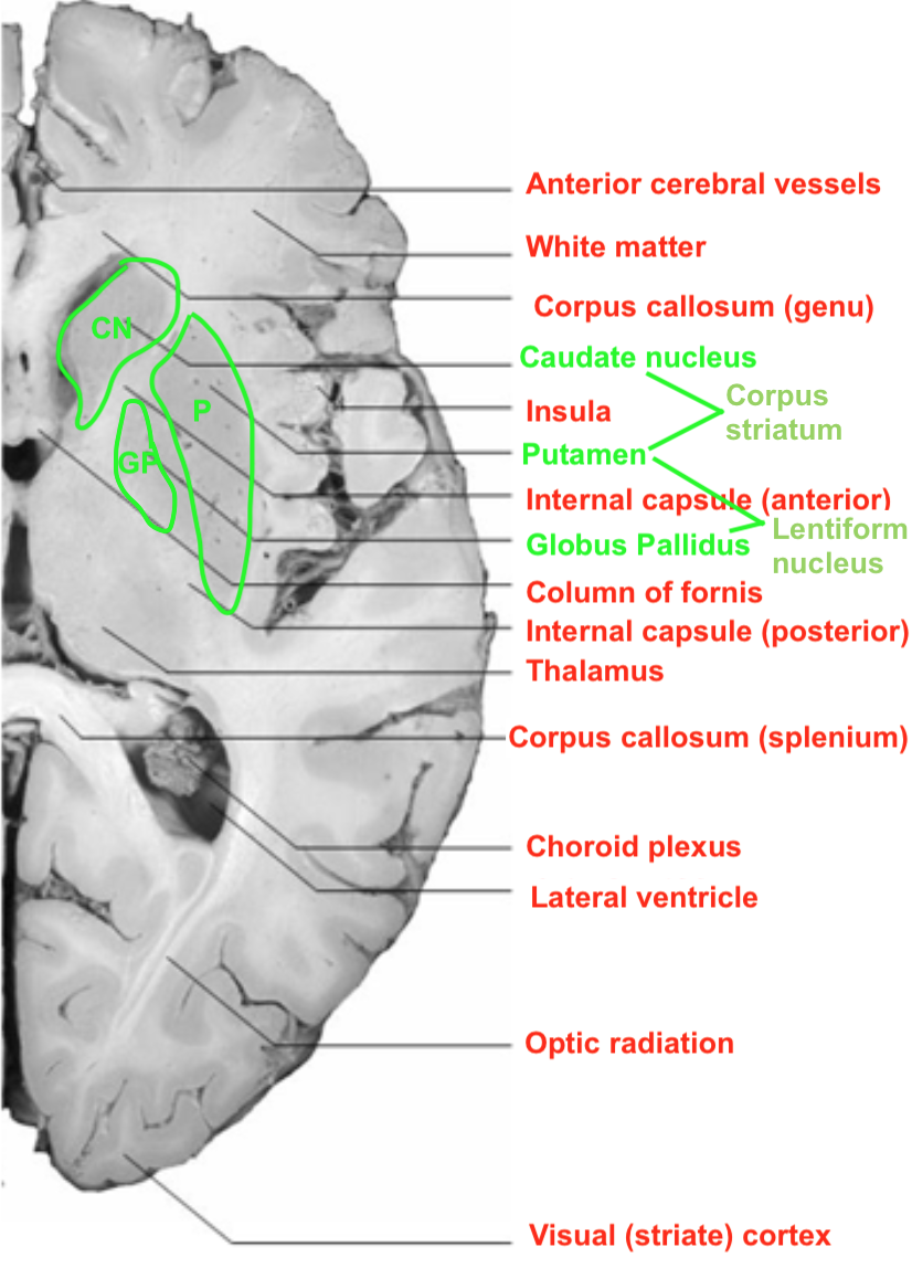
- Basal nuclei/ganglia
- MVMTS
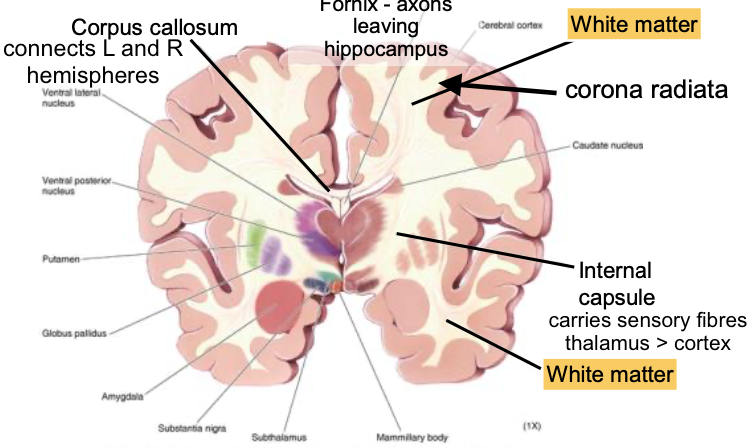
- White matter
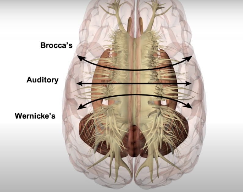
- Axons connecting different parts of brain - to&from cortex / between strutures (hidden beneath cerebral cortex)
- Tracts - 3 types:
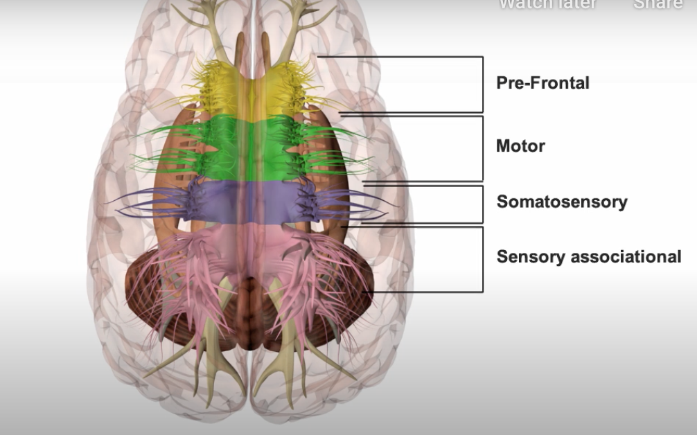
- PROJECTION: Internal Capsule - sensory fibres thalamus>cortex
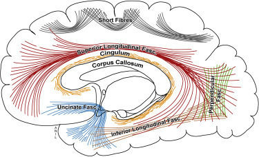
- ASSOCIATION: Short fibres, Sup & Inf Longitudinal Fasc, Cingulum, Uncinate Fasc, Perpeindicular Fasc
- COMMISURAL: Corpus Callosum, Anterior & Posterior & Hippocampal commissures
- Tracts - 3 types:
- Axons connecting different parts of brain - to&from cortex / between strutures (hidden beneath cerebral cortex)
- Cortex
- Brainstem
Blood Supply
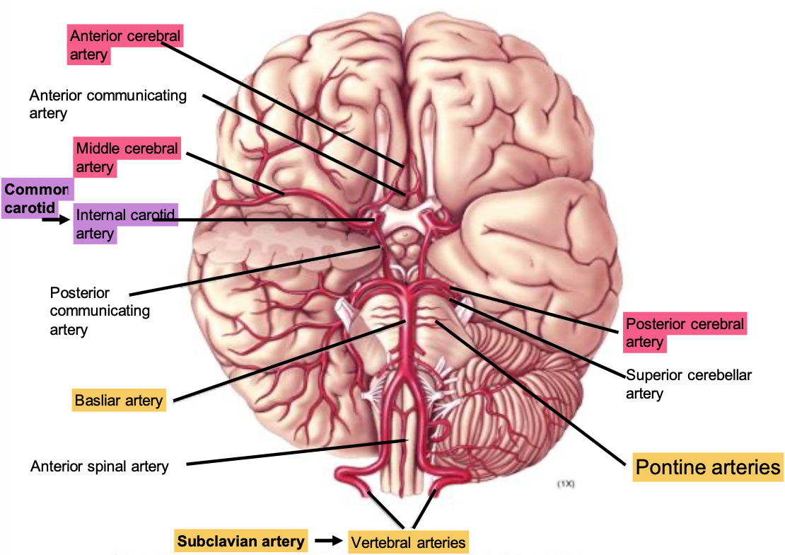
- Circle of Willis
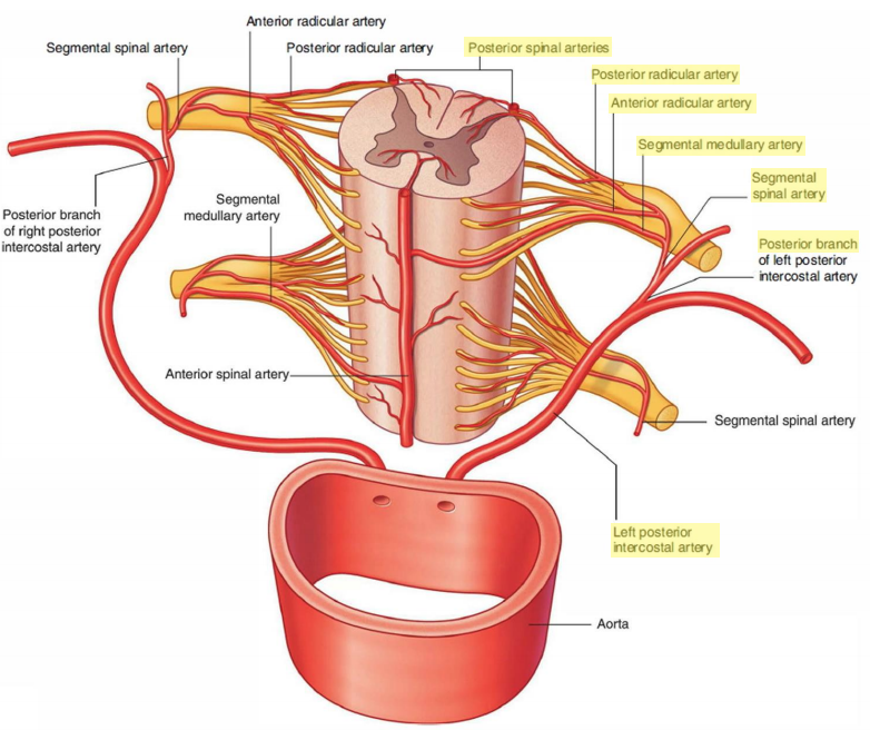
- NB. Anterior spinal artery supplies surface of spinal cord
- Occlusion > loss of pain and temp sensation. Dorsal sensory functions are relatively preserved
- Spinal blood supply
- Aorta > L/R Post intercostal artery > Post branch > Segmental spinal artery > Segmental medullary artery OR Ant/Post Radicular artery
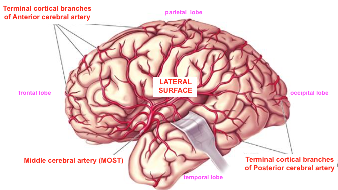
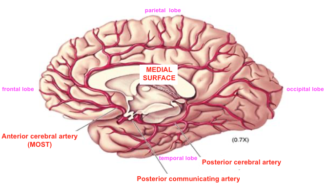
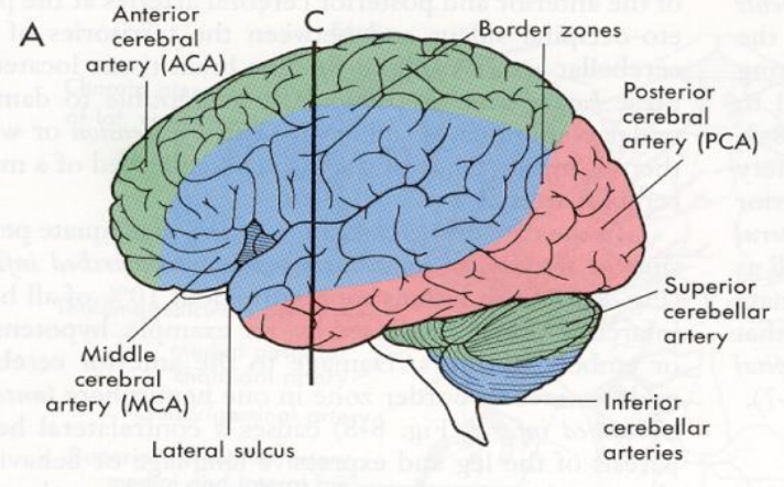
- Cerebral blood supply
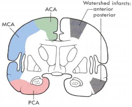
- Cerebral arteries supply:
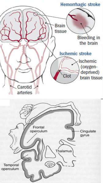
- Watershed Infarcts - if perfusion pressure is low in terminal branches (ie. where territories overlap)
- Anterior - motor & sensory cortex of LOWER LIMB
- Middle - motor & sensory cortex of UPPER LIMB & FACE, also AUDITORY CORTEX
- Most common site for ISCHAEMIC STROKE > Contralateral hemiparesis and hemisensory loss - affects FACE and UPPER + LOWER EXTREMITIES
- Think F.A.S.T. (Face, Arms, Speech, Time)
- Most common site for ISCHAEMIC STROKE > Contralateral hemiparesis and hemisensory loss - affects FACE and UPPER + LOWER EXTREMITIES
- Posterior - whole of VISUAL CORTEX
- Cerebral arteries supply:
- Cerebral blood flow
- REGULATION
- WHY NEEDED?
- TOO LOW > ischaemic damage
- TOO HIGH > ^intracranial pressure > oedema & crushing of brain tissue, shifting of structures, restricts blood flow, herniation
- Flow can be adjusted very RAPIDLY at microvascular level (as soon as 1sec after neuronal excitation)
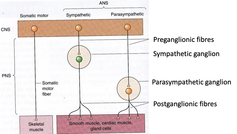
- NEURAL - symp & parasymp innervation
- METABOLIC
- Local ^metabolism > vO2 ^CO2
- vpH of ECF > VASODILATION of vascular SMOOTH MUSCLE
- Hypoxia > ^K+ > vasodilation.
- Low O2 > ADENOSINE released > vasodilation
- *[Adenosine] changes can be used as rapid detector for stroke
- Local ^metabolism > vO2 ^CO2
- LOCAL CONTROL
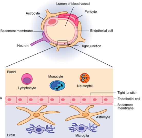
- GLUTAMATE release (main neurotransmitter for blood flow reg) > acts on astrocytes (wrap around vessels) > ^Ca2+ > activate NO-synthase > NO release > Vessels DILATE
- WHY NEEDED?
- ~15% of Cardiac output. Should be 50ml/min per 100g of brain weight.
- Functional MRI (fMRI)
- Detect rapid changes. Used with vascular markers/contrasting agents eg. GADOLINUM
- Neuronal signals cause changes in blood flow > can see this via OXYHb & DEOXYHb.
- Functional MRI (fMRI)
- REGULATION
- Circle of Willis
Formation
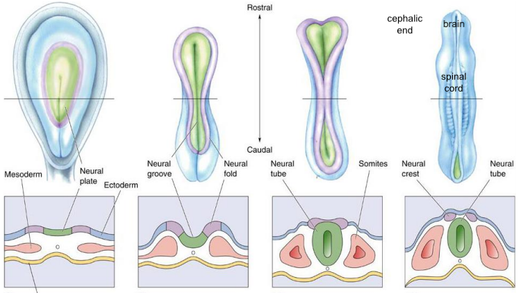
- Development of SPINAL CORD
- Development of BRAIN REGIONS
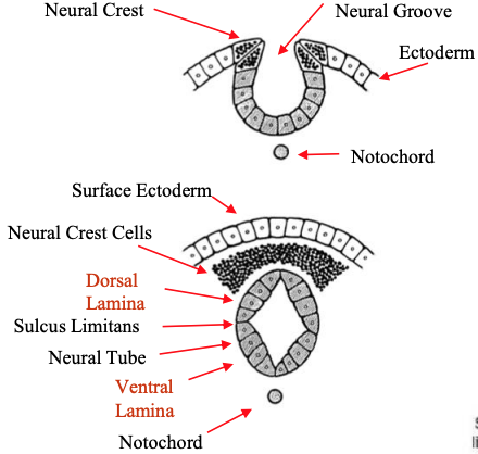
- Forms ~WEEK 3 from ECTODERMAL LAYER
- Neural groove > neural tube over several days
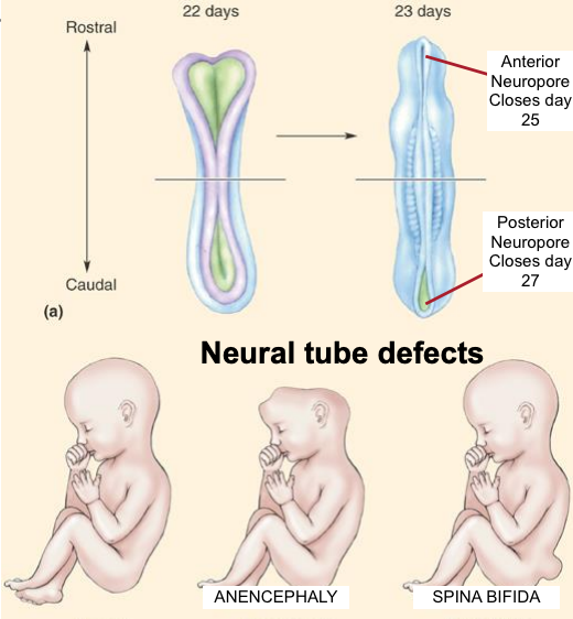
- Anterior neuropore defect > ANENCEPHALY Posterior neuropore defect > SPINA BIFIDA
- Give mothers FOLIC ACID supplements
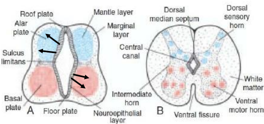
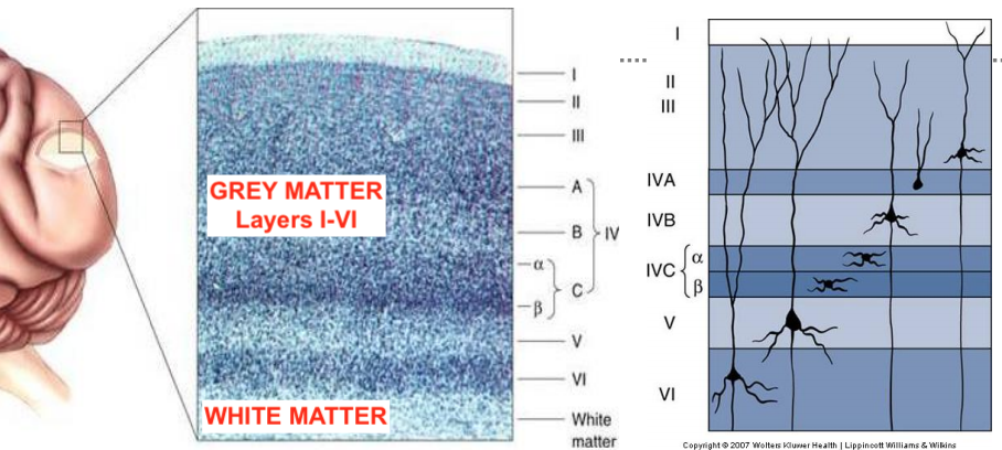
- Neuroepithelial cells > neuroblasts. Form MANTLE layer (around neuroepithelial layer) > GREY MATTER OF SPINAL CORD.
- MARGINAL layer (outermost layer) contains nerve fibres emerging from neuroblasts in mantle layer. These are MYELINATED > white appearance > WHITE MATTER OF SPINAL CORD.
- Anterior neuropore defect > ANENCEPHALY Posterior neuropore defect > SPINA BIFIDA
- Neural groove > neural tube over several days
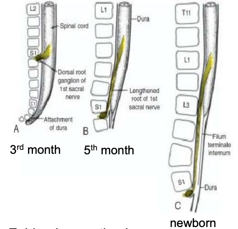
- Vertebral column & dura lengthen faster than neural tube > Spinal cord terminates L2/L3, then CAUDA EQUINA. Dural sac & subarachnoid space terminate S2. Below L2/L3, FILUM TERMINALE (threadlike extension of pia mater) attaches to coccyx > regression of spinal cord.
- No spinal cord below L3 allows EPIDURAL ANAESTHESIA & LUMBAR PUNCTURE
- Development of VENTRICULAR SYSTEM
- allows fluid to flow between lateral & 3rd ventricles
- Canal runs through brain and continues down spinal cord
- Development of CORTEX
- BIRTH = all of the neurons you will ever have.
- SYNAPSES form at a FASTEST rate in first 3 years. At 2/3yo, the brain has DOUBLE as many synapses as in adulthood. Surplus connections 'PRUNED' throughout childhood and adolescence.
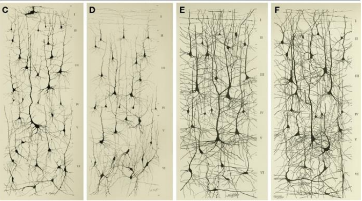
- Grey matter: neurones with different structures, all interconnected
- NO NEW neurones formed throughout life, apart from in: SUBVENTRICULAR ZONE and DENTATE GYRUS of HIPPOCAMPUS
- SYNAPSES form at a FASTEST rate in first 3 years. At 2/3yo, the brain has DOUBLE as many synapses as in adulthood. Surplus connections 'PRUNED' throughout childhood and adolescence.
- Brain size doubles in first year. By 3yo it has reached 80% of its adult volume
- More complex dendritic tree > more synapses can form > more complex brain
- BIRTH = all of the neurons you will ever have.
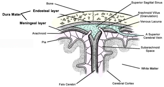
- MENINGES
- CSF
- Disorders!
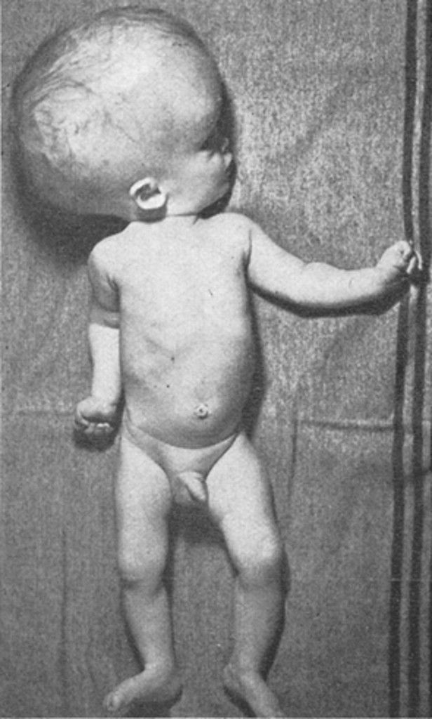
- HYDROCEPHALUS - CSF continuously produced - BLOCKAGES in circulation/drainage/production > ^intracranial pressure
- Blockage most common @ interventricular foramen & cerebral aqueduct
- in newborn > VENTRICULAR & SKULL DILATION
- in adult (cranial cavity closed) > HEADACHE/VOMITING/NAUSEA/BRADYCARDIA/HYPERTENSION/LOSS OF CONSCIOUSNESS/BRAIN STEM DYSFUNCTION
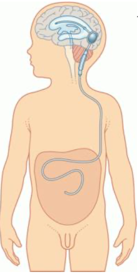
- Ttment: shunt to remove fluid. If caused by tumour > remove/shrink
- HYDROCEPHALUS - CSF continuously produced - BLOCKAGES in circulation/drainage/production > ^intracranial pressure
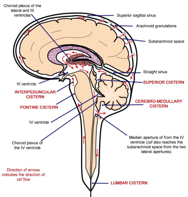
- 150ml total, 25ml in ventricles. 500ml produced per day - secreted by CHOROID PLEXUS
- CHOROID PLEXUS = capillary network separated from ventricles by choroid epithelial cells.
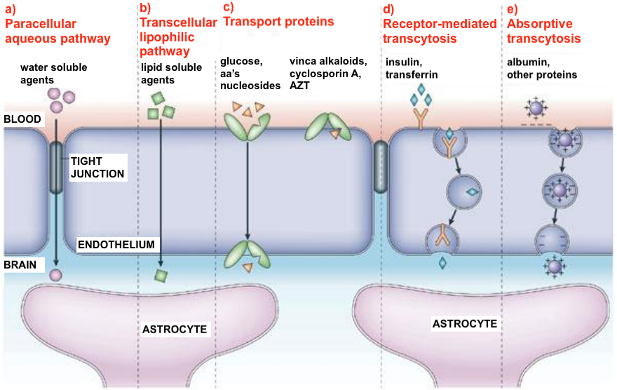
- Blood-Brain Barrier BBB
- = Endothelial cells + Tight junctions
- Fluid filters into ventricles - other substances may be transported - ie. CSF is an ultrafiltrate of blood
- NB. CP in lateral ventricles continuous with CP in 3rd ventricle
- Blood-Brain Barrier BBB
- CHOROID PLEXUS = capillary network separated from ventricles by choroid epithelial cells.
- 1) Removes WASTE products. 2) TRANSPORTS SIGNALLING molecules. 3) Makes brain BUOYANT. 4) SUPPORTS/CUSHIONS/DISTRIBUTES PRESSURE on brain.
- Disorders!
- DURA mater
- Two layers - close together apart from along sagittal midline
- MENINGEAL / PROPER / DEEP LAYER Continuous with dura of spinal cord
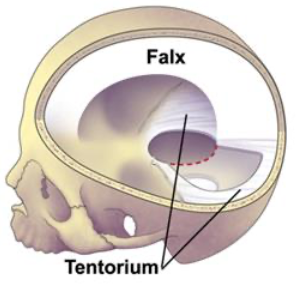
- Falx Cerebri & Tentorium Cerebelli
- Tentorium roofs over posterior cranial fossa. An opening, the 'tentorial notch', allows midbrain to pass through
- Falx Cerebri & Tentorium Cerebelli
- ENDOSTEAL / PERIOSTEAL / SUPERFICIAL LAYER Continuous with periosteum on outside of skull at foramina not cont with dura of spinal cord
- MENINGEAL / PROPER / DEEP LAYER Continuous with dura of spinal cord
- Provides protection - provides space for superior sagittal sinus > CSF venous drainage
- Disorders!
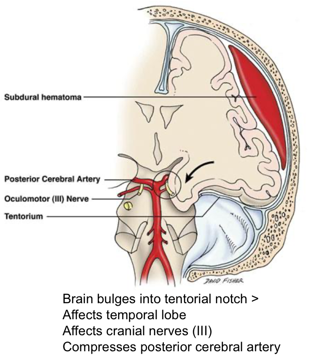
- Lesions (eg. heamatoma/tumors) cause brain to PRESS against dura (tough immovable membrane), or HERNIATE through tentorial notch.
- Two layers - close together apart from along sagittal midline
- ARACHNOID mater
- Separated from dura by subdural space (filled with film of fluid) Separated from pia by subarachnoid space (filled with CSF, BLOOD VESSELS & NERVES)
- bridges over sulci of brain
- ARACHNOID VILLI - arachnoid projects through dura into venous sinuses > form arachnoid granulations along sinuses
- 1-way valves – allow CSF to drain into sinuses > venous system REABSORPTION: greater hydrostatic pressure in subarachnoid space; greater osmotic pressure in sinus
- ARACHNOID VILLI - arachnoid projects through dura into venous sinuses > form arachnoid granulations along sinuses
- bridges over sulci of brain
- Separated from dura by subdural space (filled with film of fluid) Separated from pia by subarachnoid space (filled with CSF, BLOOD VESSELS & NERVES)
- PIA mater
- Extend into brain sulci. Covers cerebral arteries
- Disorders!
- HEADACHE - brain has no pain receptors so caused by stretch/irritation of meninges/vessels
- BACTERIAL MENINGITIS - inflammation due to Streptococcus Pneumoniae & Neisseria Meningitides. Immune response > cerebral oedema > ^internal pressure > herniation & poor blood supply
- Diagnosis: CSF examination via lumbar puncture - ^White cell count ^Protein vGlucose
- CSF
- Development of SPINAL CORD
Imagens e arquivos do mapa
Informações
- Descrição
- Year 1 - CBL Weeks 11-20 (Neuro) Mapa Mental sobre Brain Structure-Function, Blood Supply, Formation, criado por Ella Breese em 13-05-2020.
- Criado por
- Ella Breese
- Visualizações
- 0
- Avaliação