
Mapa mental
LUNG FUNCTION
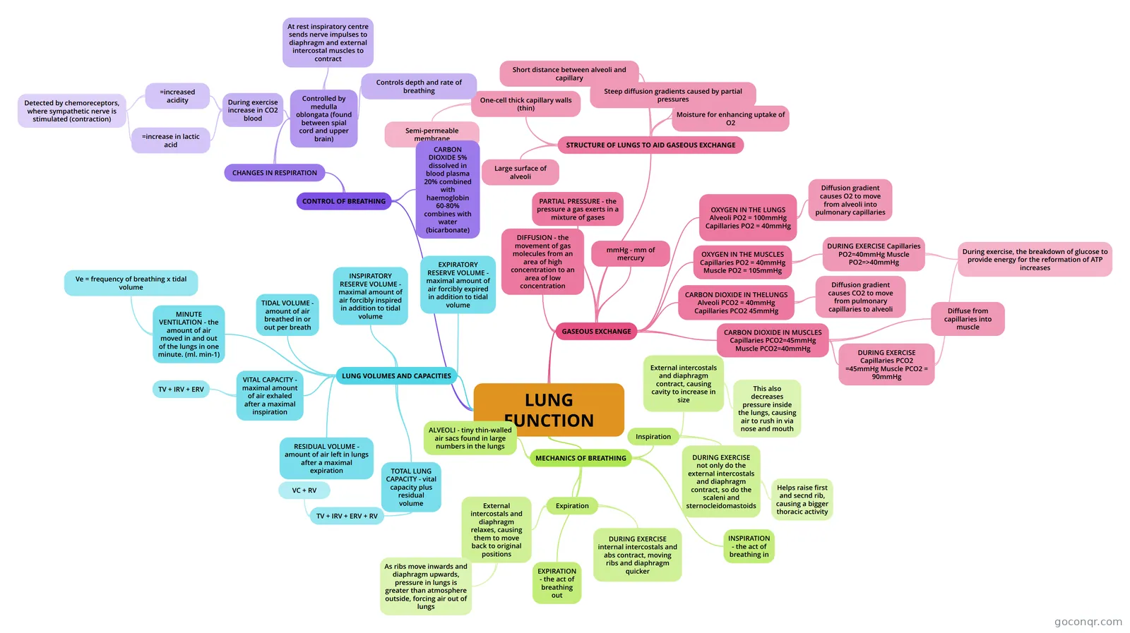
Mind map: lung function
Conteúdo do mapa
LUNG FUNCTION
MECHANICS OF BREATHING
- Inspiration
- External intercostals and diaphragm contract, causing cavity to increase in size
- This also decreases pressure inside the lungs, causing air to rush in via nose and mouth
- DURING EXERCISE not only do the external intercostals and diaphragm contract, so do the scaleni and sternocleidomastoids
- Helps raise first and secnd rib, causing a bigger thoracic activity
- External intercostals and diaphragm contract, causing cavity to increase in size
- Expiration
- External intercostals and diaphragm relaxes, causing them to move back to original positions
- As ribs move inwards and diaphragm upwards, pressure in lungs is greater than atmosphere outside, forcing air out of lungs
- DURING EXERCISE internal intercostals and abs contract, moving ribs and diaphragm quicker
- External intercostals and diaphragm relaxes, causing them to move back to original positions
- ALVEOLI - tiny thin-walled air sacs found in large numbers in the lungs
- INSPIRATION - the act of breathing in
- EXPIRATION - the act of breathing out
- Inspiration
LUNG VOLUMES AND CAPACITIES
- TIDAL VOLUME - amount of air breathed in or out per breath
- INSPIRATORY RESERVE VOLUME - maximal amount of air forcibly inspired in addition to tidal volume
- EXPIRATORY RESERVE VOLUME - maximal amount of air forcibly expired in addition to tidal volume
- VITAL CAPACITY - maximal amount of air exhaled after a maximal inspiration
- TV + IRV + ERV
- RESIDUAL VOLUME - amount of air left in lungs after a maximal expiration
- TOTAL LUNG CAPACITY - vital capacity plus residual volume
- TV + IRV + ERV + RV
- VC + RV
- TV + IRV + ERV + RV
- MINUTE VENTILATION - the amount of air moved in and out of the lungs in one minute. (ml. min-1)
- Ve = frequency of breathing x tidal volume
GASEOUS EXCHANGE
- DIFFUSION - the movement of gas molecules from an area of high concentration to an area of low concentration
- PARTIAL PRESSURE - the pressure a gas exerts in a mixture of gases
- mmHg - mm of mercury
- OXYGEN IN THE LUNGS Alveoli PO2 = 100mmHg Capillaries PO2 = 40mmHg
- Diffusion gradient causes O2 to move from alveoli into pulmonary capillaries
- OXYGEN IN THE MUSCLES Capillaries PO2 = 40mmHg Muscle PO2 = 105mmHg
- DURING EXERCISE Capillaries PO2=40mmHg Muscle PO2=>40mmHg
- During exercise, the breakdown of glucose to provide energy for the reformation of ATP increases
- DURING EXERCISE Capillaries PO2=40mmHg Muscle PO2=>40mmHg
- CARBON DIOXIDE IN THELUNGS Alveoli PCO2 = 40mmHg Capillaries PCO2 45mmHg
- Diffusion gradient causes CO2 to move from pulmonary capillaries to alveoli
- CARBON DIOXIDE IN MUSCLES Capillaries PCO2=45mmHg Muscle PCO2=40mmHg
- Diffuse from capillaries into muscle
- DURING EXERCISE Capillaries PCO2 =45mmHg Muscle PCO2 = 90mmHg
- STRUCTURE OF LUNGS TO AID GASEOUS EXCHANGE
- One-cell thick capillary walls (thin)
- Semi-permeable membrane
- Short distance between alveoli and capillary
- Steep diffusion gradients caused by partial pressures
- Large surface of alveoli
- Moisture for enhancing uptake of O2
- One-cell thick capillary walls (thin)
CONTROL OF BREATHING
- CARBON DIOXIDE 5% dissolved in blood plasma 20% combined with haemoglobin 60-80% combines with water (bicarbonate)
- CHANGES IN RESPIRATION
- Controlled by medulla oblongata (found between spial cord and upper brain)
- Controls depth and rate of breathing
- At rest inspiratory centre sends nerve impulses to diaphragm and external intercostal muscles to contract
- During exercise increase in CO2 blood
- =increased acidity
- =increase in lactic acid
- Detected by chemoreceptors, where sympathetic nerve is stimulated (contraction)
- =increased acidity
- Controlled by medulla oblongata (found between spial cord and upper brain)
Informações
- Descrição
- Lung Volumes and Capacities Gaseous Exchange Mechanics of Breathing A bit on Control of Breathing (expanded in other mind map)
- Criado por
- harmonyhennessy
- Visualizações
- 177
- Tags
- Avaliação