13666052
Prelim. Biology- Module 1 (Chapter 2): CELLS AS THE BASIS OF LIFE
Description
No tags specified
Slide Set by yeet master, updated more than 1 year ago
More
Less
|
|
Created by yeet master
almost 6 years ago
|
|
Resource summary
Slide 1
Inquiry Question: What distinguishes one cell from another?
Chapter 2, Cell structure and technologies
Slide 2
2.1 Types of cells
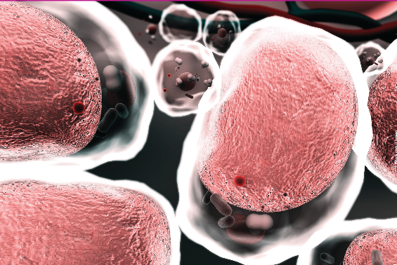
Prokaryotic cells
With the word derived from the Greek pro (before) and karyon (nucleus), prokaryotic cells range in diameter from 0.1 to 5.0 micrometers.
The four main structure that all prokaryotic cells possesses are; the cell membrane, the cytoplasm, ribosomes and genetic material.
There is no membrane surrounding the genetic material and therefore no nucleus. Most organisms that are composed of prokaryotic cells are unicellular- a single cell. Some bacterial species clump together as a colony of prokaryotic cells that work together.
Slide 3
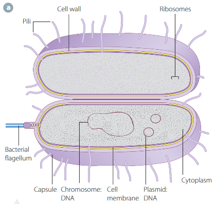
Prokaryotic organisms (or prokaryotes) can be divided into main groups: bacteria and archaea. The cells in these groups are similar in both shape and size, but differ chemically in terms of their genetic material and proteins
- cell wall: protects the cell and provides structural support
- Pili: hair like structures
- Flagella: tails that provide the cell with locomotion
Caption: : Scientific drawing of a prokaryotic cell
Slide 4
Eukaryotic cells
'Eukaryotic' is derived from the Greek eu (proper) and karyon (nucleus)
These cells range in size from 10 to 100 micrometers and are much more complex than prokaryotic cells.
Eukaryotic cells are characterized by a membrane-bound nucleus containing genetic material of the cell. All of the internal structures of these cells are membrane bound and are known as organelles. Each organelle has a specific function within the cell, and together, carry out biochemical processes and reaction, such as respiration and photosynthesis
Living organisms that contain eukaryotic cells are known as eukaryotes. These organisms can either be unicellular or multicellular.
Paramecium, amoeba, and euglena, are examples of unicellular eukaryotes.
Slide 5
Calculating total magnification
To calculate the total magnification when using a light microscope, the magnification of the ocular (eyepiece) lens is multiplied by the magnification of the objective lens being used:
total magnification = magnification of ocular lens x magnification of objective lens
- You can use a light microscope to view cells
Slide 6
2.2 Technologies in cell structure research
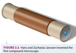
1500s: Scientists used handheld magnifying glasses to view objects of interest. In order to attain greater detail, scientists invented the first compound microscope. This microscope had two convex lenses placed at either end of a barrel and was the precursor to the light microscope
Slide 7
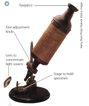
After further developments were made to the structure of these compound microscopes, such as placing them on a stand, adding focus knobs and a light source, scientists were able to determine the structure of organisms.
Robert Hooke, in the 1660s, was able to view and draw the structure in cork that led to his use of the term "cella", leading to our term "cell"
Caption: : Hooke's compound microscope
Slide 8
Modern microscopes
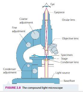
Light microscope: A light source passes through a condenser lens and then through the thin specimen. This beam of light then passes through a convex objective lens, where the image is magnified and viewed through the ocular lens. Can view both living and non-living specimens.
Compound light microscopes can produce images with a magnification (increase in size of the image) up to 1500x depending on the lenses used.
Resolution is the ability to distinguish between two separate objects. It is the smallest distance between two objects where each can be viewed separate.
For a compound light microscope, the maximum resolution is 200nm (if the distance between two objects is less, two distinct objects can be viewed)
Slide 9
Types of microscopes
Caption: : The compound light microscope
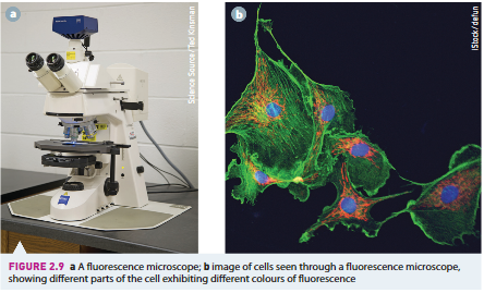
Caption: : Fluorescence
Slide 10
Fluorescence and Electron Microscopes
Fluorescence microscopes- This microscope is similar to the light microscopes but it contains extra features that enable scientists to produce images of specific parts of cells. Structures beyond the limit of resolution of a light microscope can be seen using this microscope..
The sample to be viewed is labelled with a fluorescent substance that will attach itself to the structures that the scientist wants to specifically observe. The sample is illuminated with a high-intensity source of light that causes the fluorescent substance to emit light. It is then directed through filters that separate it from surrounding light and the viewer is able to see areas of that sample that are fluorescing.
Slide 11
Electron microscopes:
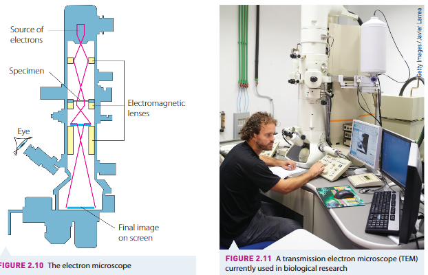
1950s; The study of microscopic organisms have been revolutionized by the development of the electron microscope.
This microscope uses an electron beam instead of light, and electromagnets instead of glass lenses. The interaction between the electrons and the object forms a view-able image on-screen.
The use of electrons instead of light provides a much greater magnification and a higher resolving power (this is due to the electrons having much shorter wavelengths than light).
The electron microscope reveals structures at not only the cellular level, but also at the sub-cellular level. Organelles have been seen for the first time, and features as small as one-tenth of a nano meter can been.
Slide 12
SEM and TEM
There are two main types of electron microscopes:
SEM (scanning electron microscope): bombards solid specimens with a beam of electrons, which causes secondary electrons to be emitted from the surface layers of the specimen. The SEM has poorer resolution (10nm) than a TEM, but gives excellent three dimensional surfaces of surfaces.
TEM (transmission electron microscope): electrons are transmitted through the specimen. The TEM produces a two-dimensional image. It is the most common form of electron microscope. It can magnify up to 1,500,000 times and has a resolution of about 2 nm.
Slide 13
Caption: : Electron microscope diagram
Slide 14
Disadvantages of electron microscopy
Although there are many advantages in using an electron microscopes, there are also some disadvantages. For example, the specimen must be placed in a vacuum for viewing, because air would interfere with the flow of electrons.
- Living tissue cannot be viewed
- Preparation of the specimen is very complicated and comes with a risk of introducing artefacts into the image. An arterfact is something that is introduced into the image that would not normally be there.
- The size, expense and maintenance costs are much larger in this type of microscope than in others.
Slide 15
Computer-enhanced technology
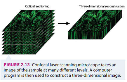
Confocal laser scanning microscopy can be used to highlight the three dimensional structure of samples being studied. A laser produces a narrow intense beam of light that is focused to a pinpoint of the sample, while all the surrounding, out of focus areas are not included in this image. An image reconstruction program puts together the data from the images taken at different levels and construct a 3D image.
Slide 16
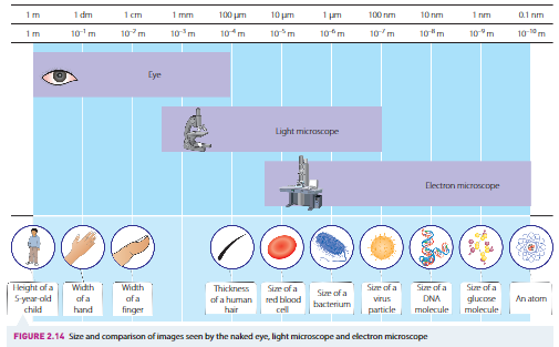
2.3 Sizes of cells
1 meter (m)=
10*2 centimeters (cm)
10*3 millimeters (mm)
10*6 micrometers or microns (pm)
10*9 nanometer (nm)
1cm = 1/100m
1mm = 1/1000m
1pm = 1/1 000 000m
!nm = 1/ 1 000 000 000m
Slide 17
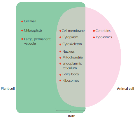
2.4 Organelles in cells
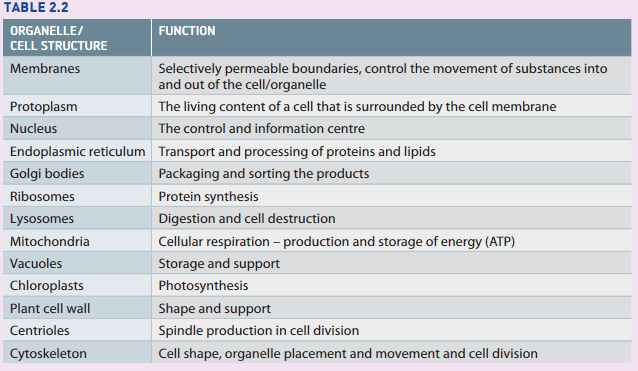
All eukaryotic cells contains membrane-bound internal structures called organelles, each with a specific function and structure. Different organelles share the common feature of having internal structures that are enclosed bu their own membrane. The membrane can either be double or single in structure.
Organelles all work together to contribute to the effective functioning of a cell as a unit. Many organelles have maximized the amount of surface area they have available to carry out their particular functions.
Some organelles (nucleus, vacoules and chloroplasts) can be seen by a light microscope, some others (mitochondria and ribosomes) can be seen only by using an electron microscope.
Slide 18
Membranes- selective boundaries
The cell membrane surrounds the cell contents in all cells and separates the cell contents from their surroundings. It controls the passage of water and other chemical substances into and out of cells.
It is a selective barrier, permitting the passage of only certain molecules into or out of cells. This property gives the cell membrane the feature of being semi permeable. Both plant and animal cells have a cell membrane. The membranes surrounding organelles are also selective in allowing only certain substances to pass between the cytoplasm and the organelle.
Slide 19
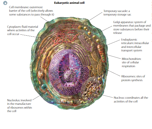
Eukaryotic cells (animal)
Slide 20
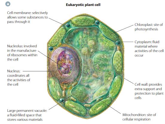
Eukaryotic cells (plants)
Slide 21
Nucleus- the control/information centre
The nucleus, appears as a large, spherical or oval structure in the cytoplasm. It is colorless, transparent and slightly more jelly-like than the rest of the cell. Most organisms have one nucleus per cell.
The nucleus stores the information needed to control all cell activities. It is therefore essential for the nucleus to be able to communicate with the surrounding cytoplasm. Electron micro graphs reveals that the nucleus is surrounded by a double nuclear membrane or nuclear envelope. These pores regulate the passage of substances between the nucleus and cytoplasm, allowing communication between them.
Slide 22
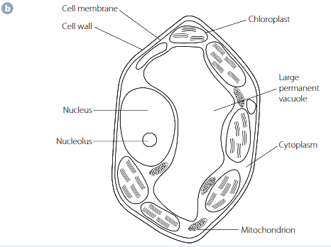
Comparing structures (plant and animals)
Slide 23
Organelles
Nucleoplasm, or nuclear sap- the liquid part of the nucleus in which the chromatin material is found. Chromatin is made up of protein and nucleic acid.
DNA (deoxyribonucleic acid), is stored in the nucleus. It is a very large chemical that holds, in a coded form, all the genetic information necessary to control the cell's function. This DNA contains the hereditary information of any organism and is passed from one generation to the next.
Chromatin- is separated into short, thick separate rod-shaped structures called chromosomes, which become visible in dividing cells.
Nucleolus- a dense, granular region commonly seen within the nucleoplasm. It contains a large amount of nucleic acid: some DNA, but mostly RNA. It is responsible for the manufacture of ribosomes, essential organelles.
Slide 24
Organelles cont.
Endoplasmic reticulum (transport and processing of proteins and lipids), a network of flattened, interconnected membranes. It provides a connection of pathways between the nucleus and the cell's environment, allowing intra-cellular transport. The immense folding of the sheets of membrane increases its surface area.
Rough ER; folds and processes proteins made by the cell and it can also synthesis lipids.
Smooth ER; the main site of lipid production, essential for membrane repair and manufacture.
Ribosomes (protein synthesis), small organelles that are made of chemicals RNA and protein, and they are the 'machinery' that carries out the genetically coded instructions of DNA to produce any proteins necessary for cell functioning and structure.
Golgi bodies (packaging and sorting the products), made up of flat membranes, and is responsible for the processing, packaging and sorting of cell products. Involves adding proteins and carbohydrates the products to package them, ready to be transported where required.
Lysosomes (digestion and destruction), special organelles found in the cytoplasm of animal cells. They are formed in the golgi body, and contain digestive enzymes that are responsible for splitting complex chemical compounds into simpler ones. It is a deliberate action by the cell to destroy old and damaged cells.
Mitochondria (cellular respiration), produces energy in the form of energy-rich molecules by the process of cellular respiration. Rod shaped but may be round, they vary in both shape and size. Mitochondria are smaller than the nucleus and chloroplasts but larger than ribosomes. They combine oxygen with sugars during the process of cellular respiration to release energy in a form (ATP) that the cell can use.
Vacuoles (storage and support), large, permanent, fluid-filled sacs in the cytoplasm of plant cells. Besides having a storage function, vacuoles play a very important role in providing support to plant cells.
Chloroplasts (photosynthesis)
Plant cell wall (shape and support)
Centrioles (spindle production of cell division)
Cytoskeleton (keeps organelles in place)
Slide 25
Summary of organelle functions
Slide 26
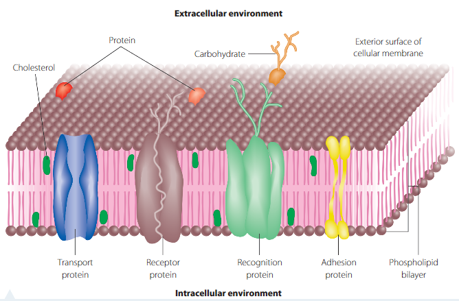
2.5 Cell membranes- gateway to cells
Fluid mosaic model- Our current accepted understanding of cell membranes is based on a model proposed in 1972 by Singer and Nicolson.
The cell membrane controls the exchange of material between the internal and external environments of the cell. It is selectively permeable. Proposes a "lipid sea" with 'many various proteins floating on and in it".
It describes the cell membrane as a double layer of lipids, a lipid bilayer, with the ability to flow and change shape, like a two-dimensional fluid. Specialised protein molecules are embedded in the lipid in various patterns like mosaic. Some of these proteins move sideways, but others are fixed in position. Both proteins and phospholipids help to control the exchange of materials between the external and internal environments.
Slide 27
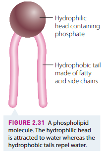
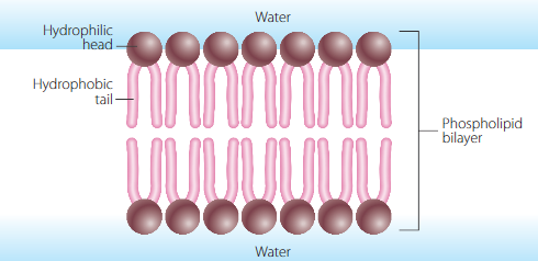
Phospholipid Bilayer- Lipid component
The 'fluid' part of the cell membrane is composed of two layers of phospholipids forming a bilayer. Each phospholipid in these layers can be represented by a head and two tails. It is not rigid in structure, but is fluid.
The phosphate group on the head makes this end hydrophillic (able to absorb and dissolve in water), otherwise known as 'water loving'. The fatty acid tails are hydrophobic (water avoiding or unable to dissolve in water).
A type of lipid called cholesterol is interspersed among the phospholipid molecules in animal cells. This makes the membrane more flexible. Membrane flexibility in plants is increased by a different lipid- phytosterol.
The lipid components of all membranes allow them to be flexible and to repair themselves, meaning that the cell can change shape and grow. Allows cells to reproduce through cell division.
Slide 29
Membrane proteins
Protein molecules are scattered throughout, and suspended in, the lipid bilayer. Some proteins penetrate all the way through the bilayer, forming channels that allow some materials to cross the membrane. Other proteins may be partly embedded in the membrane.
Some proteins are fixed in place, while others travel around freely.
Slide 30
Some proteins function as pores, or form active carrier systems or channels for transport, while other proteins have carbohydrates attached for cell recognition.
These proteins enable cell-to-cell interaction and communication, and the exchange of substances between the cell and the external environment.
- Transport proteins: acts like passageways that allow specific substances to move across the membrane. Membrane proteins are also involved in cellular communication.
- Receptor proteins: causes the cells to respond only to certain signals from substances such as hormones that bind to them, giving them instructions.
- Glycoproteins (recognition proteins): are made up of a protein molecule with a carbohydrate molecule attached. Called antigens, these proteins identify the cell, allowing the immune system to distinguish between foreign particles and the body's cells.
Want to create your own Slides for free with GoConqr? Learn more.