
Mindmap
Proteins
Inhalt der Mindmap
Proteins
Structure
- Primary: chain of amino acids determined by nucleic code
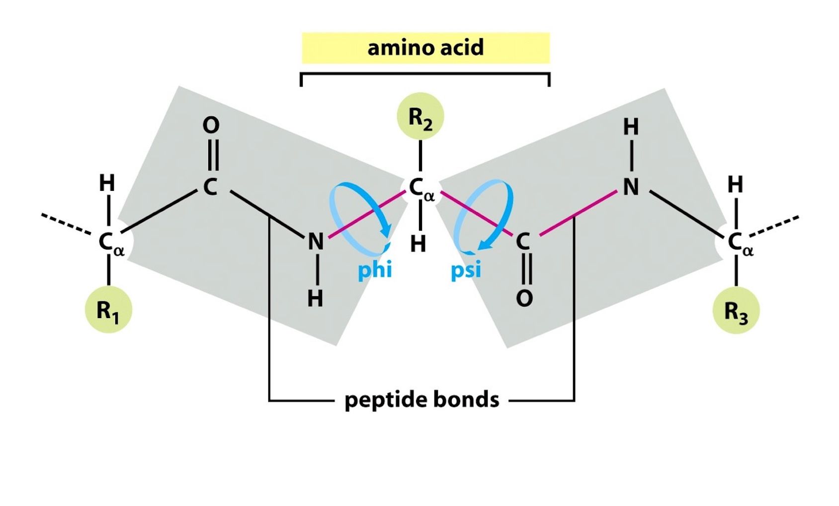
- The resonance of the double bond between the carbonyl and the peptide bond give the peptide bond a partial double bond character so there is no rotation at the peptide bond.
- Polypeptides can rotate around the central Carbon: phi and psi bonds which allows for:
- Secondary: Alpha helices or beta sheets
- Tertiary: Helices and/or beta sheets are joined by loops and turns
- Quatenary
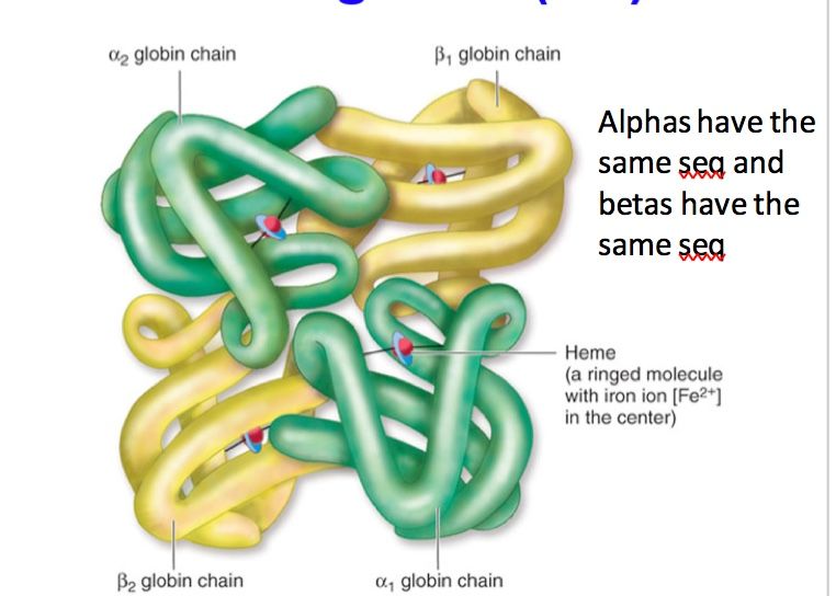
- Tetramer (2α2β)
- Transports O2 in erythrocytes
- Allostery
- As soon as the first O2 binds to one of the hemes, the other 3 globins undergo a conformational change where they don't bind 2,3-BPG anymore,
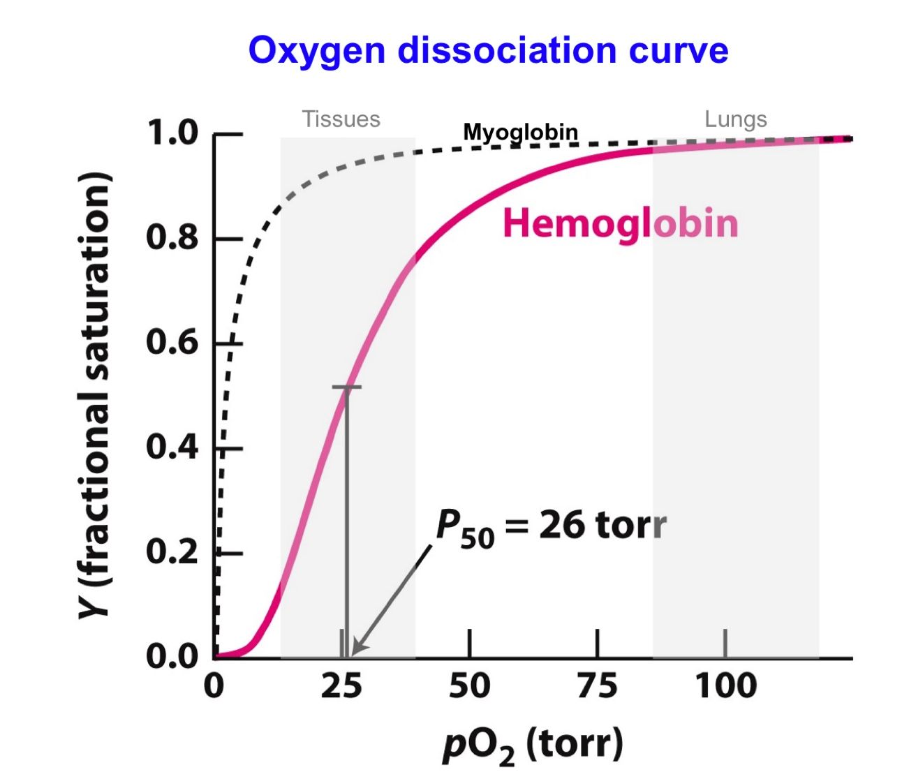
- The conformational changes increase the other 3 hemes' affinity for O2, where they bind to O2 much more readily than the first heme--gives the sigmoidal oxygen disassociation shape
- Hemoglobin's affinity for oxygen depends on the pO2 (dependent on the oxygen avail to bind).
- Hemoglobin has a high affinity of o2 at a very high pO2( in the lungs). But it begins to give up oxygen as the pO2 approaches 50torr, which makes it a good transporter.
- Hemoglobin's affinity for oxygen depends on the pO2 (dependent on the oxygen avail to bind).
- R state
- The conformational changes increase the other 3 hemes' affinity for O2, where they bind to O2 much more readily than the first heme--gives the sigmoidal oxygen disassociation shape
- Cooperativity
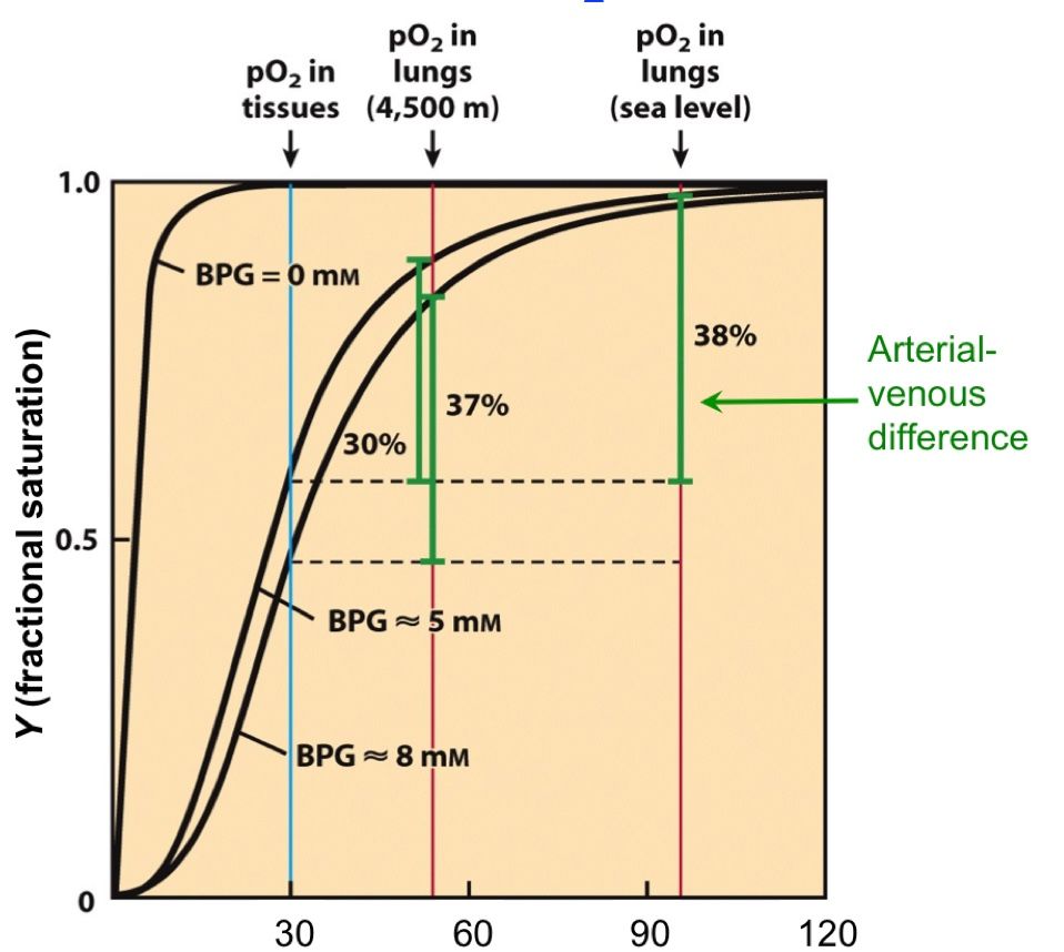
- 2, 3-BPG
- Interacts in the center of Hb in its deoxygenated state
- T state: donut
- Negative Heterotropic Effectors for O2 binding
- Fetal Hb is made of 2α2γ chains. The γ do not have the 2,3-BPG binding site. This pushes the equil to the side of T state for the mother as she is the one with the 2, 3 BPG binding and allows for offloading of O2 to the baby. Mothers also produce more 2,3-BPG during pregnancy for this reason.
- Byproduct of glycolysis; found in high quantity in tissue
- We adapt to higher elevations by producing more BPG, hence giving up more O2 than someone acclimated to sea level at any elevations.
- CO2 also modifies globin N-terminal residues which also favors the T states
- BOHR EFFECT: The increased number of protons in the muscles due to CO2 hydration, causes a conformational change favors the unloading of oxygen
- His HC3 (carboxy terminal) in the beta subunit becomes protonated due to the increased protons in the environment. This allows for a series of salt bridge formations, pushing the conformation to the T state
- High temperature means more CO2 production
- Also how it is carried back to the lungs
- BOHR EFFECT: The increased number of protons in the muscles due to CO2 hydration, causes a conformational change favors the unloading of oxygen
- CO2 also modifies globin N-terminal residues which also favors the T states
- Allosteric competitor of O2: Sigmoidal shape
- Competes with O2 in muscle tissue and in low pO2 envioronments, causes O2 to quickly disassociate from Hb, hence giving the disassociation curve a sigmoidal shape
- 2, 3-BPG
- As soon as the first O2 binds to one of the hemes, the other 3 globins undergo a conformational change where they don't bind 2,3-BPG anymore,
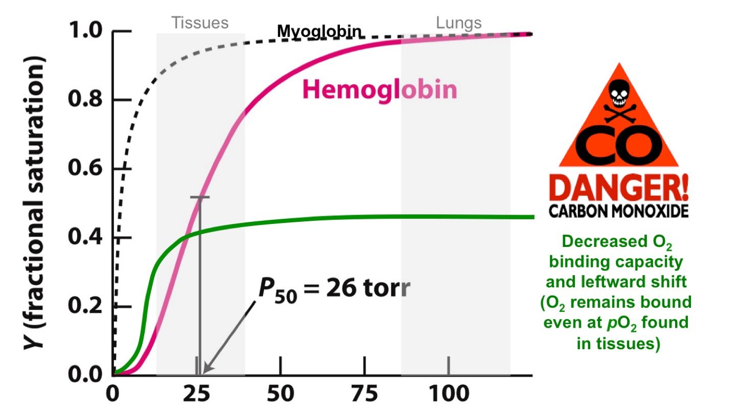
- Carbon Monoxide is a very strong competetor for Oxygen. To a point where O2 cant replace it.
- Once CO binding to 1 heme in a complex, it locks the tetramer in a non-O2 giving up form. The best treatment is just to put the person on a 100% oxygen and hope for the best. Hope for the O2 to compete.
- Multiple polypeptide chains
- Myoglobin
- Monomer
- Stores O2 in muscles
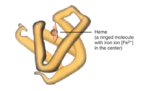
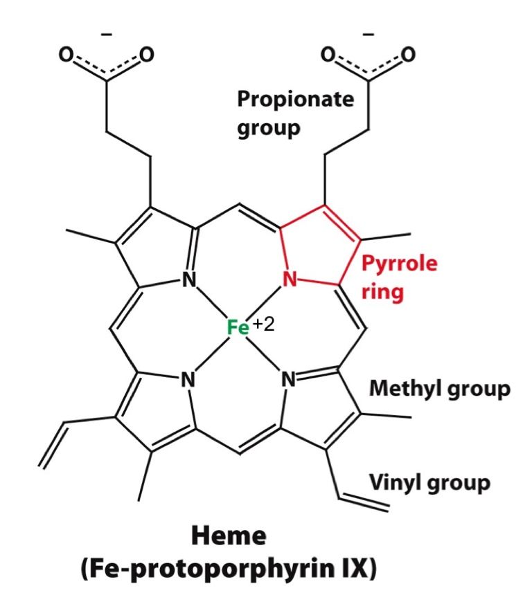
- Carries heme at the core of each globin
- Heme is originally bent with the Iron too big to fit in the middle (T state). Oxygen binding causes electronic displacement in the molecule, specifically iron, which causes it to now fit better and the heme to flatten out (R state)
- Does not give up O2 until partial pressure is <25%, which makes it a good storage unit (tissues)
- These are stabilized by weak non-covalent bonds: van der Waals forces, hydrogen bonding, electrostatic attarctions within and amongst the side and main chains of AAs
- Enthalpy undermined; Increases entropy; doesn't contribute to protein folding
- Disulfide bonds between Cys side chains stabilize 3' structures primariy in extracellular proteins
- Hydrophobic effect drives protein folding
- Hydrophobic/non polar chains are burried into the core of the molecule surrounded by the polar chains
- This frees up the Clathrin caged water surrounding the non polar chains and allows it to move around freely
- Decreases entropy and drives folding
- Quatenary
- Tertiary: Helices and/or beta sheets are joined by loops and turns
- Secondary: Alpha helices or beta sheets
- Primary: chain of amino acids determined by nucleic code
Synthesis
- Central Dogma: Once information has been passed onto the protein, it cannot be retrieived/reversed into nucleic acids again
- Transcription
- Codons are nearly universal; variations in Mit, mycoplasma, ciliate
- Mutations
- Sickle Cell Disease occurs from a missense point mutation in the beta chain of the Hb which changes 6th codon from Glutamine to Valine
- Hemoglobin
- A silent point mutation could affect regulatory machinery a protein
- A non-sense mutation usually does not produce a protein due to the non-sense mediated decay (NMD) of mRNA, if the mutated STOP is significantly far from the real STOP; if not it produces a truncated protein
- Sickle Cell Disease occurs from a missense point mutation in the beta chain of the Hb which changes 6th codon from Glutamine to Valine
- Translation
- About half of the anti-bacterials affect the process of protein translation; no anti-fungals (machinery too similar); and no anti-virals (use our machinery)
- tRNA
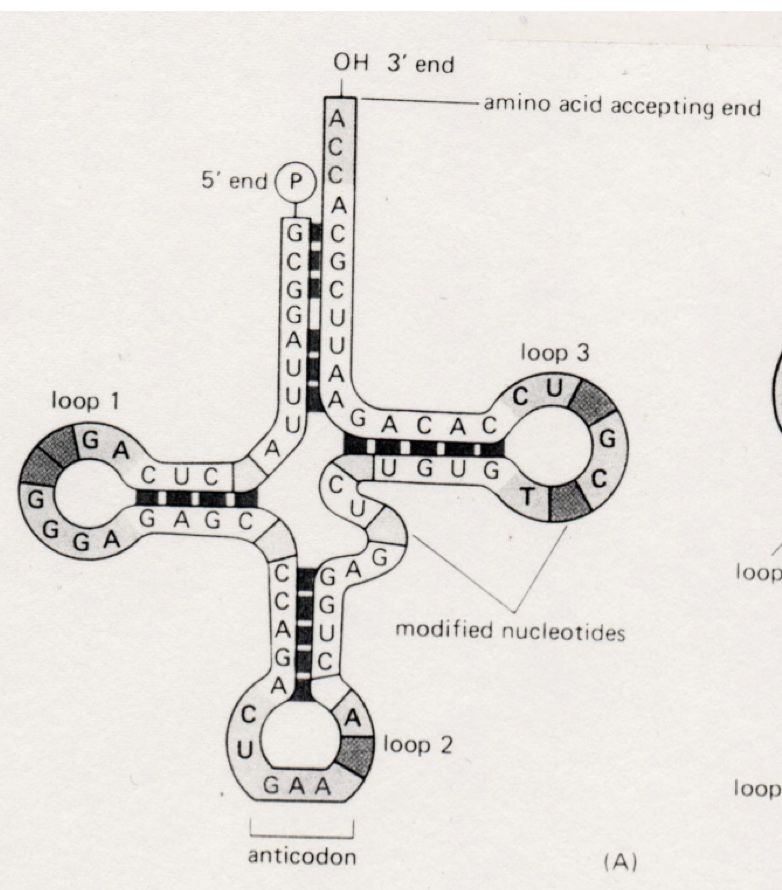
- 70-80 na long; with an anticodon; AA binding 3' and phosphorylated 5'
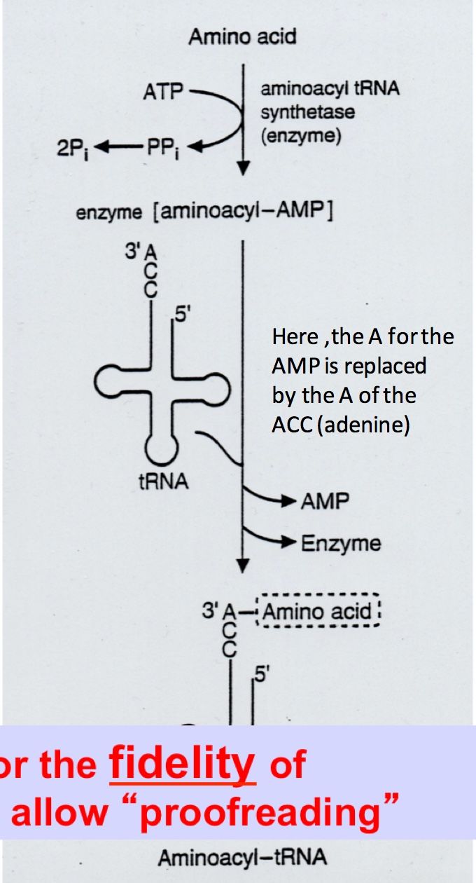
- How does it bind to AA
- This reaction exemplifies sequential linkage as it is driven by the negative free energy released by the ATP hydrolysis; -6.6kcal/mole; hence tRNA are ususally found aminoacylated
- Mupirocin (antibiotic) inhibits Ile-tRNA synthetase
- The two steps allow for the fidelity/proofreading of the process
- WOBBLE pairing
- For the 3rd bp of the mRNA, the Watson Crick rules are a bit more relaxed, allowing for 1 tRNA to bind to multiple codons: Redundancy of genetic code
- Initiation Met-tRNA
- Met-tRNAf (formyl)
- Met-tRNAi
- Initiation
- IF1-IF3
- The 16S rRNA of the 30S ribosomal subunit recognizes a Shine-Dalgarno sequence upstream of AUG
- STREPTOMYCIN (30S): causes misreading of the SD sequence
- Aminogycosides
- Gentamicin, Kanamycin, Neomycin, etc: Cause misreadings during elongation
- Aminogycosides
- STREPTOMYCIN (30S): causes misreading of the SD sequence
- The 16S rRNA of the 30S ribosomal subunit recognizes a Shine-Dalgarno sequence upstream of AUG
- eIF1-eIF5 (12)
- CAP-dependent scanning
- 40S ribosomal subunit scans the sequence starting at the CAP usually recognizing the first AUG as the initiation site
- The first AUG may not be detected if it in in an unfavorable complex, ie not flanked by the right sequence
- eIF4E is a part of a complex needed for scanning
- Upregulated by its own phosphorylation and phosphorylation of 4E-BP
- Phosphorylation upregulated by Growth factors, mitogens, hormones, Cytokines
- Upregulated by its own phosphorylation and phosphorylation of 4E-BP
- 40S ribosomal subunit scans the sequence starting at the CAP usually recognizing the first AUG as the initiation site
- CAP-dependent scanning
- AUG
- IF1-IF3
- Ribosomes
- Eukaryotes
- 60S
- Both have some long and some short rRNA
- 70S
- Both have some long and some short rRNA
- mRNA: 5'CAP----monocistronic---Poly A tail
- Circular: increases stability and translation efficiency esp when protein needs to be repeatedly synthesized
- Circularized by eIF4G and eIF4E and Poly A binding protein
- Circular: increases stability and translation efficiency esp when protein needs to be repeatedly synthesized
- 40S
- IRES: Internal Ribosomal Entry Site
- A lot of secondary structures are present upstream AUG
- GIves the DNA a conformation such that the 40S subunit lands right at AUG
- 60S subnit joins forces after
- A lot of secondary structures are present upstream AUG
- IRES: Internal Ribosomal Entry Site
- 80S
- 60S
- 50S
- 30S
- Prokaryotes
- Transcription and translation is coupled
- mRNA: 5' triphosphate---Polycistronic---hydroxy
- Prokaryotes
- 30S
- Eukaryotes
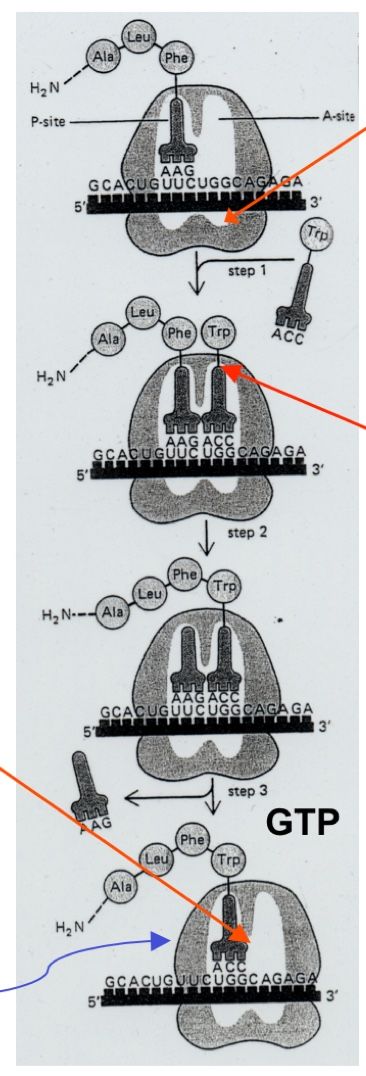
- Elongation
- EF-Tu/Ts (EF1-A,B); EF 2
- EF-Tu/TS [EF1A,B] catalyze tRNA binding to codon
- Peptidyl transferase (rRNA component) catalyzes Peptidyl transfer reaction
- EF-G [EF-2] catalyzes translocation
- Inhibitors: (50S) CLINDAMYCIN, Maclides like ERYTHROMYCIN
- Inhibitors: CHLORAMPHINICOL (50S)
- Imitates AA-tRNA but does not carry an AA; terminates elongation prematurely
- EF-G [EF-2] catalyzes translocation
- Inhibitors: TETRACYCLINS (50S)
- Peptidyl transferase (rRNA component) catalyzes Peptidyl transfer reaction
- EF-Tu/TS [EF1A,B] catalyze tRNA binding to codon
- eEF1-A,B, eEF2
- eEF1 (alpha,beta-gamma catalyzes tRNA-anticodon binding
- Peptidyl transferase catalyzes peptidyl transfer
- PUROMYCIN
- eEF2 catalyzes translocation
- Inhibitor: Diptheria toxin
- Diphtheria
- Released by C. diphtheriae infected by a phage
- Cleaved
- Fragment A is the toxin
- Catalyzes ADP-ribosylation of eF2 using NAD+
- Fragment B allows the toxin to be internalized by cells
- Fragment A is the toxin
- Diphtheria
- Alpha-Sacrin
- Inhibits tRNA-Anticodon binding and translocation
- Inhibitor: Diptheria toxin
- RICIN: from Castor beans
- Inhibits tRNA-Anticodon binding
- <1mg; kill adult, 1 bean: kill child
- Fragment B allows uptake; Fragment A depurinates 28S rRNA in 60S
- Peptidyl transferase catalyzes peptidyl transfer
- eEF1 (alpha,beta-gamma catalyzes tRNA-anticodon binding
- EF-Tu/Ts (EF1-A,B); EF 2
- Termination
- RF1-RF3 in Prokaryotes; eRF1, eRF3
- UAA, UAG, UGA
- Attract the release factor instead of a tRNA into the A site and the Amino acid is released from the P site
- Uses approximately 4ATP per AA polymerized; 1200 per avg protein
- Regulated at a General, specific, or selective level
- Ferritin (specific)
- Binds Fe2+
- Regulated at the translation level
- At low levels of Fe2+, IRE (Iron response element) binds IRE-BP, together inhibitting Fe2+ synthesis
- At high concentration, they disassociate and allow translation
- Mutations in IRE or IRE-BP that inhibit their interaction cause de-repression of ferritin
- Causing early onset cataract formation due to Ferritin accumulation
- At low levels of Fe2+, IRE (Iron response element) binds IRE-BP, together inhibitting Fe2+ synthesis
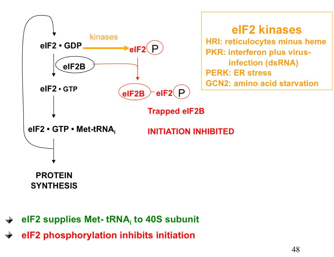
- eIF2 is needed to supply Met-tRNAi to 40S using GTP
- eIF2 can be phosphorylated by a kinase
- Now Trapped by eIF2B, which also catalyzes another reaction in the same pathway
- Initiation downregulated at a general level
- Vanishing White Matter
- Loss of function in any of the 5 subunits of eIF2B
- eIF2 kinase activated under different stresses
- HRI: In Reticulocytes that lack heme. A lot of globin accumulation (toxic); transcription needs to stop
- Interferon Plus Virus infection: activates PKR--stops transcription in cell and virus
- PERK: ER secretion doesn't work right; so translation must be inhibitted
- GCN2 activated by amino acid starvation
- (e)EF1-B follows a similar GTP-GDP cycle with EF1-B
- Now Trapped by eIF2B, which also catalyzes another reaction in the same pathway
- eIF2 can be phosphorylated by a kinase
- Ferritin (specific)
Bilder und Dateien der Mindmap
Informationen
- Beschreibung
- Biochemistry Foundations Mindmap am Proteins, erstellt von Aashka Parikh am 16/08/2017.
- Erstellt von
- Aashka Parikh
- Aufrufe
- 0
- Bewertung