
Notizen
Cardiovascular System Notes
Inhalt der Notizen (3)
Seite 1
Structures & Functions
parietal pericardium - separates heart from longs inside the thorax
visceral epicardium - outermost layer of the heart
myocardium - muscle layer of the heart; comprises 99% of the mass of the heart
endocardium - innermost layer that lines heart chambers; continuous with endothelium lining blood vessels
sinoatrial (SA) node
"pacemaker" of the heart
group of specialized cells located inside the superior wall of the right atrium
spontaneously depolarizes, creating a signal that passes to the atria and the ventricles, causing their contraction
atrioventricular (AV) node
found in the septal wall between the atria and ventricles
can take over pacing of the heart if SA node becomes dysfunctional
also spontaneously depolarizes but at a slower rate than the SA node
overdrive suppression - SA node typically paces the heart because it generates action potentials at a higher rate
automaticity - ability of the heart to self-generate a beat
rhythmicity - ability of heart to generate its own pace of beating
autonomic nervous system significantly influences rate of spontaneous depolarization
sympathetic and parasympathetic branches of autonomic nervous system innervate the SA and AV nodes, adjusting their rates of depolarization
sympathetic nervous system increases SA node depolarization rates, increasing heart rate
parasympathetic nervous system decreases SA node depolarization rates/heart rate
parasympathetic nervous system has greater control of heart rate at rest
heart rate would increase to about 100 beats per minute without its influence
electrical conductance of the heart
action potential spreads from SA node throughout the heart
travels via two methods:
internodal conducting pathway - node to node along the conducting cells
made up of 3 separate paths of conducting cells that connect SA to AV node
once it reaches the AV node, the action potential passes through the septum by conducting through the Bundle of His and into the Purkinje Fibers
signals are distributed from here throughout the ventricular myocardium
directly from cell to cell
when cells of AV node depolarize, cells surrounding the AV node also depolarize
gap junctions between cells allow electrical signals to travel quickly throughout all the individual cells of the atria
Measuring Heart Electricity
electrocardiogram (ECG or EKG) - measures electrical activity associated with the heart
tracing - waveforms seen on a monitor
consists of 3 waves:
P waves - electrical activity just before contraction of atria
QRS waves - depolarization electrical activity simulating ventricular contraction
T waves - repolarization of ventricles prior to relaxation
ECG tracings can provide lots of information:
anatomy and placement of the heart in the chest cavity
size of the chambers
rhythm of contraction
detect changes in heart function (changes in rhythm, changes in conduction pathway of electrical signals, or development of ischemic areas -- areas lacking oxygen)
P-R interval measures time from atrial to ventricular contraction
QRS wave shows electrical activity that initiates ventricular contraction or systole
S-T interval - ventricles are depolarized
tachycardia - occurs when SA node fires rapidly
atrial tachycardia - atrial rate of contraction is much higher than ventricular rate of contraction (AV node can limit number of action potentials passing from atria to ventricles)
ventricular tachycardia - ventricles continuously firing without a consistently preceding P wave
Arrhythmias - abnormal rhythms in heart rate or conduction pathway
tachycardia - heart beats too fast
bradycardia - heart beats too slow
ventricular fibrillation - ventricle beats in uncontrolled manner; does not produce a productive heart contraction; fatal if not corrected
atrial fibrillation - common arrhythmia in which multiple cells outside of the SA node spontaneously depolarize at high rates; results in atria quivering instead of beating; most blood passes into ventricles passively due to pressure differences so it's not as damaging
increases risk of thromboemboli (blood clots that move to other parts of body) and stroke
Cardiac Cycles
cardiac cycle has two distinct phases:
diastole - muscles are relaxed and chambers fill passively with blood
there is both an atrial diastole and a ventricular diastole but "diastole" commonly refers to ventricular diastole
atrial diastole
blood enters left atrium from pulmonary vein and right atrium from inferior and superior vena cava
ventricular diastole
atria are in diastolic period
ventricular pressure drops as relaxation begin
when pressure in ventricles drops below atrial pressure, AV valves open
blood passively pours into the atria and flows through the open AV valves into the ventricles
pressure builds as blood collects in ventricle
SA node depolarizes, initiating atrial systole
contraction of atrium forces more blood into the ventricle in diastole
90% of blood enters passively and 10% enters during atrial systole
systole - heart muscles contract
atrial systole helps fill the ventricles
ventricular systole pushes blood into the pulmonary artery and aorta
increasing volume of blood in ventricles increases pressure
when this pressure exceeds that in the atria, the AV valves are forced closed
closing of AV valves creates the first heart sound (lub)
atria finish systole and are in diastolic period
heart contracts in repeatable sequence; first atria, then ventricles, allowing blood to flow in one direction
isovolumetric contraction - contraction of ventricles at onset of systole when both AV and semilunar valves on either side of ventricle are closed; volume of blood in ventricles does not change but increases pressure in ventricles
when ventricular pressures exceed arterial pressures (pulmonary and aortic), pulmonary and aortic semilunar valves open to eject blood
when ventricles enter diastole, ventricular pressures fall and the valves close again
this creates the second heart sound (dub)
Heart Calculations
cardiac output - measure of the heart's efficiency; the amount of blood pumped per minute
cardiac output = heart rate (beats per minute) x stroke volume (volume of pumped blood per minute)
stroke volume affected by:
preload - amount of blood that flows into the heart immediately before it contracts
mainly dependent on flow rate of blood through the veins (venous return)
venous return increases when systemic venous pressure increases
can also increase through medications that stiffen (decrease compliance) of veins
Frank-Starling Relationship - force of heart muscle contraction will increase with an increase in end-diastolic ventricular volume
myocardial contractility - changes the force myocardial cells generate by adjusting the amount of calcium in the cytosol
increase in contractility increases the levels of cytosolic calcium
decrease in contractility has the opposite effect
more cytosolic calcium maximizes the number of crossbridges that have the potential to form
mainly affected by sympathetic branch of autonomic nervous system (greater the number of signals the myocardial cells are receiving, the higher the contractility becomes)
afterload - pressure the ventricle needs to develop to eject blood
pressure in the aorta that the heart has to overcome to eject blood
doesn't have much effect on cardiac output for most people
when arterial pressure rises about 50% above normal, it may decrease stroke volume
as heart rate increases, cardiac output increases (up to a point - at very high heart rates, heart may not have time to fill appropriately)
The Arteries
blood flow - volume of blood that moves through any vessel or tissue in a given amount of time
commonly measured in mL/min or L/min
total blood flow (or cardiac output) - amount of blood that moves through either the systemic or pulmonary vessels in one minute
blood pressure (BP) - force per unit area that blood exerts on a vessel wall
measured in millimeters of mercury (mmHg)
clinically, refers to systemic arterial pressure in the biggest arteries close to the heart, where BP is similar to that generated by the heart
BP decreases continuously along each path that blood flow can take through the vascular system
systolic blood pressure - highest arterial pressure achieved during systole
diastolic blood pressure - lowest arterial pressure reached during diastole
BP & Pulse Measurements
pulse - moving wave of pressure; matches the heart rate
sphygmomanometer - inflatable cuff used to measure BP
mean arterial pressure (MAP) - average arterial BP
1/3 of the distance between systolic and diastolic BP
MAP = diastolic BP + 1/3 (systolic BP - diastolic BP)
Factors Influencing BP
vascular resistance - friction as blood moves through a vessel
total peripheral resistance - resistance to blood flow as it moves from the aorta to the vena cavae
change in pressure = flow x resistance
resistance = change in pressure / flow
vessel lumen size
smaller the lumen, greater the resistance
most changes in lumen size are due to vasoconstriction and vasodilation of arteries and arterioles
blood viscosity - measurement of a fluid's internal resistance, which determines its ability to flow
depends on thickness or stickiness of fluid
low viscosity flows quickly
high viscosity flows slowly
mostly depends on ratio of red blood cells to blood plasma
the more red blood cells, the more viscous the blood
amount of plasma proteins can also affect viscosity
blood vessel length
direct correlation between length of blood vessel and amount of resistance
as length increases, so does resistance
resistance to blood flow increases in people who are overweight because extra fat means more miles of blood vessels to supply them with blood - common consequence is hypertension
Capillary Exchange
3 mechanisms by which substances enter and leave capillaries:
diffusion - passive movement of solute from area of high concentration to area of low concentration
transcytosis - combination of endocytosis and exocytosis necessary when the substance to be transported cannot be exposed to the cytoplasm of an intervening cell
important for moving large lipid-insoluble molecules
bulk flow - movement of fluid, ions, and other small molecules dissolved in the fluid (due to pressure gradients)
filtration - movement of fluids from arterial end of capillaries into interstitial fluid
reabsorption - movement of fluids from interstitial fluid into the capillaries at the venous end of the capillary bed
Filtration & Reabsorption
capillary exchange driven by two types of pressure:
hydrostatic pressure - caused by fluid pressing against the vessel walls or another physical barrier (same as the blood pressure measured within the capillary)
osmotic pressure - caused by presence of non-diffusing solutes within the fluid
colloid osmotic pressure - osmotic pressure created by large plasma proteins
net filtration pressure (NFP) - sum of hydrostatic and colloid osmotic pressure; can promote either filtration or reabsorption or neither (if balanced)
four pressures contribute to NFP
blood hydrostatic pressure
interstitial fluid hydrostatic pressure
blood colloid osmotic pressure
interstitial fluid colloid osmotic pressure
Starling's Law of Capillaries
Starling's law of the capillaries - movement of fluid between capillaries and interstitial fluid is due to net effect of all four pressures described above
Edema
edema - accumulation of excess fluid in the tissues
physiological causes:
standing too long without moving feet or legs
pregnancy
side effect of medication
can be a sign of serious underlying medical condition, such as heart failure
each cause leads to imbalance in rates of filtration and reabsorption during capillary exchange
Venous Return
venous return - volume of blood in systemic veins that is flowing back to the heart
skeletal muscle pump - uses muscle contractions in the legs to force venous blood upwards toward the heart
peripheral veins in legs have one-way valves that route blood away from legs and toward heart
contraction of leg muscles squeezes veins and propels blood
lack of use of skeletal muscle pump leads to lower leg edema (common in people who are bedridden or confined to a wheelchair)
respiratory pump - sequential compression and decompression of veins (operates on abdominal and thoracic veins)
downward movement of diaphragm (inhale) reduces pressure in thoracic cavity and increases pressure in abdominal cavity
the resulting compression of abdominal veins pushes blood into decompressed thoracic veins and then into the right atrium
when exhaling, pressures reverse and one-way valves prevent blood from flowing back into abdominal veins from thoracic veins
Overview of Circulatory Pathways
circulatory routes:
pulmonary circuit - right ventricle pumps blood to lungs
systemic circuit - left ventricle pumps blood to organs and tissues
blood in systemic circuit must be pumped farther than blood in pulmonary circuit
pulmonary arteries have larger diameter, thinner walls, and more elastic tissue
peak systolic pressure is much lower in right ventricle than left ventricle due to these characteristics
pulmonary circuit
flow is matched to ventilation so oxygen can move from alveoli into the bloodstream and carbon dioxide can move from bloodstream to alveoli
pulmonary trunk - originates at right ventricle
divides into right pulmonary artery and left pulmonary artery
pulmonary arteries are the only ones transporting oxygen-poor blood
pulmonary arteries divide into 3 lobar arteries in right lung and 2 lobar arteries in left lung
each supplies blood to one lung lobe
these divide into capillaries around the alveoli
pulmonary capillaries merge into venules
venules unite to become pulmonary veins
two veins leave each lung and transport oxygenated blood into left side of heart
systemic circuit
oxygen-poor blood returns to heart through superior vena cava, inferior vena cava, and coronary sinus
Seite 2
Levels of Organization
Molecular Composition of Plasma
oxygenated blood - oxygen bound to heme groups in red blood cells give blood a red pigment
deoxygenated blood - darker in color because has larger fraction of hemoglobin that isn't combined with oxygen
heme groups are part of larger hemoglobin molecules found in large concentrations within red blood cells (RBCs)
formed elements of blood include RBCs, white blood cells (WBCs), and platelets
plasma - fluid of blood with no cells
composed of water, proteins, salts, and gases
pH of blood plasma ranges from 7.35 to 7.45
specific gravity between 1.050 and 1.060; specific gravity is measure of blood density in relation to pure water (1.000)
serum - portion of plasma that doesn't have blood-clotting proteins
plasma proteins - albumin, globulins, and fibrinogen
albumin - synthesized by liver and functions to maintain colloid osmotic pressure in blood compartment
helps prevent leakage of fluid into interstitial spaces and edema
transport protein that carries small molecules, such as bilirubin, metals, hormones, and medications through blood
globulins (alpha, beta, and gamma)
alpha and beta - transport lipids and metals
gamma- antibodies released by immune cells that attack microorganisms and foreign molecules
fibrinogen - important for formation of blood clots
hypoalbuminemia
decreased levels of albumin in the blood
typically decrease secondary to liver disease, loss of albumin in urine, poor nutrition, or gastrointestinal disorders that interrupt normal nutrient absorption
medications carried by albumin may have increased concentrations of unbound drug in the plasma leading to even normal doses seeming like an overdose
Hemostasis
hemostasis - process of stopping bleeding from damaged blood vessel
3 major activities:
vasoconstriction - vascular spasm (constriction of blood vessel at site of injury
platelet aggregation - platelets stick to collagen fibers that become exposed when endothelial cell lining of blood vessels is damaged and therefore begin to plug up the hole
release secretory granules that signal binding of more platelets
process continues until enough platelets have aggregated to form a platelet plug
loose meshwork of interconnected platelets
formation of fibrin clot
thrombus (clot) forms via coagulation
coagulation - series of reactions resulting in formation of fibrin protein
fibrin protein crosslinks with other fibrin proteins and platelets
fibrinogen forms fibrin
clotting factors - plasma proteins that create a cascade of enzymatic reactions
calcium is required for steps to occur
chelating agent - inhibits coagulation by binding to calcium and making it unavailable
two pathways for coagulation:
intrinsic pathway - activated when plasma is exposed to collagen after endothelial cell injury or when blood is exposed to negatively charged surfaces such as glass
extrinsic pathway - triggered by chemicals released by damaged cells of blood vessel wall
Fibrinolysis - process by which blood clot is broken down
clot retraction - gradual removal of fibrin threads that make up the clot
kallikrein - converts plasminogen (inactive) into plasmin & plays a role in production of bradykinin
plasmin - splits fibrin polymers
bradykinin - stimulates vasodilation to help counter vasoconstrictive effects of serotonin and prostaglandins that were released by platelets during clot formation
problems in blood clotting:
decreased platelet levels
genetic defects leading to insufficient or abnormal clotting factors (hemophilias)
lack of production of clotting factors in the liver (where many are formed)
certain clotting factors require vitamin K so conditions resulting in malabsorption of fat lead to deficiency of vitamin K and increased bleeding times
thromboemboli - blood clots that break free from site of injury
get trapped within small blood vessels, blocking flow through vessels
heparin - used during surgery to inactivate thrombin
Warfarin - oral drug used for long-term reduction in clotting
Main Types of Blood Cells
red blood cells (RBCs) - also known as erythrocytes; function mainly in gas exchange
contain hemoglobin - a protein that contains iron and to which both oxygen and carbon dioxide are able to bind
white blood cells (WBCs) - also known as leukocytes
granulocytes - possess numerous granules in their cytoplasm
agranulocytes - do not have granules
platelets - smaller fragments of large cells called megakaryocytes
lack a nucleus but have a plasma membrane surrounding a cytoplasm and a few organelles
Red Blood Cells
red blood cells = RBCs = erythrocytes
contain hemoglobin (Hbg) - special protein containing iron
depressed center maximizes surface area of RBCs to facilitate O2 and CO2 exchange
flattened shape allows RBCs to travel through narrow capillaries
don't have nuclei or many other organelles to leave room for hemoglobin
this means they don't have DNA necessary to reproduce, grow, and carry out functions of maintenance and repair (which limits the lifespan of the cells -- 120 days)
hemoglobin
consists of 4 heme groups (non-protein) and 4 globin (protein) molecules
heme + oxygen creates red color of blood
each hemoglobin molecule can transport up to 4 oxygen atoms (one bound to each heme group)
1 RBC has about a quarter of a billion hemoglobin molecules!
oxyhemoglobin (HbO2) - oxygenated hemoglobin
deoxyhemoglobin (Hb) - hemoglobin no longer carrying oxygen
carbaminohemoglobin (HbCO2) - hemoglobin that has picked up carbon dioxide
bicarbonate (HCO3-) - bulk of carbon dioxide is transported in this form in the plasma
carbonic anhydrase (enzyme found in high concentrations in erythrocytes) - catalyzes conversion of carbon dioxide and water into bicarbonate
erythropoiesis - process by which bone marrow stem cells develop into erythroblasts that further differentiate into reticulocytes and then mature before being released into circulation
erythropoietin - hormone from liver and kidneys that regulates rate of RBC production
macrophages in spleen and liver remove damaged RBCs from circulation
phagocytose old RBCs
separate protein (globin) and non-protein (heme) portions of hemoglobin
protein chains further degraded into amino acids and dispersed into circulation
iron extracted from heme and stored or enters bloodstream
stored iron attaches to ferritin and hemosiderin proteins to prevent buildup of toxic free iron
iron in bloodstream attached to molecule called transferrin, this is picked up by various tissues where it is used to regenerate erythrocytes
blood groups
non-compatible blood transfusion can result in clumping together (agglutination) or destruction (hemolysis) of donated RBCs
ABO group
O allele results in no antigen being created
A and B alleles are dominant over O allele; AA or AO = A; BB or BO = B
A and B alleles are codominant; A + B = AB
antibodies (agglutinins) present in plasma
O produces anti-A and anti-B antigens (universal donor)
AB produces no antigens (universal acceptor)
A produces anti-B antigens
B produces anti-A antigens
Rh group
dominant D allele determine is person is Rh+ with wither DD, Dd, or dd
O- = no A, B, or D antigens
A+ = A antigen and D antigen
Rh- blood doesn't produce anti-D antibodies unless exposed to significant amounts of blood containing D antigen
RBC Disorders
anemia - condition in which oxygen carrying capacity of blood is reduced
iron deficiency anemia - caused by poor dietary intake of iron or blood loss; leads to failure to synthesize adequate levels of RBCs
pernicious anemia - caused by poor intestinal absorption of B12, which is necessary for RBC production; leads to failure to synthesize adequate levels of RBCs
folic acid deficiency anemia - caused by poor intake of dietary folic acid, inability to absorb folic acid, or medications that interfere with folic acid utilization; leads to failure to synthesize adequate levels of RBCs
hemolytic anemia - caused by genetic or acquired disease that alters shape of RBCs or toxins/drugs; leads to rapid degeneration of RBCs
sickle cell anemia - inherited disease leading to sickle shape in RBCs; leads to clumping of RBCs, which blocks blood vessels
thalassemias - inherited diseases that is most common in individuals from Greek, Italian, Asian, African, or Middle Eastern descent; leads to abnormal hemoglobin
WBCs
white blood cells = WBCs = leukocytes
certain leukocytes have ability to move into interstitial spaces of body tissues to attach foreign agents and protect body from infection
diapedesis - process of squeezing through pores in capillary walls to leave capillary
made possible by leukocytes' ability to rearrange cytoskeletal matrix to change their shape
5 common types:
granulocytes - nonspecific immune system responders that respond to injury and build up inflammatory response against microbes or toxins regardless of body's previous exposure to the foreign agent; have multilobulated nuclei
neutrophils
most abundant (50-70%of WBCs)
S- or C-shaped nucleus with 3-6 lobules connected via strands of chromatin
first responders during bacterial infection
chemotaxis - chemicals released by damaged cells draw neutrophils
attack via phagocytosis
granules within neutrophils release defensins (antimicrobial compounds) and lysozymes to destroy microbes
pus - culmination of destroyed neutrophils, bacteria, and dead tissue
eosinophils
bilobed nucleus linked by thin strand of chromatin
phagocytose foreign molecules, particularly in parasitic infection and allergic reactions
play a role in modulating activity of basophils
basophils
contain 2-5 U- or S-shaped lobules
granules contain histamine, serotonin, and heparin
cells release histamine during an inflammatory response to tissue damage and microbial invasion
agranulocytes - have spherical (nonlobulated) nuclei; no granules
lymphocytes
have round nucleus that fills up most of the cell
the only WBCs that reenter the bloodstream after moving into tissue to attack foreign agents
two types (both originate in bone marrow):
T lymphocytes (T cells) - grow and mature in thymus gland
attack abnormal cells in the body, such as virally infected cells, tumor cells, and donor transplant cells
B lymphocytes (B cells) - mature in bone marrow and then move to lymph nodes and similar tissue
sense and attach invading antigens (foreign substance in the body) such as toxins, viruses, or bacteria
divide into plasma cells that make antibodies aimed at neutralizing and destroying these antigens
monocytes
kidney shaped cells
have capacity to leave bloodstream and enter tissue where they mature into cells called macrophages
macrophages phagocytose microbes and tissue debris
WBC Disorders
leukocytosis - abnormally high number of mature WBCs
mononucleosis - monocyte numbers greatly increase in response to viral infection
can lead to enlarged spleen and lymph nodes
leukopenia - abnormally low levels of WBCs; commonly occurs with use of steroids like cortisone
neutropenia - abnormally low levels of neutrophils, leading to increased risk of infections that are more likely to develop into life-threatening conditions
leukemia - high number of WBCs due to cancerous proliferation of immature, nonfunctional WBCs
Platelets
platelets - primarily involved in hemostasis; lack a nucleus
platelets are smaller fragments of larger cells called megakaryocytes
platelets are repelled by endothelial lining of blood vessels under normal circumstances because endothelial cells release prostacyclin
prostacyclin levels fall during vascular injury, so platelets are able to bind to damaged area
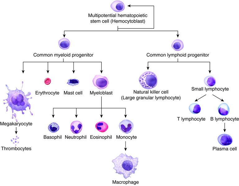
Hematopoiesis & Stem Cells
hematopoiesis (formation of blood cells) occurs in red bone marrow of body
all the different types of blood cells can be traced back to stem cells in bone marrow
myeloid progenitor - most mature in myeloid tissues of red bone marrow in skull, vertebrae, & rib cage
RBCs
granular WBCs (neutrophils, eosinophils, and basophils)
platelets
monocytes
lymphoid progenitor - typically mature in lymphoid tissues throughout body, like spleen, tonsils, and lymph nodes
T and B lymphocytes
Cells of the Vessels
endothelial cells - line the inside of any surface that comes in contact w/ blood (inside of heart and every blood vessel)
vascular endothelium - line blood vessels
endocardium - endothelium on interior surfaces of heart chambers
only one layer thick
functions:
mechanical-chemical stimulation - sense changes in local chemical/mechanical environment and signal underlying muscle cells
immune response - cytokines released by immune system activate endothelial cells to promote association with circulating immune cells; pores open between endothelial cells to allow immune cells to leave lumen of vessel and invade tissue
forming new blood vessels (angiogenesis)
vascular endothelial growth factor (VEGF) - stimulates endothelial cells to divide
cells break down extracellular matrix and form new blood vessels
preventing clot formation (thrombus prevention)
smooth muscle cells - form layer outside of endothelial cells
provide strength to blood vessels
allow for blood vessel adaptation (vasoconstriction and vasodilation)
adjust contraction based on sensed stretch or the concentration of various chemical factors
Extracellular matrix (ECM) of vessels
all vessels except capillaries have one or more layers of connective tissue
connective tissue layers provide structure, support, and determine how easily vessels with passively expand
tunica externa (adventitia) - outer layer of connective tissue
internal elastic lamina - only found in some vessels; found between endothelial layer and smooth muscle layer
elastin - gives blood vessel ability to deform and return to original form when internal or external pressures are applied
collagen - found in tunica externa and provides stiffness to blood vessel to prevent rupture
hematocrit (hct) = fraction of blood composed of RBCs
Muscle Cells of the Heart
3 types of cells that make up the myocardium:
pacemaker cells (nodal cells) - found within sinoatrial (SA) and atrioventricular (AV) nodes
conducting cells - carry action potentials at high rate from SA to AV node and then from AV node to the rest of the ventricular cells
myocardial cells - 99% of the mass of the heart; function is to contract
Vascular Tissue
3 vessel wall layers called tunics:
tunica interna - forms inner lining of blood vessels
endothelium in a continuous sheet
supported by basement membrane composed largely of collagen fibers providing strength and flexibility
basement membrane anchors endothelium to outermost component of tunica interna (internal elastic lamina)
internal elastic lamina - sheet of elastic fibers with numerous openings to allow movement of solutes between layers
tunica media
composed of smooth muscle and connective tissue layers
smooth muscle cells oriented to encircle the lumen of the vessel
smooth muscle cells regulate size of vessel lumen based on stimulus of associated neurons, circulating hormones, or local signaling molecules
connective tissue composed of elastic fibers interspersed within muscle cell layers and a separate sheet-like layer (external elastic lumina)
tunica externa - outermost layer
composed mostly of collagen fibers
contains many nerves and (in larger vessels) a system of vessels that supply the vessel tissues called the vasa vasorum
The Heart
heart made of 2 pumps connected by series of tube-like vessels that create circulatory system
pumps separated by thick wall called the septum
valves of heart prevent blood from flowing in opposite direction
AV valves - separate atria from ventricles
tricuspid valve - separates right atrium from right ventricle
has 3 flaps (cusps)
on vetricular side, has long muscular cords (chordate tendinae) extending from inferior side of each cusp
attach to cusp on one end and papillary muscle on the other end
papillary muscles prevent valves from inverting back into atria during ventricular contraction
ventricular pressure causes flap to close
bicuspid valve (mitral valve) - separates left atrium from left ventricle
semilunar valves - located between ventricles and arteries
pulmonary semilunar valve (pulmonary valve) - remains closed when ventricle is filling and opens during ventricular contraction because pressure is greater inside ventricle
aortic semilunar valve (aortic valve)
pericardium - fluid-filled membranous structure that separates heart from lungs; made up of visceral pericardium (outer epicardial layer of heart) and parietal pericardium
heart has 3 layers:
endocardium - innermost layer forming barrier between blood and muscle
myocardium - thickest layer formed of cardiac muscle
epicardium - thin layer of cells covering outside of heart
cardiac tamponade - infection of pericardium that leads to increased pressure exerted on exterior of heart and prevents heart from filling fully
Blood Vessels
arteries -> arterioles -> capillary networks -> venules -> veins
aorta is divided into:
ascending aorta
coronary arteries branch from here
aortic arch
brachiocephalic trunk - right upper limb, head, neck
right subclavian artery - right upper limb, brain, spinal cord, neck, shoulder, thoracic viscera and wall, scapular muscles
right vertebral artery - posterior part of brain
axillary artery - passes through armpit
brachial artery - passes through upper arm
radial artery - passes through lateral forearm
ulnar artery - passes through medial forearm
right common carotid artery - head and neck
left common carotid artery - head and neck
left subclavian artery - similar branching to right subclavian artery
thoracic aorta - supply structures of the thorax
abdominal aorta - supply structures of the abdomen and lower limbs
bifurcates into right and left common iliac arteries
brain has special arterial circle (circle of Willis)
provides collateral pathways to ensure uninterrupted blood supply to brain if some arteries are blocked
arterial supply of thorax and abdomen has both visceral (serving the organs) and parietal (serving the wall structures) branches
Arteries
arteries can be classified by function:
elastic arteries (conducting arteries) - vast capacity for expansion and retraction
arteries with largest diameters in the body are elastic arteries
temporarily store blood by expanding and slowly recoiling
muscular arteries (distributing arteries) - higher percentage of muscle cells (thick tunica media)
Aging in Circulatory System
arteriosclerosis - conditions that make the arteries less elastic (hardening of the arteries); increases resistance to blood flow
calcific arteriosclerosis - calcium deposition in walls of vessels
senile arteriosclerosis - degenerative loss of elastic fibers with age
atherosclerosis - formation of plaques on arterial walls making them stiffer
plaques are made of fatty material containing cholesterol
may eventually be replaced with connective tissue and deposits of calcium
plaques can rupture, initiating a blood clot at the site
high levels of LDL ("bad" cholesterol) can lead to development of atherosclerosis
congestive heart failure - caused by impaired pumping efficiency in the heart
Arterioles/Capillaries/Venules
arterioles - tunica interna and tunica externa are thin; tunica media has 1-2 layers of muscle cells
rich supply of sympathetic nerve fibers
arterioles constrict when they receive a high number of signals from the sympathetic nervous system, and vice versa when they receive less signals
hormonal signals and local signaling molecules can also lead arterioles to constrict/dilate
capillaries - form extensive networks throughout the entire body
have only a tunica interna to facilitate rapid exchange of gases and solutes
3 types of capillaries:
continuous capillary
lowest permeability rate
common in muscle, nervous, and connective tissues
smooth tubes with narrow intercellular clefts between endothelial cells
basement membrane is whole and without pores
small molecules like glucose and oxygen can readily pass between cells, but larger molecules cannot
fenestrated capillary
have numerous fenestrae (windows) - plasma membranes from opposite sides of a cell adhere and exclude cytoplasm
membranes of fenestrations are composed of material more porous than typical phospholipid bilayer
basement membrane has openings or pores that increase permeability of vessels
allow for faster solute exchange but still exclude larger molecules from passing through
found in areas requiring rapid absorption or filtration, such as small intestines, kidneys, and choroid plexuses of brain
sinusoid capillary
limited to areas where blood cells move in and out of the bloodstream (found where blood cells are formed -- red bone marrow -- and where large proteins enter the bloodstream -- liver)
endothelial cells are widely separated and contain large pores without membrane coverings
basement membrane is sparse or missing
venules - much thinner walls than arteries and arterioles
arteries retain shape without blood but veins collapse
Veins
tunica interna and tunica media are quite thin
both internal and external elastic laminae are absent or very thin
capable of great expansion to hold variable volume of blood
venous valves - augment venous return; most commonly found in limbs
formed by extensions of tunica interna that create flaps in the vessel lumen
valves close when blood in a vein tries to move backward
venous sinuses - large-diameter veins with extremely thin walls and no smooth muscle
dense connective tissue surrounding the vessel provides support
collection points for oxygen-poor blood
includes coronary sinus of heart and dural sinuses of brain
anastomoses - connecting channels between veins
portal veins - drain one capillary network and transfer blood to another organ or tissue to empty into another capillary network
Seite 3
Homeostasis
autoregulation - tissue's ability to meet metabolic needs by automatically altering its blood flow
Hormonal Regulation
renin-angiotensin-aldosterone system
decrease in blood pressure or renal blood flow stimulates cells in kidney to secrete renin
pathway produces hormone angiotensin II, which increases BP:
vasoconstriction - leads to increased peripheral vascular resistance
causes kidney tubules to reabsorb sodium
high plasma angiotensin also triggers higher production of aldosterone by adrenal cortex
acts on kidneys to increase reabsorption of sodium ions
increased Na+ leads to enhanced water reabsorption by kidneys and causes total blood volume to rise, elevating BP
epinephrine and norepinephrine - sympathetic stimulation promotes release of these hormones by adrenal medulla
increase heart rate (chronotropy)
increase force of contraction (inotropy) - this increases stroke volume
these two factors combined increase cardiac output
increase in CO will lead to increase in arterial pressure
epinephrine/norepinephrine also constrict arterioles and veins in abdominal organs and skin while dilating vessels in heart and skeletal muscle
this results in increased blood flow to muscles during physical activity
antidiuretic hormone (ADH) - prompted by decreased blood volume or dehydration
produced in hypothalamus and stored in posterior pituitary
causes kidney to retain water
retention leads to increased blood volume and increasing preload
at very high levels, it causes peripheral blood vessels to constrict to help raise BP
atrial natriuretic peptide (ANP) - secreted by myocytes of atria
decreases BP:
dilates blood vessels
reduces blood volume by increasing excretion of salt
Informationen
- Beschreibung
- TEAS A&P (Cardiovascular System) Notiz am Cardiovascular System Notes, erstellt von Arielle Solcher am 05/03/2019.
- Erstellt von
- Arielle Solcher · Mär 2019
- Aktualisiert
- 12 Mär 2019
- Aufrufe
- 0
- Bewertung