
Mind map
Left-Sided Heart Failure
Map content
Left-Sided Heart Failure
Main Causes (American Heart Association, 2017))
- Coronary Artery Disease
- The coronary arteries become narrowed or blocked, which reduces blood flow to the heart (Heart & Stroke Foundation, 2018)
- High Blood Pressure
- The Blood pressure in the arteries is elevated and the heart must work harder than normal to pump blood through the vessels (Heart & Stroke Foundation, 2018)
- Previous Heart Attacks (myocardial infarction)
- Depending on how long the blood supply is cut off during a MI, the damage can be mild, severe or cause lifelong problems (Heart & Stroke Foundation, 2018)
- Other Causes (Heart & Stroke Foundation, 2018)
- Excessive Use of Alcohol & Drugs
- Diabetes
- Being obese or overweight
- High Blood Cholesterol
- Heart Valve Disease
- Myocarditis
- Heart muscle disease of unknown causes
- Other Medical Conditions; thyroid diseases or anemia
- Coronary Artery Disease
Classification
- The Left ventricle of the heart supplies most of the heart's pumping power and is the largest chamber making it essential for normal function (American Heart Association, 2017)
- Left-sided HF is the most common form of initial HF and results from left ventricular dysfunction. This causes blood to back up through the left atrium and into the pulmonary veins. This increased pulmonary pressure causes fluid extravasation from the pulmonary capillary bed into the interstitium and the the alveoli, which is then manifested as pulmonary congestion and edema (Lewis 2014)
- There are two types of Left-sided HF (American Heart Association, 2017)
- Heart Failure with reduced ejection fraction (HFrEF), also called systolic failure:
- The left ventricle loses its ability to contract normally, which means the heart cannot pump with enough force to push enough blood into circulation
- Heart Failure with Preserved ejection fraction (HFpEF), also called diastolic failure:
- The Left ventricle loses its ability to relax normally due to the muscle becoming stiff. This makes it so the heart cannot fill properly with blood during the resting period between each beat
- Heart Failure with reduced ejection fraction (HFrEF), also called systolic failure:
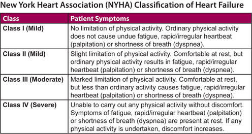
- The Severity of Heart failure based on symptoms is classified using the New York Heart Association Functional Classification (American Heart Association, 2017)
- Figure 1: (Hamilton Cardiology Associates, 2018)
- There are two types of Left-sided HF (American Heart Association, 2017)
- Left-sided HF is the most common form of initial HF and results from left ventricular dysfunction. This causes blood to back up through the left atrium and into the pulmonary veins. This increased pulmonary pressure causes fluid extravasation from the pulmonary capillary bed into the interstitium and the the alveoli, which is then manifested as pulmonary congestion and edema (Lewis 2014)
- The Left ventricle of the heart supplies most of the heart's pumping power and is the largest chamber making it essential for normal function (American Heart Association, 2017)
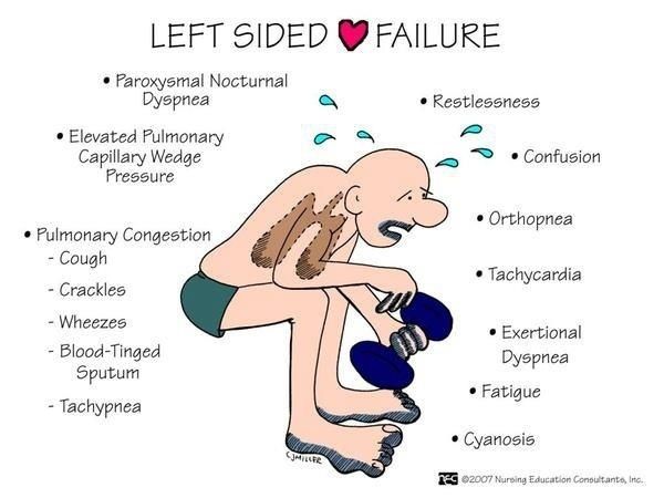
Manifestations (Lewis, 2014; McMaster Pathophysiology Review, 2018)
- Signs
- LV heaves
- Cheyne-strokes respirations
- Pulsus alternans
- alternating pulses: strong - weak
- Increased Heart Rate
- Crackles
- Pulmonary edema
- S3 and S4
- Decreased PaO2, slight increased PaCO2
- Poor O2 exchange
- Cardiac "asthma"
- Coarse ronchi and wheezing caused by compression of conduction airways by pulmonary congestion
- Mitral Regurgitation Murmur
- cardiomegaly
- chronically increased workload and excessive volume cause ventricular dilation and hypertrophy
- PMI displaced inferiorly and posteriorly
- LV hypertrophy
- Symptoms
- Fatigue
- Dyspnea
- shallow respirations < or = 32-40/min
- Orthopnea
- SOB in recumbent position
- Dry Hacking cough
- Pulmonary Edema
- Nocturia
- Paroxysmal nocturnal dyspnea
- Confusion and/or impaired memory
- Decreased Urine Output
- Chest pain/pressure
- Palpitations
- Cachexia
- Figure 3: (Google images, 2016)
- Signs
Diagnostic Studies (Lewis, 2014)
- Brain natriuretic peptide
- a biomancer used by left ventricle when there is excessive pressure due to hear failure
- < 100 = no heart failure; 100-300 = HF present
- >300 = mild: >600 = moderate: >900 = severe HF
- < 100 = no heart failure; 100-300 = HF present
- a biomancer used by left ventricle when there is excessive pressure due to hear failure
- Chest x-ray
- if there is any congestion, enlarged
- echocardiogram
- Nuclear stress test
- Health History & Physical Examination
- CBC, Cardiac enzymes, thyroid function studies, and LFT
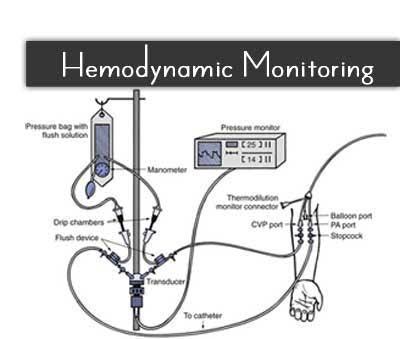
- Hemodynamic Monitoring
- Figure 5: (Google images, 2017)
- Figure 4: (Google images, 2015)
- Brain natriuretic peptide
Treatment & Managment
- Administering Medications (Lewis, 2014; Lilley, Collins, & Synder, 2014)
- Decrease preload
- Diuretics
- Side effects
- Allergic reaction, low or high K+, ototoxicity, nephrotoxicity
- Nursing Consideration
- Assess client's vitals especially BP, Kidney function tests, allergies, sigs of ototoxicity , K+ and Na
- Side effects
- Diuretics
- Decrease afterload
- ACE inhibitors and ARBS
- Side effects
- nagging cough, low BP, high K+, allergic reactions, mood changes, angioedema, fatigue, dizziness, headache
- Nursing Consideration
- Assess for allergies, BP, electrolytes, especially K+, Kidney function tests
- Side effects
- ACE inhibitors and ARBS
- Reduce anxiety
- Morphine
- Side effects
- Low RR, sedation, nausea, vomiting, constipation, hypotension, allergic rxns such as itching and rash
- Nursing Consideration
- Assess for anxiety, allergic rxn, signs of respiratory depression, encourage clients to eat food that are high in fiber, monitor clients vitals, encourage turning and deep breathing
- Side effects
- Morphine
- Improve cardiac function
- eg digoxin
- side effects
- GI upset, headache, Colored vision, halo vision, flickering lights low K+, low Mg, high Ca, narrow therapeutic index
- Nursing consideration
- narrow therapeutic index so monitor Blood levels, K+, HR, BP, cardiac rhythms, signs of toxicity, avoid foods high in fiber, weigh daily
- side effects
- eg digoxin
- Decrease preload
- Collaborative Care (Lewis, 2014; Pasoz-Lopez et al., 2011)
- High fowler's position
- Administer O2 as required
- Cardiac monitoring and O2 sat
- Monitor BP, HR, RR, Urinary output q1h
- Monitoring IABP, PAOP, CO
- Weighing client's daily
- Na restriction to 2-3g
- Fluid restriction to 2-3 l
- Regular activity and rest
- Vaccination Flu+ pneumonia
- Smoking cessation and limiting ETOH
- Administering Medications (Lewis, 2014; Lilley, Collins, & Synder, 2014)
Client-Centered Care (Robbio, 2018)
- Understand that nursing is more than performing technical skills (RNA0, 2015)
- Establish an authentic relationship that is based on trust and respect (Messner, 1993)
- Value the uniqueness of every client (Robbio, 2018)
- Embody holistic and caring attitude (Cara, 2003)
- Implement Watson's theory of Human Caring such as adopting caring consciousness (Cara, 2003)
- Recognize that clients are the expert of their lives (Robbio, 2018)
- Support client's healthcare decisions (Robbio, 2018)
- Adopt a non-judgemental attitude (Robbio, 2018)
- Involve the family in the care (RNAO, 2015; Robbio, 2018)
- Figure 6: (Google images, 2018)
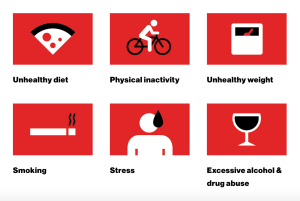
Risk Factors (Heart & Stroke Foundation, 2018)
- Unhealthy Eating
- Unhealthy Weight
- Not enough exercise
- Smoking (Tabacco use)
- Too much alcohol
- Birth control and Hormone Replacement Therapy (HRT)
- Recreational Drug Use
- Stress
- Figure 2: (Heart & Stroke Foundation, 2018)
Images and files in the map

Information
- Description
- Nursing Mind Map on Left-Sided Heart Failure, created by Violet Biskup on 30/10/2018.
- Created by
- Violet Biskup
- Views
- 20
- Rating