253749
Descripción
Mapa Mental por Emma Jones, actualizado hace más de 1 año
|
|
Creado por Emma Jones
hace más de 10 años
|
|
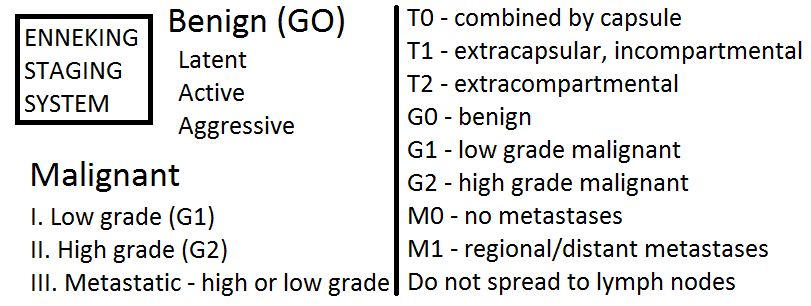
Soft tissue
tumours:
- Muscle, fat, fibrous tissue, blood vessels, peripheral nerves
- Benign lesions
- Lipoma
- Benign adipose tissue tumour, usually subcutaneous,
sites where adipose tissue is normally present
- Middle/late adult life
- Well circumscribed, resemble mature fat
- Subtypes: fibrolipoma, chondroid lipoma, spindle
cell lipoma, pleomorphic lipoma, angiolipoma
- Benign adipose tissue tumour, usually subcutaneous,
sites where adipose tissue is normally present
- Ganglia
- In vicinity of joints, sometime tendon sheaths
- Myxoid softening and cystic degeneration
of tissues of joint capsule or tendon sheath
- Dorsal hand, wrist, foot, ankle, knee, spine -->
not lined by true synovium, do not communicate
- In vicinity of joints, sometime tendon sheaths
- Lipoma
- Aggressive benign
- Fibromatoses
- Well differentiated fibroblastic proliferations, infiltrative
growth pattern, aggressive, repeated local recurrence
- Classification
- Deep/musculo-aponeurotic/desmoid tumours
- 5-10cm, firm,
grey, scar-like
- Spindle cells, fibroblastic,
collagen stroma, infiltrative growth
- 5-10cm, firm,
grey, scar-like
- Superficial: Dupytron's contracture
- Deep/musculo-aponeurotic/desmoid tumours
- Well differentiated fibroblastic proliferations, infiltrative
growth pattern, aggressive, repeated local recurrence
- Fibromatoses
- Pseudosarcomatous
- Nodular fasciitis
- Young adults --> arms, trunk, neck
- Rapid growth of tender mass, associated with
fascia --> infiltration of subcutis, muscle, fascia
- Spindle cell proliferation, stroma rich with ground substance,
infiltrative, mitotically active, no malignant potential
- Young adults --> arms, trunk, neck
- Nodular fasciitis
- Liposarcoma
- Adult --> thigh, retroperitoneum
- Large, well-circumscribed, unencapsulated
- Low grade: myxoid,
well-differentiated
- High grade: round cell, pleomorphic
- Adult --> thigh, retroperitoneum
- Synovial sarcoma
- 5-10% of soft tissue
sarcomas, high grade
- Young adults, M>F, 80% knee/ankle
- Other sites: head, neck,
retroperitoneum, mediastinum
- Enlarging mass, pain,
infrequent systemic symptoms
- Well circumscribed, pink/grey
- Monophasic or biphasic,
poor differentiation
- Biphasic: carcinomatous & sarcomatous
- Biphasic: carcinomatous & sarcomatous
- t(x;18)
- Poor prognosis
- 5-10% of soft tissue
sarcomas, high grade
- Diagnosis takes into account
history, location, radiological
apperance --> differential diagnosis
- Prebiopsy planning required -->
inappropriate biopsy leads to wrong
diagnosis or healing complications
- Fine needle aspiration
- Diagnostic closed core biopsy
- Open biopsy/curettage
- Formal resection
- Open biopsy creates complication, but less invasive
procedures might not obtain enough tissue for diagnosis
- Fine needle aspiration
- Diagnosis of
chromosomal
abnormality
- Prebiopsy planning required -->
inappropriate biopsy leads to wrong
diagnosis or healing complications
Recursos multimedia adjuntos
¿Quieres crear tus propios Mapas Mentales gratis con GoConqr? Más información.