
Flashcards
microbiology

Cards in this deck (50)
- 1Classification of bacteriaThe physical difference of bacteria relates to their: size, shape, cell wall structure and hence staining charecteristics- genetic difference produces different metabolic features and different surface molecules- so they have different surface properties.
- 2Classification of bacteria- prokaryotesProkaryotes vary a lot in size- smallest = archea- the size of the smallest cells is limited by size of molecules needed for life.
- 3staphyloccocus aureus
- 4streptococcus pyogenes

- 5rest of bacteria images

- 6classification by shapea genus of a bacteria has 3 main shapes- shape sometimes is indicated in its name
Bacillus or rod shaped-Eschericha; bacillus
Coccus or spherical- staphyloccocus; streptococcus
Spiral or corkskrew- spirillum - 7Further identification...According to the way bacteria groups- may be single- helicobacter, in pairrs eg Diplococcus pneumoniae or in chains eg Streptococcus or in clusters eg Staphylococcus
- 8Classiication by gram stain reactionGram stain allows people to distinguish between gram positive and gram negative bacteria
- 9Difference between gram positive and gram negative bacteriaThe cell wall of all bacteria is a 3 dimensional network of polysaccharides and polypeptides, known as peptidoglycan or murein- cross linking of these molecules provides strength- gives cell its shape- protects against swelling or bursting caused by osmotic uptake of water- +ve has this basic cell wall structure whereas gram negative has an extra layer of lipopolysaccharide
- 10So why does +ve and -ve stain differentlyGram positive bacteria have cell walls with a thicker layer of peptidoglycan/murein (than Gram-negative bacteria), which retains the crystal violet/iodine complex within their cells when washed with alcohol - staining purple.
- 11more on difference between +ve and -ve- staining/ positiveOn treatment with alcohol, the Gram negative cell walls lose their outer lipopolysaccharide membrane, and the thin inner peptidoglycan layer is left exposed, this means that the crystal violet/iodine complexes are washed from the gram-negative cell along with the outer membrane - they stain red with the counterstain safranin
Absence of outer layer means +ve can bind stain efficiently but does make them more succeptible to penicillin and enzyme lysozyme - 12more on difference between +ve and -ve- negativeGram negative's cell wall is supplemented with large molecules of lipopolysaccharide that protect cell- exclude dyes like crystal violet
lipopolysaccharide protects peptidoglycan below- more resistant to penicillin- needs different antibiotics which interfere with cells ability to make proteins - eukaryotic cells also make proteins but protein making machinery is different- unaffected by antibiotics - 13Lysozome effectBacteria constantly makes/ breaks chemical links in cell walls- lysozyme hydrolyses these bonds holding peptidoglycan molecules together
- 14Penicillin effectPrevents bonds interlinking peptidoglycan molecules from forming- significant when bacteria make new cell walls when they divide- makes the cell walls weak and easy to collapse- water uptake by osmosis bursts the cell
- 15gram staining

- 16gram positive vs negative structure

- 17chemicals used in gram staining stepsCrystal violet- basic dye- binds to peptidoglycans so all bacteria stain purple
Lugol's iodine- Mordant- Binds crystalviolet to peptidoglycan more strongly
Acetone alcohol- Decolouriser- removes unbound crystal violet and lipopolysaccharide- -ve loses stain goes colourless, +ve stays purple
Sutamin- counter stain- -ve- red, +ve- stays purple - 18Conditions necessary for culturing bacteria in labBacteria reproduce asexually by binary fission and can be cultured on a nutrient rich agar jelly or in a nutrient broth. These growth media must include: ● A suitable carbon source, usually glucose or lactose ● A suitable nitrogen source such as ammonium or amino acids ● A source of sulphur and phosphorous ● Vitamins, minerals and growth factors ● A suitable pH ● The growth media should be incubated at a suitable temperature – 25oC ● Different species vary in their requirements and usually grow over a range of temperature and pH values, with an optimum within the range
- 19nutrientsglucose/ lactose- glucose= easier to break down- monomer-
carbon- needed for making up organisms eg proteins, lipids, carbohydrates- carbon mostly provided in organic form (glucose not CO2- bacteria adapted to living on organic (living or once living material) - 20Nutrients pt 2nitrogen needed for proteins, DNA, RNA- usually given in organic form (proteins) or inorganic form (nitrogen gas or ammonia)
growth factors- essential substances that the bacteria can't synthesize itself from simpler substances- needed in tiny amounts- vitamins to make enzymes, mineral salts, amino acids - 21The effect of oxygen●Obligate aerobe require oxygen for metabolism
●Obligate anaerobe metabolism is inhibited in the presence of oxygen.
●Facultative anaerobes grow best in the presence of oxygen, but can respire anaerobically if they need to - 22How to classify bacteria- oxygenObligate aerobes- bacteria are clustered at top of tube as they need oxygen for respiration and growth (O2 diffuses from air into growth medium)
Obligate anaerobes- bacteria are at bottom of tube, away from their source of oxygen
Facultative anaerobes- bacteria is found throughout tube but mainly toward top can grow without O2 but grow best with it - 23Aseptic techniques are used to prevent...Contamination of the environment by the microorganisms being handled. Contamination of the bacterial cultures by unwanted microorganisms from the environment
- 24Aseptic TechniqueAseptic TechniquesAseptic techniques are used to prevent: ➔Contamination of the environment by the microorganisms being handled. ➔Contamination of the bacterial cultures by unwanted microorganisms from the environment.● Use a petri dish of sterile nutrient agar jelly.● Open the culture bottle of the bacteria.● Flame the neck of the culture bottle.● Use a sterile pipette or syringe.● Place 0.1 ml of culture onto the agar.● Flame the spreader.● Allow spreader to cool.● Distribute culture evenly over the surface of the agar.● Place all apparatus into disinfectant after use.● Autoclave all instruments to sterilise them
- 25Aseptic technique reasonshands washed before/ after exp- remove micro organisms
surface cleaned before/ after exp- kill microbes
autoclave or pressure cooker- sterilise glassware
sterile petri dishes of nutrient agar kept close until needed/ lid held over dish during inoculation-prevent entry of contaminants
wire loop flamed before use- kill unwanted microbes
innoculated dish taped- prevent entry/ exit of microbes - 26Sterilisation of equipmentAll equipment and growth media which come into contact with microorganisms being cultured (grown) must be sterile. Examples of sterilisation include: ● Passing metal transfer tools (such as inoculating loops) through a roaring/blue Bunsen flame until they glow red. ● Using pre-sterilised petri dishes. ● Sterilising any glassware used under high pressure and high temperature (121oC) in an autoclave for 15 minutes. ● Heating the nutrient agar used for the plates in an autoclave to sterilise it before pouring it into the plates and letting it set
- 27Pouring sterile agar plate using aseptic techniqueTo prepare a sterile agar plate you would need a sterile Petri dish, molten nutrient agar which has been autoclaved, Bunsen burner and tape. Open the culture bottle cap using your little finger and do not place the cap or bottle on the bench. Flame the neck of the bottle in a blue Bunsen flame. Work close to the Bunsen flame as the updraft helps prevent contamination. Open the sterile Petri dish lid at an angle. Pour in the molten nutrient agar and close lid immediately. Swirl gently to remove air bubbles. Secure lid with tape. Inoculating a set nutrient agar plate
- 28Innoculating nutrient agar plate with bacteria growing in milkA – Sterilise the inoculating loop in a roaring blue Bunsen flame until it glows red. B – Dip the inoculating loop into the milk sample. C – Hold the Petri dish lid at an angle to reduce contamination by microorganisms in the air. Use the loop to spread the droplet of milk across the surface of the agar in a zig-zag pattern while rotating the plate. D – Tape the lid shut to secure it in place; the lid should not be completely sealed as anaerobic conditions encourage pathogen growth. Incubate the plate at 25oC – never 37oC as this would also encourage pathogen growth
- 29Lag PhaseVery Low or no reproduction
Cells begin synthesizing enzymes, DNA etc needed for cell division - 30Log PhaseExponential growth ● Resources are plentiful + optimal conditions. ● Enzymes and factors for growth and division are available● The population will rapidly increases●No limiting factors (surplus of resources)● Rate of reproduction >> rate of mortality
- 31Stationary phaseDue to: ● a decline in food supply● a build up of waste products● an increase in competition (intraspecific or interspecific).● Rate of Reproduction = Rate of Mortality
- 32Death phase● Nutrients are exhausted● Toxic excretory products accumulate. ● Population numbers decline rapidly until no living microorganisms remain.● Rate of mortality >>> (greater than) rate of reproduction
- 33Bacterial growth curve

- 34Counting bacteria and estimating population sizeBacteria grown in liquid culture (nutrient broth) can be counted directly (by counting each cell) or indirectly by measuring turbidity (cloudiness of the culture medium) with a colorimeter. Direct counts:Total counts include both living and dead cells. Viable counts only count living or actively growing cells and therefore underestimates population size.
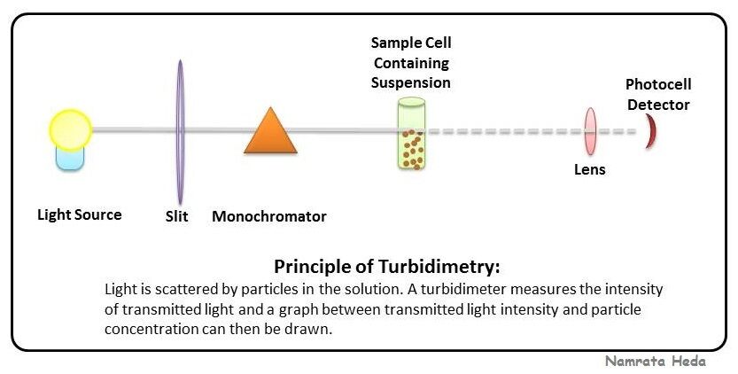
- 35Measuring number of bacteria - turbidimetryTurbidity- cloudiness of a liquid
A colorimeter is used to measure the cloudiness of a culture.● This method will measure both living and dead cells.● The more cells – the more cloudy (increase in turbidity).● Find absorbance of suspension- then read a standard graph of light absorbance plotted against no. bacteria cells - 36Turbidimetry diagram
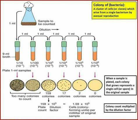
- 37One cell one colony theoryA colony is a cluster of cells (all clones) arising from a single bacterium by asexual reproduction or fungal spore. ➔One cell - one colony theory
- 38Dilution method reasonViable counts count cells which are able to grow into visible colonies on an agar plate. Population density of the sample may be too high to count; if this is the case a dilution technique is used.
- 39Serial dilution/ dilution plating method1. Fill 5 test tubes with 9 cm3 sterile water using a sterile pipette.
2. Add 1 cm3 of your sample to the first tube; this is a 1 in 10 dilution or 10 -1.
3. Mix the 10 -1 dilution thoroughly and pipette 1 ml into the second test tube; this is a 1 in 100 or 10 -2 dilution.
4. Repeat this process until you reach a 1 in 10 000 dilution (10 -4).
5. Plate each dilution on nutrient agar using aseptic techniques - 40Serial dilution/ dilution plating method pt 2● Plates with good separation (isolated)of serially diluted individual bacterial colonies - colonies are easily counted on such a plate.● Too many to count - merged colonies (overlapping) cause non-reliable results for statistical analysis● Too few colonies to provide reliable results Population in 1cm3 of original culture = No. of colonies x Dilution factor
- 41Serial dilution/ dilution plating method - calculations/ limitationsPopulation in 1cm3 of original culture = No. of colonies x Dilution factor
This number is usually an underestimate as it does not include dead or non-viable bacteria- we cannot be sure that each colony has grown from a single bacterium (bacterial cells may have been clumped together). - 42Serial dillution/ dillution plating diagram
- 43Importance of temperature/ PHOptimum temperature- Allows proteins in the cell to function- enzymes/ allows diffusion of substances in cell for metoballic processes
PH- enzymes for biochemical reactions which cause growth. - 44More definitionsAseptic technique- Labratory practice that maintains sterility in apparatus and prevents contamination of equipment and environment
Gram stain: A method of staining cell walls of bacteria as an aid to their identification
Pathogen- An organism that’s causes disease in their host
Colony- cluster of cells, or clone- arises from single bacterium or fungal spore by asexual reproduction - 45MathsDillution= 1/concentration
Population= no.colonies x dilution factor/ volume of sample eg (1cm^3)
Dillution factor eg concentration of 10^-1= factor of 10 - 46Q: The average number of cells in the field of view was found to be 156 and the radius of the field of view was 0.09mm. Calculate the number of cells in 100mm2. The area of microscopic field of view = πr 2 where π is 3.14 and r is the radius of the field of viewAccept any answer in the range 613 000 - 624 000 = 3 marks Accept in standard form
If incorrect award 2 marks for either
100/(0.09 × 0.09) × 3.14 ( π) × 156
100/0.025(43) × 156
If incorrect award 1 mark for area calculation 0.09 × 0.09 × 3.14 (π) - 47Using a haemocytometerMore accurate method of colony counting- uses a specialised microscope slide called haemocytometer- can’t distinguish between living and dead- total count
- 48How to describe serial dillutionSerial dilution involves the process of taking a sample and diluting it through a series of standard volumes of sterile diluent, which can either be distilled water or 0.9 % saline.
Then, a small measured volume of each dilution is used to make a series of pour or spread plates.- then give example - 491) Why are animal cells not affected by penicillin 2) what May bacteria not be able to be cultured1) Don’t have cell wall
2) May be: murualistic- needs other species, be intracellular parasites, have very particular growth requirements, have long generation time, be poisoned by media components - 50Another way growth media can be estimatedRegularly measuring diameter of bacteria colony spreading from central point over surface of growth medium
Information
- Description
- A level Biology Flashcards on microbiology, created by Darcey Griffiths on 07/08/2024.
- Created by
- Darcey Griffiths
- Views
- 18
- Tags
- Rating