
Flashcards
Cardiovascular - General knowledge flashcards
Cards in this deck (53)
- 13 components of the circulatory systemblood, blood vessels, heart
- 23 types of blood vesselsArteries
Veins
Capillaries - 3ArteriesCarry blood away from the heart, divide into smaller vessels called arterioles, carry oxygenated blood, with the exception of the pulmonary artery
- 4VeinsCarry blood to the heart, divide into smaller vessels called venules, carry deoxygenated blood, with the exception of the pulmonary vein
- 5CapillariesTiny vessels that link arteries and veins, thin walls aid diffusion to cells
- 6Tough, inelastic protein (fibre) in arteries and veinsCollagen (prevents walls from over-expansion)
- 7Middle layer in arteries and veinsMuscle and elastic fibre
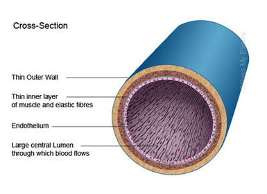
- 8Inner single layer of living cells surrounding the lumen/hollow core (epi = above, so under/inside = ?)Endothelium
- 9—Vein
- 10—Artery

- 11Capillary walls are ___________ and are made of ______permeable ; single layer of endothelium cells
- 12Blood pressureThe force the blood exerts against the wall of a blood vessel
- 13Function of valvesTo prevent the backflow of blood
- 14systoliccontraction
- 15diastolicrelaxation
- 16average systolic pressure110 -140 mm Hg
- 17average diastolic pressure75-80 mm Hg
- 18Location of the heartBetween the two lungs (slightly to the left side of the chest) just above the diaphragm in the thoracic cavity.
- 19Muscle of the heartCardiac muscle
- 20Double membrane surrounding the heartPericardium
- 21Fluid in the pericardiumPericardial fluid
- 22What type of pump is the heart?A double pump
- 23Which ventricle is thickest?Left (pumps blood all around the body)
- 24Wall that divides the heartInterventricular septum
- 25Four chambers of the heartTwo atria
Two ventricles - 26Thickness of walls in the atriaThin
- 27Tough chords / heart stringsTendons (chordae tendinae)
- 28Tendons are attached to the heart wall by projections calledpapillary muscles
- 29Valve on the right side of the heartTricuspid valve
- 30Valve on the left side of the heartBicuspid valve
- 31Valve that allows blood to flow into the pulmonary arteryPulmonary semilunar valve
- 32Valve that allows blood to flow into the aortaAortic semilunar valve
- 33Deoxygenated blood enters the heart through theVenae Cavae
- 34Deoxygenated blood flows out of the heart to the lungs through thepulmonary artery
- 35Oxygenated blood enters the heart through thepulmonary veins
- 36The oxygenated blood flows out of the heart and around the body through theaorta
- 37The cardiac muscle is supplied with blood from thecoronary/cardiac arteries branching from the aorta at the point where it leaves the heart, just beyond the semilunar valve
- 38two circuitsPulmonary circuit
Systemic circuit - 39Pulmonary circuitthe blood is pumped to the lungs to lose carbon dioxide and gain oxygen and is then returned to the heart
- 40Systemic circuitheart - body - heart
- 41Heartbeat is controlled by thePacemaker
- 42What is the pacemaker and where is it located?A small bundle of specialised tissue located close to the entry of the superior vena cava within the right atrium wall
- 43The pacemaker sends outregular electrical impulses
- 44AV nodeatrio-ventricular node
- 45function of the pacemakerto control the rate of the heartbeat
- 46A record of the electrical activity of the heartECG (electrocardiogram)
- 47Nerves connecting the pacemaker to the brain (one reduces rate of heartbeat, the other increases)Medulla oblongata
- 48SA node / pacemakerSino-atrial node
- 49noise made when the bicuspid and tricuspid valves closelub
- 50noise made when the semilunar valves closedub
- 51filling phasediastole (approx 0.4 secs)
-relaxation (passive) - 52emptying phasesystole (approx 0.4 secs)
1. Atrial systole (0.1 secs)
2. Ventricular systole (0.3 secs) - 53Exerciseincreases heart rate which strengthens the cardiac muscle making it more efficient at pumping blood, this improve the oxygen supply to the cardiac muscle and reduces blood pressure
Information
- Description
- Basic revision for the heart and blood vessels structure and function
- Created by
- Antonia Smith
- Views
- 7
- Tags
- History
-
- Created by eimearkelly3 · Dec 15, 2015
- Copied by Antonia Smith · Dec 15, 2015
- Rating