
Flashcards
Abdomen Procedure
Cards in this deck (123)
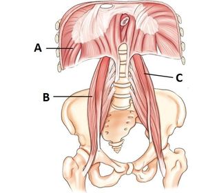
- 1—A. diaphragm
B. right psoas muscle
C. left psoas muscle - 2—A. right psoas
B. left psoas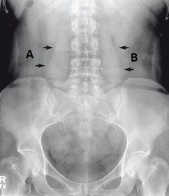
- 3—A. spleen
B. splenic flexure
C. descending colon
D. sigmoid colon
E. rectum
F. anus
G. vermiform/appendix
H. cecum
I. ileocecal valve
J. ascending colon
K. transverse colon
L. hepatic flexure
M. liver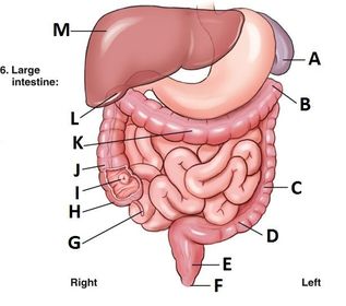
- 4—A. liver
B. spleen
C. stomach
D. pancreas
E. small intestine
F. large intestine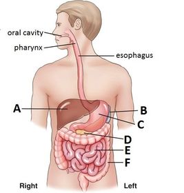
- 5—A. duodenum
B. jejunum
C. ileum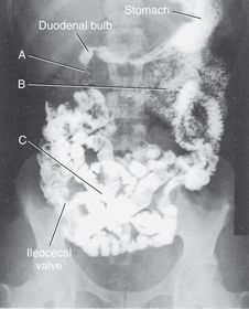
- 6—A. duodenal bulb
B. duodenum
C. jejunum
D. stomach
E. ileum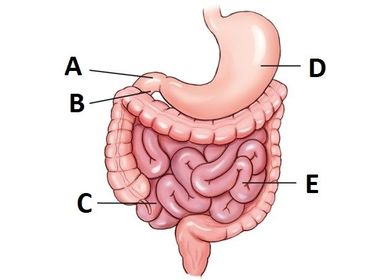
- 7—A. liver
B. spleen
C. pancreas
D. gallbladder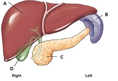
- 8—A. mesentery
B. small bowel
C. peritoneal cavity
D. parietal pleura
E. visceral pleura
F. left kidney
G. aorta
H. inferior vena cava
I. right kidney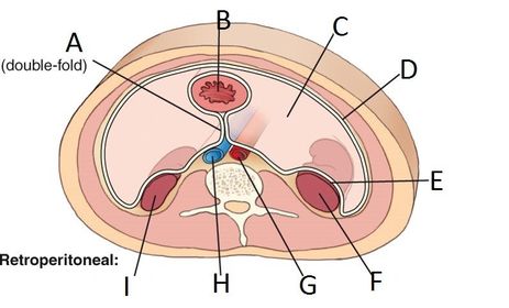
- 9—A. lesser omentum
B. greater omentum
C. peritoneal cavity
D. parietal peritoneum
E. visceral peritoneum
F. stomach
G. mesentery
H. small bowel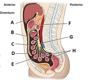
- 10—A. liver
B. stomach
C. greater omentum (pulled up over chest)
D. greater omentum
E. transverse mesocolon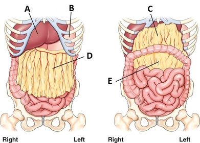
- 11—A. RUQ (right upper quadrant)
B. LUQ (left upper quadrant
C. RLQ (right lower quadrant)
D. LLQ (left lower quadrant)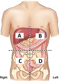
- 12—1. right hypochondriac
2. epigastric
3. left hypochondriac
4. right lateral (lumbar)
5. umbilical
6. left lateral (lumbar)
7. right inguinal (iliac)
8. pubic (hypogastric)
9. left inguinal (iliac)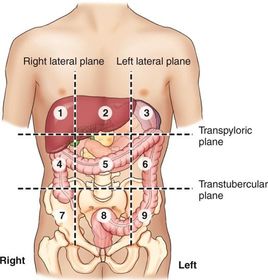
- 13—1. xiphoid process T9-10
2. inferior costal margin L2-3
3. iliac crest L4-5
4. ASIS
5. greater trochanter
6. symphysis pubis
7. ischial tuberosity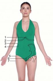
- 14—1. xiphoid process T9-10
2. inferior costal margin L2-3
3. iliac crest L4-5
4. ASIS
5. greater trochanter
6. symphysis pubis
7. ischial tuberosity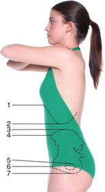
- 15—A. iliac crest
B. ASIS
C. ischial tuberosity
D. symphysis pubis
E. greater trochanter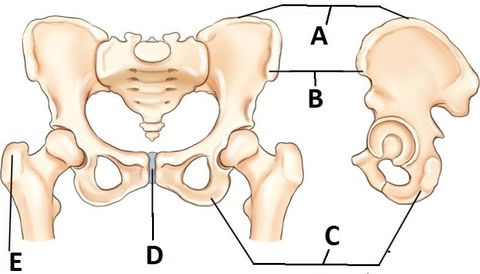
- 16Which organs are RETROPERITONEAL?SAID PUCKER:
suprarenal
aorta
inferior vena cava
duodenum
pancreas
ureter
colon (ascending/descending)
kidney
esophagus
rectum - 17Which organs are INTRA PERITONEAL?GLISS:
gallbladder
liver
intestines (ileum, jejunum, cecum, transverse, sigmoid)
stomach
spleen - 18Which organs are INFRA PERITONEAL?URL:
urinary bladder
reproductive organs
lower rectum - 19Which structures are located in the RUQ?liver
gallbladder
right kidney, adrenal gland, and ureters
head of the pancreas
duodenum - 20Which structures are located in the RLQ?ascending colon
cecum
ileum
vermiform (appendix) - 21Which structures are located in the LUQ?spleen
stomach
left kidney, adrenal gland, and ureter
tail of the pancreas - 22Which structures are located in the LLQ?jejunum (2/3)
sigmoid colon
descending colon - 23What are the routine abdomen procedures for a KUB?AP Supine
- 24What are the routine abdomen procedures for an acute abdominal series? (3-way)AP Supine
AP Erect
PA Erect Chest - 25What are the principal exposure factors for abdominal radiographs?- Analog/Digital kV (70-80)
- Short exposure time
- adequate mAs for sufficient density - 26Abdominal exposures are taken on ________, however some clinical sites take exposures on a small _______ to ensure the diaphragm is in the image.Expiration
Inspiration - 27movements such as breathing are classified as _____.Voluntary Movements
- 28movements like peristaltic action of the bowel are classified as ____.Involuntary Movements
- 29Which personal history questions should be asked before beginning an examination?1. Where is your abdominal pain?
2. When/where did the pain start?
3. Any previous films/surgeries?
4. LMP/Chance of pregnancy?
5. Any bowel problems, N/V? - 30How should the patient be prepared for an abdominal procedure?1. All clothing is removed
2. Opaque objects removed
3. Hospital gown on
4. pillow for head, clean linen on table
5. Cover patient for warmth/modesty - 31What should you do to set up the room before bringing the patient in?1. Set up technique
2. Align your tube, table/wall bucky
3. IR size and IR markers - 32When do you shield for an abdominal procedure?MALES: shield at upper edge of symphysis pubis
FEMALES: Shield ovaries/uterus only if it will not obstruct the desired anatomical view - 33What are some special/additional abdominal procedures?- PA Prone
- AP Lateral Decub
- AP Erect
- Dorsal Decub (lateral) - 34When would you perform this projection/position?AP SUPINE:
- viewing abdominal pathology
- bowel obstructions
- neoplasms
- calcifications
- ascites
- scout image for contrast studies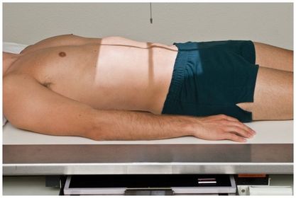
- 35—A. Liver
B. 12th Rib
C. Right Psoas
D. Sacrum
E. Coccyx
F. Symphysis Pubis
G. Left Femur
H. Obturator Foramen
I. Left Iliac Crest
J. L4 transverse process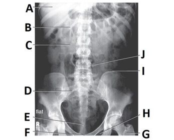
- 36Evaluation Criteria & TechniqueAP SUPINE:
SID 40 in
IR 14x17 lengthwise
Analog/Digital 70-80 kV
Grid
CR @ level of Iliac Crests perpendicular to midsagittal plane
- no motion/rotation
- must see symphysis pubis, psoas muscles, margins of kidneys and liver
- long scale contrast (kV) and sufficient exposure (mAs)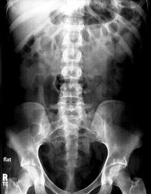
- 37Why would you perform this projection/position?PA PRONE:
- viewing pathology of the abdomen
- bowel obstructions
- neoplasms
- calcifications
- ascites
- scout imaging for contrast studies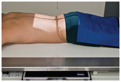
- 38Evaluation criteria & TechniquePA PRONE:
SID 40 in
IR 14x17 in legnthwise
Analog/Digital 70-80 kV
Grid
CR @ Iliac Crests perpendicular to midsagittal plane
- no motion/rotation
- kidneys and lower liver margins visible
- symphysis pubis visible
- long scale contrast (kV) adequate exposure (mAs)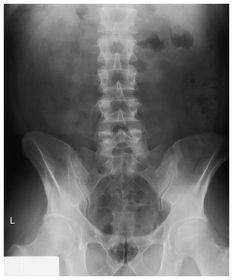
- 39When would you perform this projection/position?LEFT LATERAL DECUBITUS:
- viewing abdominal masses
- demonstrate air/fluid levels
- demonstrates intraperitoneal air
- best demonstrates intraperitoneal air in the RUQ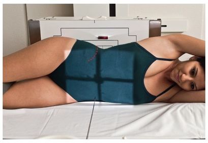
- 40Evaluation Criteria & TechniqueLEFT LATERAL DECUBITUS:
SID 40 in
IR 14x17 in crosswise
Analog/Digital 70-80 kV
Grid
CR horizontal & perpendicular to midsagittal plane, 2 in (5 cm) above Iliac Crests
- must view diaphragm, both sides ot the body, and both iliac wings
- no rotation/motion
- slightly less overall desnsity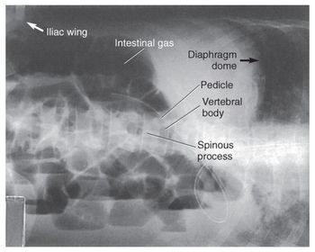
- 41The patient should stay up on his(her) side for a minimum of ___ min before left lateral decub exposure.5 min
- 42Ideally, the patient would be on his/her side for ______ before taking left lateral decub exposure.10-20 min
- 43Which markers would you use for a left lateral decubitus?mark which side is down
- 44When would you perform this projection/position?AP ERECT ABD:
- viewing abdominal masses
- demonstrating air/fluid levels
- demonstrates accumulations of intraperitoneal air under the diaphragm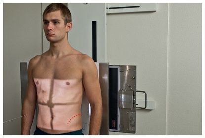
- 45Evaluation Criteria & TechniqueAP ERECT ABD:
SID 40 in
IR 14x17 in lengthwise
Analog/Digital: 70-80 kV
Grid
CR horizontal centered @ 2 in (5 cm) above Iliac Crests (perpendicular to midcoronal plane)
- bilateral diaphragm and iliac wings should be visible
- no rotation/motion
- ribs and gas bubbles should appear sharp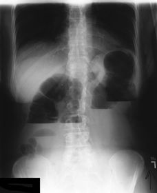
- 46If you cannot palpate the iliac crests for an AP erect ABD, you should collimate the top light border to the _____.Axillae
- 47A patient comes to Radiology for an acute abdominal series. The patient is ambulatory. Which position/projection should be done first?Always do the ERECT positions/projections first if patients are ambulatory.
- 48When would you perform this projection/position?DORSAL DECUBITUS (RIGHT LATERAL):
- aneurysms
- calcification of aorta or other vessels
- umbilical hernias
- abnormal masses
- accumulations of gas or air/fluid levels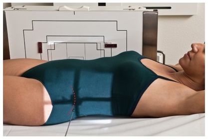
- 49Evaluation Criteria & TechniqueDORSAL DECUBITUS (RIGHT LATERAL)
SID 40 in
IR 14x17 crosswise
Analog/Digital 70-80 kV
Grid
CR perpendicular to the midsagittal plane centered @ 2 in (5 cm) above the iliac crests
- diaphragm and iliac wings visible
- no motion/rotation
- lumbar vertebrae may appear 50% underexposed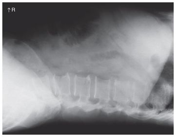
- 50Which markers would you use for the dorsal decubitus position?use the arrow to mark which side of the patient is DOWN
- 51When would you perform this projection/position?RIGHT LATERAL
- umbilical hernias
- abnormal masses
- location of foreign bodies
- viewing the prevertebral regions for possible aneurysms of aorta or calcifications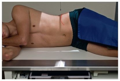
- 52Evaluation Criteria & TechniqueRIGHT LATERAL:
SID 40 in
IR 14x17 lengthwise
Analog/Digital 80-90 kV
Grid
CR perpendicular to table and midsagittal plane centered @ level of iliac crest
- diaphragm and lower abdomen visible
- no rotation/motion
- lumbar vertebrae may appear 50% underexposed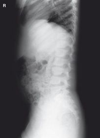
- 53Which projections/positions are included in a 3-way acute abdominal series?AP SUPINE
AP ERECT or LATERAL DECUB
PA CHEST - 54Why would you perform a lateral decub instead of an AP erect in the acute abdominal series?If the patient is too weak or ill to stand.
- 55Why would you perform an acute abdominal series?- diagnosing Ileus
- Ascites
- Perforated hollow viscus
- Intra-abdominal masses
- Post-op (abd surgeries) - 56The 2-way acute abdominal series includes the ________ and the ________ positions/projections.AP Supine
AP Erect or Left Lateral Decubitus - 57What's wrong with this image?- diaphragm is cut off
- CR is centered too low (RUQ cut off)
- collimation is not adequate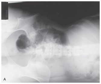
- 58What's the repeatable error on this image?- patient has pants on
- navel ring
there's a stent placed in the right ureter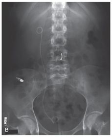
- 59What's the repeatable error in the image?- symphysis pubis is cut off
there are stones in the right kidney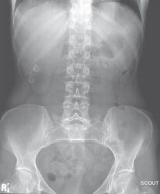
- 60What are the errors apparent on this image?- collimation and CR are too low
- diaphragm not entirely visible
- motion
- no marker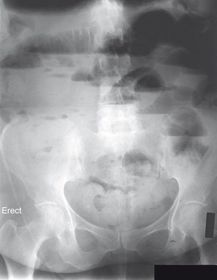
- 61How can you tell if contrast has been injected vs. ingested?INJETED: shows up in vessels, vasculature, and organs
INGESTED: shows up in bowels and intestines as it moves through the system - 62Which topographic landmarks may be institutionally prohibited from being palpated?Ischial Tuberosity
Symphysis Pubis - 63How is rotation determined on abdominal radiographs?If one iliac wing is longer/wider than the other, the patient is rotated.
- 64How can you tell which way a patient is rotated?The side rotated closest to the IR shows up wider/longer:
patient is in LPO, patient's left iliac wing is wider than the right - 65For a KUB, the ______ must be shown on the radiographSymphysis Pubis
- 66For an AP Erect, the _____ and _____ must be shown on the radiographDiaphragm
outline of the kidneys - 67When/why would you use positioning sponges for ABD procedures?When the patient is recumbent
In order to center the patient to the IR - 68What are the clinical indications for this projection/position?PEDIATRIC AP SUPINE KUB
- pathology of the abdomen
- evaluate gas patterns, soft tissue, and possible calcifications
- other anomalies or diseases of the abd.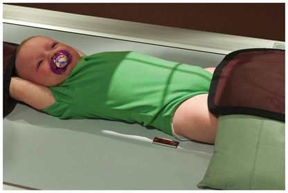
- 69Evaluation Criteria & TechniquePEDIATRIC AP SUPINE KUB
SID 40 in
IR size determined by patient
Analog (newborn-18): 65-75 kV
Digital (neonate-18): 70-80 kV
Grid if 10 cm or larger
CR @ 1 in (2.5 cm) above umbilicus for young children, and center @ iliac crest for older children and teens
- make exposure when baby is most still
- no rotation
- vertebral column is aligned
- collimate as closely as possible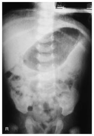
- 70What are the clinical indicators for this projection/position?PEDIATRIC AP ERECT ABDOMEN:
- pathology of abdomen
- possible intestinal obstruction
- demonstrates air/fluid levels
- free intraperitoneal air
- part of 3-way abdomen series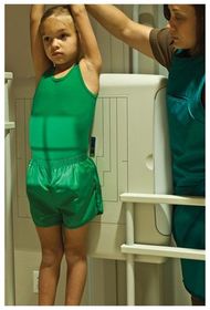
- 71Evaluation Criteria & TechniquePEDIATRIC AP ERECT:
SID 72 in (only use 72 SID for upright PEDS ABD - the rest use 40 SID)
IR sized by patient size
Analog (newborn - 18): 65-75 kV
Digital (newborn - 18): 70-80 kV
Grid if 10 cm or larger
CR @ 1 in (2.5 cm) above umbilicus (newborns & infants) or center @ 1-2 in above iliac crests (older children & teens)
- entire contents of abdomen shown
- no rotation/motion - vertebral column aligned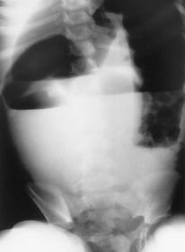
- 72What are the clinical indicators for this projection/position?PEDIATRIC LEFT LATERAL DECUBITUS
- air/fluid levels
- free air in abdomen
- possible calcifications, masses, or other anomalies
- demonstrates prevertebral region of abdomen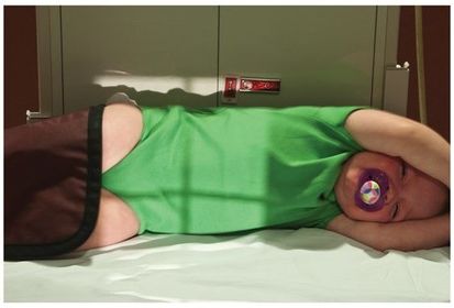
- 73Evaluation Criteria & TechniquePEDIATRIC LEFT LATERAL DECUBITUS
SID 40 in
IR sized by patient size
Analog (newborn - 18) 65-75 kV
Digital (neonate - 18) 70-80 kV
Grid if 10 cm or larger
CR horizontal @ 1 in (2.5 cm) above umbilicus for newborns and small children
CR horizontal @ 1 - 2 in above iliac crests for older children and teens
- diaphragm and iliac wings are included
- no rotation/motion
- 4 bordered collimation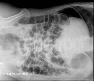
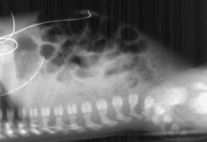
- 74When would you perform this position?When clinically indicated instead of a Left Lateral PEDS Decubitus
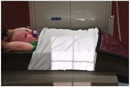
- 75Evaluation Criteria & TechniqueSame as Left Lateral PEDS Decubitus
- 76Extra _____ or _____ under the back and buttocks are used for _____ patients along with extra blankets for comfort.padding
sponges
geriatric - 77Sonography has become the method of choice to view the ______ to look for ______gallbladder
gall stones - 78Small neoplasms are diagnosed using ___ and ___ because they can be discriminated better with these modalities.CT
MRI - 79______ is the modality of choice to view GI motilityNuclear Medicine
- 80When placing your markers, always mark which side is _____ for abdomen decubitus positions, mark which side is ____ for chest positionsDOWN
UP - 81When performing a left lateral decub, which pathology are you trying to view?air/fluid levels
- 82A non-mechanical bowel obstruction is also known as _____Ileus
- 83The mesentery connects the ______ to the _______ abdominal wallsmall intestines
posterior - 84the omentum is a double folded peritoneum that connects the _____ to another organ.STOMACH
- 85the lesser omentum connects the stomach to the _____liver
- 86the greater omentum connects the stomach to the _____ _____transverse colon
- 87the 2-way acute abdominal series includes the _____ and _____AP Supine
Left Lateral Decubitus - 88the 3-way acute abdominal series includes the _____ , ______ and the ______AP Supine
AP Erect
PA Erect Chest
* Left Lateral Decubitus may be used as an extra view - 89Gonadal shielding for females involves placing the top of the shield at or slightly above the level of the _____ with the bottom at the ____ ____ .ASIS
symphysis pubis - 90Why is the LEFT lateral decub preferred over the RIGHT lateral decub?it best visualizes free intraperitoneal air in the area of the liver in the RUQ away from the gastric bubble in the LUQ
- 91What is the alternate position you could use if the patient cannot stand for an AP Erect?Left Lateral Decubitus
- 92What's the best way to reduce INVOLUNTARY motion on a radiograph?use the shortest exposure time possible
- 93Which of the following structures is not visible on a properly exposed KUB? A. Kidneys B. Liver margins C. Pancreas D. Lumbar spinous processesC. Pancreas
- 94Which decub position best demonstrates umbilical hernias, possible aneurysms, or calcifications of the aorta?Dorsal Decubitus
- 95Which projection/position in the 3-way abdominal series best demonstrates free air under the diaphragm?PA Erect Chest
- 96What is the recommended overlap when using two crosswise images for an AP Supine of a broad hypersthenic patient?1-2 inches, or 3 fingers
- 97Which scale of contrast is recommended for visualization of the abdominal structures on an abdominal x-ray?Long Scale Contrast
- 98An opening to an organ that is closed is called ______Atresia
- 99"mega colon" - rhythmic contractions that are absent in large intestines is also termed _________Hirschsprung's Disease
- 100the slipping (telescoping) of one part of intestine into another is called _______ . It is most common in young pediatric patients.Intussusception
- 101A hereditary condition in which the bones are soft, fragile, and break easily is called ______ ______ .Osteogenesis Imperfecta
- 102The narrowing or blocking of the pylorus that results in repeated forceful vomiting is known as ______Pyloric Stenosis
- 103an abnormal accumulation of fluid in the peritoneal cavity is known as ______ .Ascites
- 104free air or gas in the peritoneal cavity is called _______pneumoperitoneum
- 105the most common cause of mechanically based obstruction in which a fibrous band of tissue interrelates with the intestine, creating a blockage is called ______ _____Fibrous Adhesions
- 106A chronic inflammation of the intestinal wall that results in bowel obstruction is known as ____ ____ .Crohn's Disease
- 107The twisting of a loop of intestine that creates an obstruction is called _______Volvulus
- 108A non-mechanical bowel obstruction that is frequently caused by a lack of intestinal motility is termed ______Ileus
- 109Chronic inflammation of the colon that occurs primarily in young adults is called ______ _____ .Ulcerative Colitis
- 110When would you need to overlap your images?If the patient is hypersthenic and you need to use the IR crosswise, you'll need 2 images to fit in all the anatomy
- 111For AP Supine, PA Prone, Right or Left Lateral Dedubitus, and AP/PA Erect positions the CR is perpendicular to the _____ plane of the body.Midsagittal
- 112For Dorsal or Ventral Decubitus, Erect or Supine Right or Left Lateral positions, the CR is perpendicular to the ________ plane.Midcoronal
- 113Which projections/positions might magnify certain organs on the radiograph?Obliques
PA Prone (organs are compressed from lying down) also magnifies kidneys - 114TRUE OR FALSE: You should place your marker directly on the image receptor vs. the table or wall board.FALSE:
always put the marker in the field of collimation on the table or wall board - 115Which positions MUST demonstrate the Psoas muscles and the symphysis pubis on the radiographs?KUB
(AP Supine) - 116Which positions MUST show the diaphragm on the radiograph?AP Erect
all Decubitus positions - 117Why does the patient need to have an empty bladder for an ABD XR?Urine in the bladder can obstruct the view of the pathology
- 118Which projections/positions require the tube to be horizontal to the floorany Decub
any Erect - 119Which projection/positions have the CR centered directly at the level of the iliac crests?Lateral Recumbent
AP Supine
PA Prone - 120Which projections/positions have the CR centered 2 in (5 cm) above the iliac crests?AP Erect
All Decubitus (laterals, ventral, & dorsal) - 121What are 3 positioning techniques for patients on a cart when you have to do a lateral decubitus?1. arms above the head
2. knees bent
3. hips and shoulders superimposed
4. patient can be on a sponge to raise them off the cart so the IR views all of the abdomen - 122What is the evaluation criteria for pediatric abdomen radiographs?vertebral column centered
must view diaphragm -> pubic symphysis
no rotation/motion
optimal exposure factors
markers - 123Why would you use manual technique for pediatric exams vs. automatic exposure (phototiming)you want shortest exposure time as possible
high mA as possible
kV range differs depending on size of child
Information
- Description
- ARRT Radiologic Procedures I Flashcards on Abdomen Procedure, created by RadTech Fairy on 27/09/2016.
- Created by
- RadTech Fairy
- Views
- 8
- Rating