
Flashcards
Cells and Tissue Types
Cards in this deck (139)
- 1Epithelial Tissue Description- Covers body surface
-Lines hallow organs, tubes, cavities and ducts
-Form glands - 2Epithelial Tissue MAJOR Functions-Protection
-Filtration
-Secretion and absorption
-Excretion - 3Connective Tissues DescriptionCells in a matrix of fibres and ground substance (incl. blood and bone)
- 4Connective Tissue MAJOR Functions-Protects and supports
-Binds organs together
-Stores energy
-Transport (blood) - 5Muscle Tissue DescriptionContractile cells
(muscle fibres) - 6Muscle Tissue MAJOR FunctionsMovement
- 7Key Functions of Epithelial TissueSelective barriers - limiting or aiding transfer
Secretory - onto a free surface
Protection - especially from abrasion - 8Epithelium Cells......are arranged in continuous sheets (single or multiple layers)
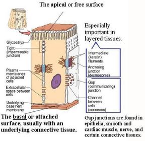
...held together by a variety of cell junctions - 9What are the different junctions found in epithelium?-Tight junction
-Adherens junction
-Desmosome
-Hemidesmosome
-Gap junctions
(take note of location) - 10What is the function of the TIGHT JUNCTION?-Located on the apical surface to prevent fluids diffusing into cells
-Maintain polarity - 11What are the 2 PROTEINS associated with TIGHT JUNCTIONS?Occludins & claudins
- 12What is the function of the ADHERENS JUNCTION?Belt adjacent cells.
Cadherins span the gap...
Catenin links cadherins to ACTIN (microfilaments in cell cytoplasm) - 13What is the function of the DESMOSOME?Resist shearing forces e.g. bind muscle cells.
Cadherins spans the gap - join cell surface to keratin. - 14Role of KERATIN in the DESMOSOME...Network of fibres = transportation mechanism
- 15What is the FUNCTION of GAP JUNCTIONS?Direct connection between cells, allows cells to communicate; send chemical signals between one another.
- 16The channel in a GAP JUNCTION is formed by...6 connexINS molecules (protein)
DIFFERENT from the connexONS - 17What is the FUNCTION of the HEMIDESMOSOME?Connect the epithelium to the basement membrane.
Links cellular INTERMEDIATE FILAMENT (Keratin) to basement membrane. - 18What are two things that INTEGRIN protein binds to in the HEMIDESMOSOME?1. Laminin in the basement membrane
2. Keratin intermediate filament in
cytoplasm - 19The B____ M_____ separates the epithelium from other tissuesBasement membrane
- 20The basement membrane is composed of ...1. Basal lamina (secreted by the epithelial cells)
2. Reticular lamina (produced by cells of the underlying connective tissue, known as FIBROBLASTS) - 21Basal laminaSecreted by the
epithelial cells containing collagen,
laminin, other proteoglycans,
glycoproteins - 22Reticular laminaProduced by
cells of the underlying connective
tissue known as fibroblasts
containing fibrous proteins such as
fibronectin, collagen - 23Functions of the BASAL LAMINA1. Supports overlying epithelium
2. Provides surface along which epithelial cells migrate during growth & wound healing
3. Acts as a physical barrier - 24Epithelium contain N____ but no B____ V____ = AvascularContain NERVES but no BLOOD VESSELS
- 25Exchange of nutrients and wastes takes place by......diffusion from vessels in the connective tissue
- 26Two types of EPITHELIAL TISSUE:1. Covering and lining epithelia
2. Glandular epithelia - 27Covering and lining epithelia1. Forms the outer covering of the skin and some internal organs
2. Forms the inner lining of blood vessels, ducts & blood cavities and interior of the RESPIRATORY, DIGESTIVE, URINARY & REPRODUCTIVE systems - 28Glandular epitheliaConstitute the secretory portions of glands
- 29Glandular epithelia (Classifications)1. Endocrine gland
2. Exocrine gland - 30Endocrine GlandSecretes directly into the blood, via traversing interstitial fluid
e.g. Pituitary gland - 31Exocrine glandsecretes into ducts that empty onto the surface of a covering or lining epithelia
e.g. Sweat and salivary glands - 32Connective Tissue is composed of...CT = ECM + Cells
- 33ECM - Extracellular Matrix - is composed of...ECM = GS + Fibres
- 34GS - Ground SubstanceWater + protein core + polysaccahrides = ground (jelly) substance
PROTEOGLYCAN aka GAGS - 35What is the function of GAGs?1. Draws in H2O = hydration
2. Makes a jelly substance: lubrication of joints & shock absorbtion - 36Cell types present in CONNECTIVE TISSUE:1. Fibroblasts
2. Adipocytes (fat cells) - 37Where are fibroblasts located?Widely distributed in connective tissues; migratory
- 38What is the function of the fibroblasts?Secretes components of the matrix (fibres and ground substance)
- 39Where are adipocytes located?Under the skin and around organs
- 40What is the function of adipocytes?Store fats, triglycerides
- 41Types of PROTEIN FIBRES in ECM:1. Collagen Fibres (THICK)
2. Reticular fibres
3. Elastic fibres - 42CT: Collagen Fibres- Very strong but flexible to resist pulling forces
-Features vary in different tissues
-Most abundant protein - 43CT: Reticular Fibres- Composed of collagen in fine bundles with a strong coating of GLYCOPROTEIN
- Made by fibroblasts
- Gives strength and support
-Thinner branching; spreads through tissue - 44CT: Elastic Fibres- Thinner than collagen fibres
- Fibrous network
- Protein elastin surrounded by GLYCOPROTEIN (fibrillin) for strength
- Can stretch 150% (found in skin and lungs, blood vessels) - 45Marfan Syndrome (Elastic fibres)- Hereditary defect in elastic fibres, mutation in gene for fibrillin
- Phenotype: long-limbed, tall, chest deformity, weakened heart valves - 46Other cells found in solid CONNECTIVE TISSUES:1. Macrophages
2. Mast cells
3, Plasma cells
4. Leucocytes - 47CT: MacrophagesPhagocytic cells
1. FIXED form: Alreolar tissue, spleen
2. WANDERING form: sites of inflammation/infection - 48CT: Mast CellsProduce histamine that dilates blood vessels
FOUND: beside blood vessels - 49CT: Plasma CellsFrom blymphocytes, which produce anti-bodies.
FOUND: in many CT but especially in gut and lung, salivary gland, lymphnodes, spleen, red bone marrow - 50CT: LeucocytesWhite blood cells; migrate out from blood
- 51What are the classifications for CONNECTIVE TISSUE?1. Embryonic
2. Mature - 52Embryonic Connective Tissue1. Gives rise to all other connective tissues.
2. Consists of connective tissue cells
(mesenchymal cells) in a semi-fluid ground
substance containing reticular fibres - 53What are the classifications of Connective Tissue?1. Embryonic
2. Mature - 54Types of Mature Connective Tissues1. Connective tissue proper
2. Fluid connective tissue
3. Supporting connective tissue - 55Types of Connective Tissus ProperLoose & Dense
- 56Types of Fluid Connective TissueBlood & Lymph
- 57Types of Supporting Connective TissueCartilage & Bone
- 58Loose Connective Tissue TypesAreolar CT
Adipose CT
Reticular CT
Loose means: many cells and fewer fibres - 59Dense Connective Tissue TypesDense Regular CT
Dense Irregular CT
Dense Elastic CT - 60Cartilage Connective Tissue Types1. Hyaline Cartilage
2. Elastic
3. Fibrocartilage - 61Bone/Osseus Connective Tissue TypeCompact
- 62Loose CT: Areolar1. Most common form of CT
2. Widely distributed around almost every structure like packing material
STRENGTH, ELASTICITY & SUPPORT - 63What are the 3 types of fibres which make up the loose areolar connective tissue?1. Collagen
2. Reticular
3. Elastic - 64Loose CT: Adipose1. Adipocytes dominant
2. Derived from fibroblasts; central triglyceride droplet
3. Found with Areolar connective tissue
4. White adipose & brown adipose
INSULATION, ENERGY SOURCE & TEMP. CONTROL - 65Loose CT: Reticular1. Reticular fibres are the main structural part of tissue - delicate network
2. Forms a scaffolding for other cells in several organs i.e. bone marrow, lymph nodes - 66Dense CT: Regular1. Regularly arranged collagen (not living tissue)
2. Shiny white colour
3. Slow healing tendons, ligaments, aponeuroses
ATTACHMENT - 67Dense CT: Irregular1. Collagen fibres are not arranged in parallel bundles
2. Comprises a large portion of the dermis
3. Makes up submucosa in digestive tract - 68Dense CT: Elastic1. Bundles of proteins (elastin) found in ECM
2. Can stretch up to 1.5x its length and snap back
3. Produced by fibroblasts and smooth muscle arteries - 69Cartilage CT: Hyaline1. Abundant
2. Relatively weak - resilient gel; fibres are present but not obvious
FLEXIBILITY & MOVEMENT
Found: respiratory cartilage - nose, bronchi, trachea - 70Cartilage CT: Elastic1. Present in outer ear and epiglottis
2. Principal protein: elastin
3.. Withstand bending
SUPPORT & MAINTAIN SHAPE - 71Cartilage CT: Fibrocartilage1. Cartilaginous tissue (& fibrous tissue)
2.
FLEXIBILITY, TOUGHNESS & ELASTICITY
Found: Pubic symphysis (aid in birth), bone interface (transition from soft tendon to un/calcified fibrocartilage to bone) - 72Bone/osseous Tissue Types1. Compact
2. Spongy - 73Bone Tissue: Compact1. Contains osteons
2. Stores calcium & phosphorus
PROTECTION & SUPPORT - 74Bone Tissue: Spongy1. Lacks osteons
2. Stores triglycerides (yellow marrow) and produces blood cells (red marrow) - 75What are the four cell types found in bone?1. Osteogenic cells
2. Osteoblasts
3. Osteocytes
4. Osteoclasts - 76Bone Cells: Osteogenic CellsMESENCHYMAL stem cells that develop, become trapped = become OSTEOBLASTS
- 77Bone Cells: OsteoblastsBone-forming cells
Become trapped in ECM = become OSTEOCYTES - 78Bone Cells: OsteocytesMature bone cells
Maintain bone tissue
Exchange of nutrients and wastes
Have gap junctions - 79Bone Cells: OsteoclastsLarge MULTINUCLEATED cells formed the fusion of blood monocytes.
BREAK DOWN BONE - 80Unit of compact bone?Osteons - aligned along lines of stress
- 814 Osteon Components1. Lamellae
2. Lacunae
3. Canaliculi
4. Central (Haversian) canal - 82Osteon: LamellaeConcentric rings of mineral salts for hardness
Collagen for tensile strength - 83Osteon: LacunaeSmall spaces between the lamellae that contain OSTEOCTYES
- 84Osteons: CanuliculiMinute canals
- 85Osteons: Central Haversian canalContains blood vessels and nerves
- 86Liquid Connective Tissue Types1. Blood
2. Lymph - 87Liquid CT: BloodConsists of blood plasma (liquid ECM) and formed elements
- 88Formed elements of blood1. Erythrocytes (red cells)
2. Leukocytes (white cells)
3. Platelets - 89Elements of Blood: ErythrocytesTransport O2 and CO2
- 90Elements of Blood: Leukocytes1. Neutrophils & monocytes
2. Basophils
3. Eosinophils
4. Lymphocytes - 91Neutrophils & monocytesPhagocytic, engulfing bacteria
- 92BasophilsRelease substances that intensify inflammatory reaction
- 93Eosinophils1. Effective against certain parasitic worms
2. Acute allergic response - 94LymphocytesImmune response
- 95Elements of Blood: PlateletsClotting; from megakaryocytes in red marrow
- 96Muscle CellsElongated cells aka muscle fibres
Energy from hydrolysis of ATP = force
Contracts - produces body heat , maintains posture, & produces movement - 97Muscle Tissue Types1. Skeletal muscle
2. Cardiac muscle
3. Smooth muscle - 98Skeletal Muscle1. Attached to bones via tendons
2. Conscious controlled CONTRACTIONS i.e voluntary EXCEPT posture - 99What is the structure of Skeletal Muscle?Striated - highly organised myofibrils
Cylindrical fibres (=cells)
Long cells
Multinucleate - 100What is the function of skeletal muscle?1. Motion
2. Posture
3. Heat
4. Protection - 101Smallest skeletal muscleSTAPEDIUS 1.25mm
In the ear, prevents hypercausis - 102Longest skeletal muscleSARTORIUS 60cm
Hip: flexor, abductor, lateral rotator
Knee: flexor - 103Skeletal Muscle: Myofibrils locationFill the cytoplasm (sarcoplasm) of the muscle fibre, extending the entire length within the cell
- 104What are myofibrils composed of?MYOFILAMENTS
1. Thick filaments (MYOSIN)
2. Thin filaments (ACTIN) - 105The arrangement of myofilaments within the myofibril -Myofilaments are arranged in compartments called sarcomeres (
- 106Connective tissue of skeletal muscle:1. Epimysium - surrounds anatomical muscle
2. Perimysium - fascicles
3. Endomysium - muscle fibres ("cells") - 107Cardiac muscle1. Located in the heart
2. Involuntary control of contractions - 108Cardiac muscle structure:Striated
Branched
Single central nucleus
Intercalated disks - 109Cardiac muscle : Intercalated disks......is how fibres join end to end.
...contain desmosomes and gap junctions - 110Smooth Muscle1. In the walls of hollow internal structures e.g. blood vessels, intestines, skin
2. Involuntary contraction control - 111Smooth muscle structureNON-STRIATED
Single central nucleus - 112Nervous TissueEssential component to the nervous system
- 113The subdivisions of the nervous system...1. CNS - brain and spinal cord
2. PNS - all nervous tissue outside CNS - 114Nervous system helps to...1. Maintain homeostasis (+ endocrine)
2. Initiate voluntary movements
3. Perception, behaviour and memory - 115Functions of the nervous system:1. Sensory
2. Integrative
3. Motor - 116Sensory function of the nervous system:Detection of internal and external stimuli and transfer to CNS
- 117Integrative function of the nervous system:Analysis and storing of info
- 118Motor function of the nervous systemStimulation of effectors (e.g. muscle and glands) through PNS
- 119Nervous Tissue Cell Types:1. Neurons (nerve cells)
2. Neuroglia (supportive cells) - 120Neuron structure1. Longest cells in the body
2. Short cell body, branched dendrites & single axon - 121Types of Neurons1. Multipolar
2. Bipolar
3. Unipolar
4. Anaxonic - 122Multipolar Neurons- 2 or more dendrites
-single axon
-all motor neurons are this class
-longest neurons (i.e. spinal cord - toe muscle)
- 123Bipolar Neurons- 1 dendritic process
-1 axon
-Cell body between axon and dendrite
-Rare and small
-Relay info from special sense organs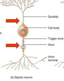
- 124Unipolar Neurons- dendrites and axon are continuous
-cell body off to one side
-most sensory nerves
-very long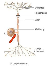
- 125Anaxonic Neuron- rare and function poorly understood
-cannot distinguish axons from dendrites
-brain and special sense organs
- 126Neuroglia1. Do not propagate action potentials BUT can COMMUNICATE
2. Can divide within the mature nervous system
3. Smaller but more numerous than neurons - 127Neuroglia LocationFound in both the CNS and PNS
(makes up half the volume of CNS ... "glue") - 128What are the functions of neuroglia?1. Physical structure of NT
2. Repair NT framework
3. Phagocytosis
4. Nutrient supply to neurons
5. Regulate interstitial fluid in neural tissue - 129Types of CNS Neuroglia1. Astrocytes
2. Ogliodendrocytes
3. Microglia
4. Ependymal cells - 130CNS: Astrocytes Structure1. Star shaped
2. Largest most numerous
3. Syncytium network - 131CNS: Astrocytes Functions1. Support and repair (microfilaments & scar)
2. Communicate w/ neurons via gliotransmitters
3. Maintain environment e.g. ion regulation
4. Maintain blood-brain barrier via endothelium, influence permeability - 132CNS: Ogliodendrocyte FunctionForms insulating multilayered MYELIN SHEATH (protein lipid layer) around CNS axons = accelerates action potentials
- 133CNS: Microglia FunctionPhagocytic = protection
- 134CNS: Ependymal Cells LocationLine CSF-filled ventricles in the brain and central canal of spinal cord
(or wherever CSF is found) - 135CNS: Ependymal Cells Function1. Produce cerebrospinal fluid (CSF)
- 136CNS: Ependymal Cells Structure1. Single layer of predominantly cuboidal cells
...have cilia (flow)
...have microvilli (sampling) - 137Types of PNS Neuroglia1. Schwann cells
2. Satellite cells - 138PNS: Schwann Cell FunctionForms insulating myelin sheath around one axon at a time OR can support multiple axons and surround non-myelinated axons
NB: Ogliodendrocyte equi. - 139PNS: Satellite Cells FunctionSurround neuron cell bodies - support and fluid exchange
NB: Astrocyte equi.
Information
- Description
- Biology (Cells and Tissues) Flashcards on Cells and Tissue Types, created by Mia Brown on 25/03/2017.
- Created by
- Mia Brown
- Views
- 6
- Rating