
Mind map
Cardiovascular system
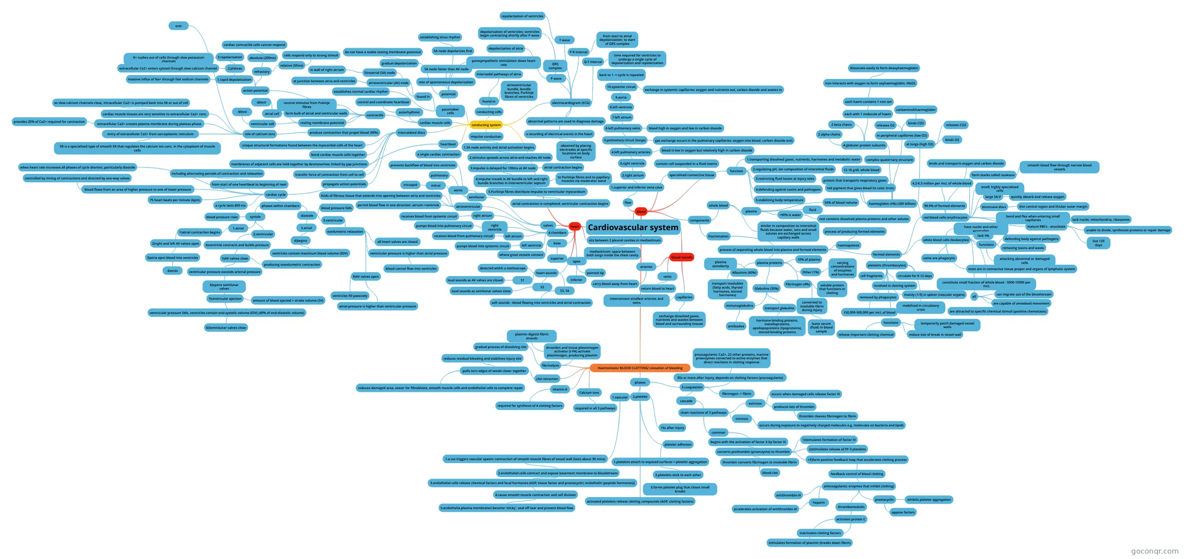
Mind map: Cardiovascular system
Map content
Cardiovascular system
heart
- 4 chambers
- right atrium
- receives blood from systemic circuit
- right ventricle
- pumps blood into pulmonary circuit
- left atrium
- receives blood from pulmonary circuit
- left ventricle
- pumps blood into systemic circuit
- right atrium
- base
- superior
- where great vessels connect
- apex
- inferior
- pointed tip
- sits between 2 pleural cavities in mediastinum
- mediastinum: space between both lungs inside the chest cavity
- valves
- atrioventricular
- tricuspid
- mitral
- folds of fibrous tissue that extends into opening between atria and ventricles
- permit blood flow in one direction: atrium->ventricle
- semilunar
- pulmonary
- aortic
- prevents backflow of blood into ventricles
- atrioventricular
- conducting system
- heartbeat
- a single cardiac contraction
- cardiac cycle
- from start of one heartbeat to beginning of next
- including alternating periods of contraction and relaxation
- phases within chambers
- systole
- 1.atrial
- 1)atrial contraction begins
- 2)right and left AV valves open
- 3)atria eject blood into ventricles
- 4)ends
- 3)atria eject blood into ventricles
- 2)right and left AV valves open
- 1)atrial contraction begins
- 2.ventricular
- 4)ventricle contracts and builds pressure
- 5)AV valves close
- producing isovolumetric contraction
- ventricular pressure exceeds arterial pressure
- 6)opens semilunar valves
- 7)ventricular ejection
- amount of blood ejected = stroke volume (SV)
- ventricular pressure falls, ventricles contain end-systolic volume (ESV) (40% of end-diastolic volume)
- 8)Seminlunar valves close
- 7)ventricular ejection
- 6)opens semilunar valves
- 5)AV valves close
- 4)ventricle contracts and builds pressure
- blood pressure rises
- 1.atrial
- diastole
- 2.atrial
- 4)begins
- ventricles contain maximum blood volume (EDV)
- 4)begins
- 3.ventricular
- isovlumetric relaxation
- all heart valves are closed
- ventricular pressure is higher than atrial pressure
- blood cannot flow into ventricles
- 9)AV valves open
- ventricles fill passively
- atrial pressure is higher than ventricular pressure
- isovlumetric relaxation
- blood pressure falls
- 2.atrial
- systole
- 75 heart beats per minute (bpm)
- a cycle lasts 800 ms
- blood flows from an area of higher pressure to one of lower pressure
- controlled by timing of contractions and directed by one-way valves
- when heart rate increases all phases of cycle shorten, particularly diastole
- from start of one heartbeat to beginning of next
- cardiac muscle cells
- autorhythmic
- control and coordinate heartbeat
- contractile
- produce contraction that propel blood (99%)
- role of calcium ions
- extracellular Ca2+ crosses plasma membrane during plateau phase
- provides 20% of Ca2+ required for contraction
- entr
- entry of extracellular Ca2+ from sarcoplasmic reticulum
- SR is a specialized type of smooth ER that regulates the calcium ion conc. in the cytoplasm of muscle cells
- cardiac muscle tissues are very sensitive to extracellular Ca2+ conc.
- as slow calcium channels close, intracellular Ca2+ is pumped back into SR or out of cell
- extracellular Ca2+ crosses plasma membrane during plateau phase
- role of calcium ions
- form bulk of atrial and ventricular walls
- receive stimulus from Pukinje fibres
- resting membrane potential
- ventricular cell
- -90mV
- atrial cell
- -80mV
- ventricular cell
- action potential
- 1.rapid depolatization
- massive influx of Na+ through fast sodium channels
- 2.plateau
- extracellular Ca2+ enters cytosol through slow calcium channel
- 3.repolarization
- K+ rushes out of cells through slow potassium channels
- refractory
- absolute (200ms)
- cardiac contractile cells cannot respond
- relative (50ms)
- cells respond only to strong stimuli
- absolute (200ms)
- 1.rapid depolatization
- produce contraction that propel blood (99%)
- autorhythmic
- pacemaker cells
- establishes normal cardiac rhythm
- found in
- Sinoatrial (SA) node
- in wall of right atrium
- atrioventricular (AV) node
- at junction between atria and ventricles
- Sinoatrial (SA) node
- potential
- gradual depolarization
- do not have a stable resting membrane potential
- rate of spontaneous depolarization
- SA node faster than AV node
- SA node depolarizes first
- establishing sinus rhythm
- parasympathetic stimulation slows heart rate
- gradual depolarization
- conducting cells
- found in
- internodal pathways of atria
- atrioventricular bundle, bundle branches, Purkinje fibres of ventricles
- found in
- impulse conduction
- 1.SA node activity and atrial activation begins
- 2.stimulus spreads across atria and reaches AV node
- 3.impulse is delayed for 100ms at AV node
- atrial contraction begins
- 4.impulse travels in AV bundle to left and right bundle branches in interventricular septum
- to Purkinje fibres and to papillary muscles via moderator band
- 5.Purkinje fibres distribute impulse to ventricular myocardium
- atrial contraction is completed; ventricular contraction begins
- 3.impulse is delayed for 100ms at AV node
- 2.stimulus spreads across atria and reaches AV node
- 1.SA node activity and atrial activation begins
- electrocardiogram (ECG)
- a recording of electrical events in the heart
- obtained by placing electrodes at specific locations on body surface
- abnormal patterns are used to diagnose damage
- P wave
- depolarization of atria
- QRS complex
- depolarization of ventricles; ventricles begin contracting shortly after P wave
- T wave
- repolarization of ventricles
- P-R interval
- from start to atrial depolarization; to start of QRS complex
- Q-T interval
- time required for ventricles to undergo a single cycle of depolarization and repolarization
- a recording of electrical events in the heart
- intercalated discs
- unique structural formations found between the myocardial cells of the heart
- bond cardiac muscle cells together
- membranes of adjacent cells are held together by desmosomes; linked by gap junctions
- transfer force of contraction from cell to cell
- propagate action potentials
- heartbeat
- heart sounds
- detected withh a stethoscope
- S1
- loud sounds as AV valves are closed
- S2
- loud sounds as semilunar valves close
- S3, S4
- soft sounds - blood flowing into ventricles and atrial contraction
- 4 chambers
blood
- flow
- 1.superior and inferior vena cava
- 2.right atrium
- 3.right ventricle
- 4.left pulmonary arteries
- 5.pulmonary circuit (lungs)
- gas exchange occurs in the pulmonary capillaries: oxygen into blood, carbon dioxide out)
- 6.left pulmonary veins
- 7.left atrium
- 8.left ventricle
- 9.aorta
- 10.systemic circuit
- exchange in systemic capillaries: oxygen and nutrients out, carbon dioxide and wastes in
- back to 1. = cycle is repeated
- 10.systemic circuit
- 9.aorta
- 8.left ventricle
- blood high in oxygen and low in carbon dioxide
- 7.left atrium
- blood is low in oxygen but relatively high in carbon dioxide
- 5.pulmonary circuit (lungs)
- 4.left pulmonary arteries
- 3.right ventricle
- 2.right atrium
- 1.superior and inferior vena cava
- specialized connective tissue
- contain cell suspended in a fluid matrix
- function
- 1.transporting dissolved gases, nutrients, hormones and metabolic water
- 2.regulating pH, ion composition of interstitial fluids
- 3.restricting fluid losses at injury sites
- 4.defending against toxins and pathogens
- 5.stabilizing body temperature
- components
- whole blood
- plasma
- fluid
- 55% of blood volume
- >90% is water
- rest contains dissolved plasma proteins and other solutes
- similar in composition to interstitial fluids because water, ions and small solutes are exchanged across capillary walls
- plasma
- fractionation
- process of separating whole blood into plasma and formed elements
- plasma proteins
- 10% of plasma
- Albumins (60%)
- plasma osmolarity
- transport insolubles (fatty acids, thyroid hormones, steroid hormones)
- Globulins (35%)
- immunoglobulins
- antibodies
- transport globulins
- hormone-binding proteins, metalloproteins, apoliopoproteins (lipoproteins), steroid-binding proteins
- immunoglobulins
- Fibrinogen (4%)
- soluble protein that functions in clotting
- converted to insoluble fibrin during injury
- leave serum (fluid) in blood sample
- Other (1%)
- varying concentrations of enzymes and hormones
- haemopoiesis
- process of producing formed elements
- formed elements
- red blood cells (erythrocytes
- haemoglobin (Hb) (280 billion)
- red pigment that gives blood its color (iron)
- protein that transports respiratory gases
- 12-18 g/dL whole blood
- complex quaternary structure
- 4 globular protein subunits
- 2 alpha chains
- 2 beta chains
- each with 1 molecule of haem
- each haem contains 1 iron ion
- iron interacts with oxygen to form oxyhaemoglobin, HbO2
- dissociate easily to form deoxyhaemoglobin
- iron interacts with oxygen to form oxyhaemoglobin, HbO2
- each haem contains 1 iron ion
- 4 globular protein subunits
- in peripheral capillaries (low O2)
- releases O2
- binds CO2
- carbaminohhaemoglobin
- at lungs (high O2)
- binds O2
- releases CO2
- 99.9% of formed elements
- 4.2-6.3 million per mcL of whole blood
- small, highly specialized cells
- large SA:V
- quickly absorb and release oxygen
- biconcave discs
- thin central region and thicker outer margin
- form stacks called rouleaux
- smooth blood flow through narrow blood vessels
- binds and transports oxygen and carbon dioxide
- bend and flex when entering small capillaries
- mature RBCs - anucleate
- lack nuclei, mitochondria, ribosomes
- unable to divide, synthesize proteins or repair damage
- live 120 days
- haemoglobin (Hb) (280 billion)
- white blood cells (leukocytes)
- have nuclei and other organelles
- lack Hb
- functions
- defending body against pathogens
- removing toxins and waste
- attacking abnormal or damaged cells
- most are in connective tissue proper and organs of lymphatic system
- constitute small fraction of whole blood - 5000-10000 per mcL
- all
- can migrate out of the bloodstream
- are capable of amoeboid movement
- are attracted to specific chemical stimuli (positive chemotaxis)
- some are phagocytic
- platelets (thrombocytes)
- cell fragments
- involved in clotting system
- circulate for 9-12 days
- removed by phagocytes
- mainly (1/3) in spleen (vascular organs)
- 150,000-500,000 per mcL of blood
- mobilized in circulatory crisis
- functions
- release important clotting chemical
- temporarily patch damaged vessel walls
- reduce size of break in vessel wall
- red blood cells (erythrocytes
- plasma proteins
- process of separating whole blood into plasma and formed elements
- whole blood
- Haemostasis/ BLOOD CLOTTING/ cessation of bleeding
- phases
- 1.vascular
- 1.a cut triggers vascular spasm: contraction of smooth muscle fibres of vessel wall (lasts about 30 mins)
- 2.endothelial cells contract and expose basement membrane to bloodstream
- 3.endothelial cells release chemical factors and local hormones (ADP, tissue factor and prostacyclin; endothelin (peptide hormones))
- 4.cause smooth muscle contraction and cell division
- 5.endothelia plasma membranes become 'sticky', seal off tear and prevent blood flow
- 4.cause smooth muscle contraction and cell division
- 3.endothelial cells release chemical factors and local hormones (ADP, tissue factor and prostacyclin; endothelin (peptide hormones))
- 2.endothelial cells contract and expose basement membrane to bloodstream
- 1.a cut triggers vascular spasm: contraction of smooth muscle fibres of vessel wall (lasts about 30 mins)
- 2.platelet
- platelet adhesion
- 1.platelets attach to exposed surfaces = platelet aggregation
- 2.platelets stick to each other
- 3.forms platelet plug that closes small breaks
- 2.platelets stick to each other
- 1.platelets attach to exposed surfaces = platelet aggregation
- activated platelets release clotting compounds (ADP, clotting factors)
- 15s after injury
- platelet adhesion
- 3.coagulation
- 30s or more after injury, depends on clotting factors (procoagulants)
- procoagulants: Ca2+, 22 other proteins, inactive proenzymes converted to active enzymes that direct reactions in clotting response
- fibrinogen -> fibrin
- cascade
- chain reactions of 3 pathways
- extrinsic
- occurs when damaged cells release factor III
- produces lots of thrombin
- thrombin cleaves fibrinogen to fibrin
- intrinsic
- occurs during exposure to negatively charged molecules e.g. molecules on bacteria and lipids
- common
- begins with the activation of factor X by factor III
- converts prothombin (proenzyme) to thrombin
- thrombin converts fibrinogen to insoluble fibrin
- blood clot
- 1)stimulates formation of factor III
- 2)stimulates release of PF-3 platelets
- =3)form positive feedback loop that accelerates clotting process
- feedback control of blood clotting
- anticoagulants: enzymes that inhibit clotting)
- antithrombin-III
- heparin
- accelerates activation of antithrombin-III
- thrombomodulin
- activates protein C
- inactivates clotting factors
- stimulates formation of plasmin (breaks down fibrin)
- activates protein C
- prostacyclin
- inhibits platelet aggregation
- oppose factors
- anticoagulants: enzymes that inhibit clotting)
- feedback control of blood clotting
- thrombin converts fibrinogen to insoluble fibrin
- converts prothombin (proenzyme) to thrombin
- begins with the activation of factor X by factor III
- extrinsic
- chain reactions of 3 pathways
- 30s or more after injury, depends on clotting factors (procoagulants)
- 1.vascular
- Calcium ions
- required in all 3 pathways
- vitamin K
- required for synthesis of 4 clotting factors
- clot retraction
- pulls torn edges of vessel closer together
- reduces residual bleeding and stabilizes injury site
- reduces damaged area, easier for fibroblasts, smooth muscle cells and endothelial cells to complete repair
- pulls torn edges of vessel closer together
- fibrinolysis
- gradual process of dissolving clot
- thrombin and tissue plasminogen activator (t-PA) activate plasminogen, producing plasmin
- plasmin digests fibrin strands
- phases
- flow
blood vessels
- arteries
- carry blood away from heart
- veins
- return blood to heart
- capillaries
- interconnect smallest arteries and veins
- exchange dissolved gases, nutrients and wastes between blood and surrounding tissues
- arteries
Information
- Description
- Mind Map on Cardiovascular system, created by Gillian C on 05/09/2019.
- Created by
- Gillian C
- Views
- 0
- Rating