
Mind map
Exchange and Transport
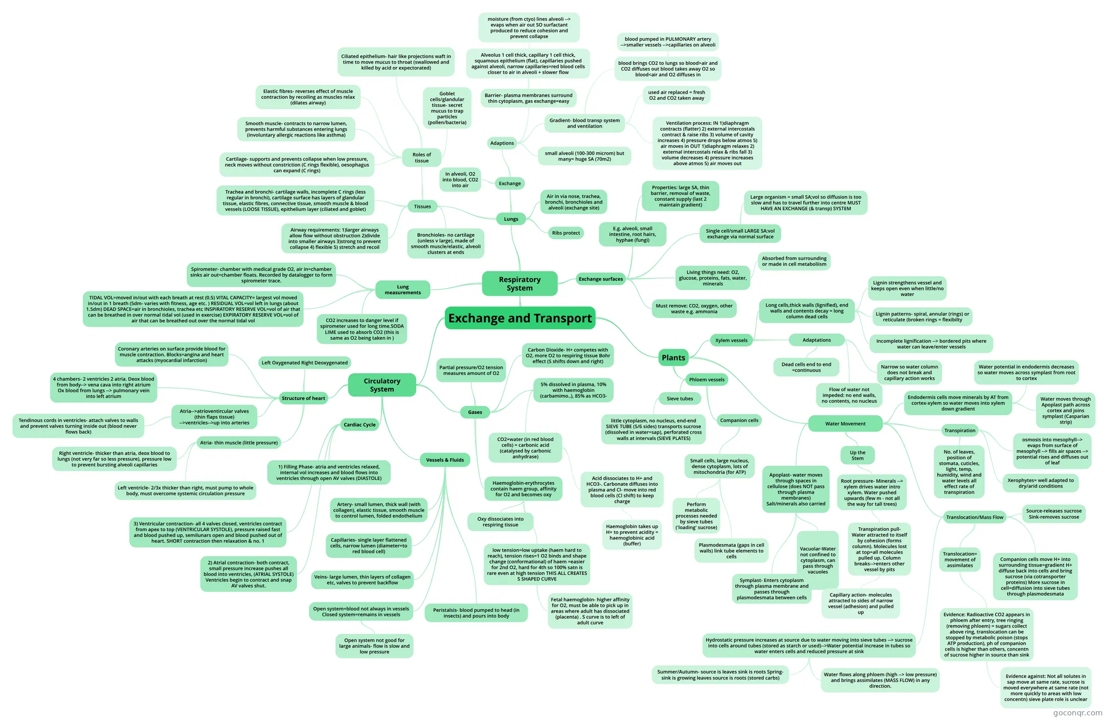
Mind map: Exchange and transport
Map content
Exchange and Transport
Circulatory System
- Cardiac Cycle
- 1) Filling Phase- atria and ventricles relaxed, internal vol increases and blood flows into ventricles through open AV valves (DIASTOLE)
- 2) Atrial contraction- both contract, small pressure increase pushes all blood into ventricles, (ATRIAL SYSTOLE) Ventricles begin to contract and snap AV valves shut.
- 3) Ventricular contraction- all 4 valves closed, ventricles contract from apex to top (VENTRICULAR SYSTOLE), pressure raised fast and blood pushed up, semilunars open and blood pushed out of heart. SHORT contraction then relaxation & no. 1
- Structure of heart
- Left Oxygenated Right Deoxygenated
- Coronary arteries on surface provide blood for muscle contraction. Blocks=angina and heart attacks (myocardial infarction)
- 4 chambers- 2 ventricles 2 atria. Deox blood from body--> vena cava into right atrium Ox blood from lungs --> pulmonary vein into left atrium
- Atria-->atrioventircular valves (thin flaps tissue) -->ventricles-->up into arteries
- Tendinous cords in ventricles- attach valves to walls and prevent valves turning inside out (blood never flows back)
- Atria- thin muscle (little pressure)
- Right ventricle- thicker than atria, deox blood to lungs (not very far so less pressure), pressure low to prevent bursting alveoli capillaries
- Left ventricle- 2/3x thicker than right, must pump to whole body, must overcome systemic circulation pressure
- Gases
- Haemoglobin-erythrocytes contain haem group, affinity for O2 and becomes oxy
- Oxy dissociates into respiring tissue
- low tension=low uptake (haem hard to reach), tension rises=1 O2 binds and shape change (conformational) of haem =easier for 2nd O2, hard for 4th so 100% satn is rare even at high tension THIS ALL CREATES S SHAPED CURVE
- Fetal haemoglobin- higher affinity for O2, must be able to pick up in areas where adult has dissociated (placenta) . S curve is to left of adult curve
- Partial pressure/O2 tension measures amount of O2
- Carbon Dioxide- H+ competes with O2, more O2 to respiring tissue Bohr effect (S shifts down and right)
- 5% dissolved in plasma, 10% with haemoglobin (carbamimo..), 85% as HCO3-
- CO2+water (in red blood cells) = carbonic acid (catalysed by carbonic anhydrase)
- Acid dissociates to H+ and HCO3-. Carbonate diffuses into plasma and Cl- move into red blood cells (Cl shift) to keep charge
- Haemoglobin takes up H+ to prevent acidity = haemoglobinic acid (buffer)
- Acid dissociates to H+ and HCO3-. Carbonate diffuses into plasma and Cl- move into red blood cells (Cl shift) to keep charge
- CO2+water (in red blood cells) = carbonic acid (catalysed by carbonic anhydrase)
- Haemoglobin-erythrocytes contain haem group, affinity for O2 and becomes oxy
- Vessels & Fluids
- Open system=blood not always in vessels Closed system=remains in vessels
- Open system not good for large animals- flow is slow and low pressure
- Peristalsis- blood pumped to head (in insects) and pours into body
- Artery- small lumen, thick wall (with collagen), elastic tissue, smooth muscle to control lumen, folded endothelium
- Veins- large lumen, thin layers of collagen etc, valves to prevent backflow
- Capillaries- single layer flattened cells, narrow lumen (diameter=to red blood cell)
- Open system=blood not always in vessels Closed system=remains in vessels
- Cardiac Cycle
Respiratory System
- Exchange surfaces
- Living things need: O2, glucose, proteins, fats, water, minerals
- Absorbed from surrounding or made in cell metaboliism
- Must remove: CO2, oxygen, other waste e.g. ammonia
- Single cell/small LARGE SA:vol exchange via normal surface
- Large organism = small SA;vol so diffusion is too slow and has to travel further into centre MUST HAVE AN EXCHANGE (& transp) SYSTEM
- Properties: large SA, thin barrier, removal of waste, constant supply (last 2 maintain gradient)
- E.g. alveoli, small intestine, root hairs, hyphae (fungi)
- Living things need: O2, glucose, proteins, fats, water, minerals
- Lungs
- Tissues
- Airway requirements: 1)larger airways allow flow without obstruction 2)divide into smaller airways 3)strong to prevent collapse 4) flexible 5) stretch and recoil
- Trachea and bronchi- cartilage walls, incomplete C rings (less regular in bronchi), cartilage surface has layers of glandular tissue, elastic fibres, connective tissue, smooth muscle & blood vessels (LOOSE TISSUE), epithelium layer (ciliated and goblet)
- Bronchioles- no cartilage (unless v large), made of smooth muscle/elastic, alveoli clusters at ends
- Roles of tissue
- Elastic fibres- reverses effect of muscle contraction by recoiling as muscles relax (dilates airway)
- Smooth muscle- contracts to narrow lumen, prevents harmful substances entering lungs (involuntary allergic reactions like asthma)
- Cartilage- supports and prevents collapse when low pressure, neck moves without constriction (C rings flexible), oesophagus can expand (C rings)
- Ciliated epithelium- hair like projections waft in time to move mucus to throat (swallowed and killed by acid or expectorated)
- Goblet cells/glandular tissue- secret mucus to trap particles (pollen/bacteria)
- Air in via nose, trachea, bronchi, bronchioles and alveoli (exchange site)
- Ribs protect
- Exchange
- In alveoli, O2 into blood, CO2 into air
- Adaptions
- small alveoli (100-300 microm) but many= huge SA (70m2)
- Barrier- plasma membranes surround thin cytoplasm, gas exchange=easy
- Alveolus 1 cell thick, capillary 1 cell thick, squamous epithelium (flat), capillaries pushed against alveoli, narrow capillaries=red blood cells closer to air in alveoli + slower flow
- moisture (from ctyo) lines alveoli --> evaps when air out SO surfactant produced to reduce cohesion and prevent collapse
- Gradient- blood transp system and ventilation
- blood brings CO2 to lungs so blood>air and CO2 diffuses out blood takes away O2 so blood<air and O2 diffuses in
- blood pumped in PULMONARY artery -->smaller vessels -->capillaries on alveoli
- used air replaced = fresh O2 and CO2 taken away
- Ventilation process: IN 1)diaphragm contracts (flatter) 2) external intercostals contract & raise ribs 3) volume of cavity increases 4) pressure drops below atmos 5) air moves in OUT 1)diaphragm relaxes 2) external intercostals relax & ribs fall 3) volume decreases 4) pressure increases above atmos 5) air moves out
- blood brings CO2 to lungs so blood>air and CO2 diffuses out blood takes away O2 so blood<air and O2 diffuses in
- Tissues
- Lung measurements
- TIDAL VOL=moved in/out with each breath at rest (0.5) VITAL CAPACITY= largest vol moved in/out in 1 breath (5dm- varies with fitness, age etc. ) RESIDUAL VOL=vol left in lungs (about 1.5dm) DEAD SPACE=air in bronchioles, trachea etc INSPiRATORY RESERVE VOL=vol of air that can be breathed in over normal tidal vol (used in exercise) EXPIRATORY RESERVE VOL=vol of air that can be breathed out over the normal tidal vol
- Spirometer- chamber with medical grade O2, air in=chamber sinks air out=chamber floats. Recorded by datalogger to form spirometer trace.
- CO2 increases to danger level if spirometer used for long time.SODA LIME used to absorb CO2 (this is same as O2 being taken in )
- Exchange surfaces
Plants
- Xylem vessels
- Long cells,thick walls (lignified), end walls and contents decay = long column dead cells
- Lignin strengthens vessel and keeps open even when little/no water
- Lignin patterns- spiral, annular (rings) or reticulate (broken rings = flexibilty
- Incomplete lignification --> bordered pits where water can leave/enter vessels
- Adaptations
- Dead cells end to end =continuous
- Narrow so water column does not break and capillary action works
- Flow of water not impeded: no end walls, no contents, no nucleus
- Long cells,thick walls (lignified), end walls and contents decay = long column dead cells
- Phloem vessels
- Sieve tubes
- little cytoplasm, no nucleus, end-end SIEVE TUBE (5/6 sides) transports sucrose (dissolved in water=sap), perforated cross walls at intervals (SIEVE PLATES)
- Companion cells
- Small cells, large nucleus, dense cytoplasm, lots of mitochondria (for ATP)
- Perform metabolic processes needed by sieve tubes ('loading' sucrose)
- Plasmodesmata (gaps in cell walls) link tube elements to cells
- Small cells, large nucleus, dense cytoplasm, lots of mitochondria (for ATP)
- Sieve tubes
- Water Movement
- Apoplast- water moves through spaces in cellulose (does NOT pass through plasma membranes) Salt/minerals also carried
- Vacuolar-Water not confined to cytoplasm, can pass through vacuoles
- Symplast- Enters cytoplasm through plasma membrane and passes through plasmodesmata between cells
- Endodermis cells move minerals by AT from cortex-xylem so water moves into xylem down gradient
- Water potential in endodermis decreases so water moves across symplast from root to cortex
- Water moves through Apoplast path across cortex and joins symplast (Casparian strip)
- Up the Stem
- Root pressure- Minerals --> xylem drives water intro xylem. Water pushed upwards (few m - not all the way for tall trees)
- Transpiration pull- Water attracted to itself by cohesion (forms column). Molecules lost at top=all molecules pulled up. Column breaks-->enters other vessel by pits
- Capillary action- molecules attracted to sides of narrow vessel (adhesion) and pulled up
- Transpiration pull- Water attracted to itself by cohesion (forms column). Molecules lost at top=all molecules pulled up. Column breaks-->enters other vessel by pits
- Root pressure- Minerals --> xylem drives water intro xylem. Water pushed upwards (few m - not all the way for tall trees)
- Transpiration
- osmosis into mesophyll--> evaps from surface of mesophyll --> fills air spaces --> potential rises and diffuses out of leaf
- No. of leaves, position of stomata, cuticles, light, temp, humidity, wind and water levels all effect rate of transpiration
- Xerophytes= well adapted to dry/arid conditions
- Translocation/Mass Flow
- Translocation= movement of assimilates
- Evidence: Radioactive CO2 appears in phloem after entry, tree ringing (removing phloem) = sugars collect above ring, translocation can be stopped by metabolic poison (stops ATP production), ph of companion cells is higher than others, concentn of sucrose higher in source than sink
- Evidence against: Not all solutes in sap move at same rate, sucrose is moved everywhere at same rate (not more quickly to areas with low concentn) sieve plate role is unclear
- Evidence: Radioactive CO2 appears in phloem after entry, tree ringing (removing phloem) = sugars collect above ring, translocation can be stopped by metabolic poison (stops ATP production), ph of companion cells is higher than others, concentn of sucrose higher in source than sink
- Source-releases sucrose Sink-removes sucrose
- Companion cells move H+ into surrounding tissue=gradient H+ diffuse back into cells and bring sucrose (via cotransporter proteins) More sucrose in cell=diffusion into sieve tubes through plasmodesmata
- Hydrostatic pressure increases at source due to water moving into sieve tubes --> sucrose into cells around tubes (stored as starch or used)-->Water potential increase in tubes so water enters cells and reduced pressure at sink
- Water flows along phloem (high --> low pressure) and brings assimilates (MASS FLOW) in any direction.
- Summer/Autumn- source is leaves sink is roots Spring- sink is growing leaves source is roots (stored carbs)
- Translocation= movement of assimilates
- Apoplast- water moves through spaces in cellulose (does NOT pass through plasma membranes) Salt/minerals also carried
- Xylem vessels
Information
- Description
- A-Level Biology (Cells, exchange and transport) Mind Map on Exchange and Transport, created by beccy.booster on 04/05/2015.
- Created by
- beccy.booster
- Views
- 8
- Rating