
Mind map
Biological Molecules
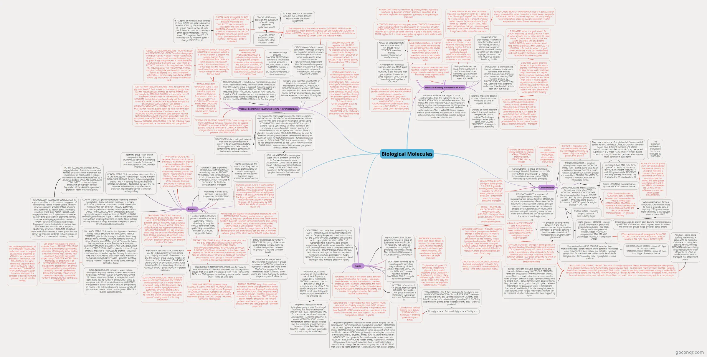
Mind map: Biological molecules
Map content
Biological Molecules
Molecular Bonding + Properties of Water
- Monomers = small molecules that bind together to form POLYMERS. Polymer = a large molecule made from many small molecules joined together, called MONOMERS
- Condensation reaction = a reaction that occurs when two molecules are JOINED together, REMOVING water. Hydrolysis reaction = a reaction that occurs when a molecule is split into two smaller molecules, with the ADDITION of water
- HYDROGEN BOND = a weak interaction (IF) that occurs wherever molecules contain a slightly negative O, F or N, bonded to a slightly positively charged HYDROGEN atom (the bond occurs between SEPARATE molecules' H and -ve atom)
- Biological Molecules are made from mainly C, H, O and N (may have other elements eg. S). Some are MONOMERS, which join to form POLYMERS
- COVALENT BOND (non-metals) = a chemical bond formed between 2 non-metals, in which 2 atoms share a pair of electrons to achieve stability eg. water (dot-and-cross or displayed formulas). Can form MULTIPLE covalent bonds! eg. double
- IONIC BOND = a chemical bond formed between a metal and a non-metal that involves DONATING an electron from one atom to another, forming IONS. The ions are held by ELECTROSTATIC ATTRACTION eg. NaCl (dot-and-cross diagrams - make sure draw brackets around each ion + put charge
- Almost all CONDENSATION reactions occur when 2 -OH groups REACT TOGETHER - reaction involves breaking/formation of covalent bonds
- Condensation + hydrolysis reactions LINK and SPLIT apart biological molecules. MONOMERS are the units that join together. 2 monomers joined together = DIMER, lots of molecules joined together = POLYMER
- Biological molecules, such as carbohydrates, proteins and nucleic acids form POLYMERS (carbohydrates - monomer = MONOSACCHARIDES, polymer = POLYSACCHARIDES) (Proteins - monomer = AMINO ACIDS, polymer = POLYPEPTIDES/PROTEINS). (Nucleic acids - monomer = NUCLEOTIDES, polymer = DNA/RNA) Lipids DON'T form polymers
- In a water molecule, the oxygen is more electronegative than the hydrogens so exerts a GREATER ATTRACTION to the shared electrons. This makes the water molecule POLAR as oxygens are slightly negative and hydrogens are slightly positive. This means HYDROGEN BONDS form between water molecules. This is WEAKER than a covalent bond. In some polymers, thousands of H bonds form between monomer chains (helps stabilise biological molecules' structures)
- Polarised molecules dissolve in polar solvents, non-polar molecules dissolve in organic solvents
- Functions of water: reactant, solvent, transport medium, temperature control, habitat. The hydrogen bonding in water gives it some ANOMALOUS PROPERTIES, allowing it to perform its functions
- 1. LIQUID: Water molecules constantly move so continuously make/break H bonds. H bonds between water molecules make it more difficult for molecules to break away + form a gas (other molecules eg. H2S = gases at room temp). Even w/ H bonds, water has a LOW VISCOSITY (can flow easy). As its liquid at room temp, it can provide habitats, form a part of tissues, provide reaction mediums + transport mediums
- 2. DENSITY: Water becomes denser as it gets colder until 4 degrees. From then, because of its polar nature, water molecules align themselves in a regular lattice structure (molecules held apart). This makes ice less dense than water + means aquatic organisms are insulated from extreme cold, and they have an environment to live in, as ice will float to the top + prevent the whole pond from freezing
- 3. SOLVENT: water is a good solvent for POLAR molecules. Eg. in NaCl, the +ve H part of water attracts the -ve solute ions (Cl-) and the -ve O part of water attracts the +ve solute ions (Na+). The ions become SURROUNDED by water molecules, which keep them separated, so they DISSOLVE + a SOLUTION is formed. As water is a good solvent, molecules + ions can move around and react in water, molecules + ions can be TRANSPORTED when dissolved
- 2. DENSITY: Water becomes denser as it gets colder until 4 degrees. From then, because of its polar nature, water molecules align themselves in a regular lattice structure (molecules held apart). This makes ice less dense than water + means aquatic organisms are insulated from extreme cold, and they have an environment to live in, as ice will float to the top + prevent the whole pond from freezing
- 1. LIQUID: Water molecules constantly move so continuously make/break H bonds. H bonds between water molecules make it more difficult for molecules to break away + form a gas (other molecules eg. H2S = gases at room temp). Even w/ H bonds, water has a LOW VISCOSITY (can flow easy). As its liquid at room temp, it can provide habitats, form a part of tissues, provide reaction mediums + transport mediums
- 4. COHESION: Hydrogen bonding gives water COHESION (molecules of water pulled together). This also happens on the surface of water (SURFACE TENSION) - water molecules more attracted to each other than the air - surface of water contracts + gives it the ability to RESIST FORCE applied to it -> mean water pulled up xylem + pond skaters walk on water
- 5. HIGH SPECIFIC HEAT CAPACITY: Water molecules held together tightly by H bonds so a lot of energy is required to increase their KE + temperature (SHC = amount of energy needed to raise the temperature of 1kg of water by 1 degree - 4.2kJ) - so NO rapid water temperature changes - means aquatic organisms have stable environments + living things have stable temps for reactions
- 6. HIGH LATENT HEAT OF VAPORISATION: Due to H bonds, a lot of energy is needed for water molecules to break away + evaporate, due to HEAT ENERGY. So, water helps to COOL living things + keep temperature stable eg. sweat evaporation + water evaporation in plants (takes heat energy w/ it
- 6. REACTANT: water is a reactant eg. photosynthesis, hydrolysis reactions. eg. digestion of starch, proteins + lipids. Role as a reactant = important for digestion + synthesis of large biological molecules
Carbohydrates
- Carbohydrates = a group of molecules containing C, H and O. 'Hydrated carbons' (for every C, there are 2 H's and 1 O - CH2O). Some carbohydrates are part of OTHER molecules eg. nucleic acids, glycolipids
- Functions of carbohydrates: energy SOURCE eg. glucose, energy STORAGE eg. starch/glycogen, STRUCTURAL unit eg. cellulose
- Main carbohydrate groups: monosaccharides (MONOMER), disaccharides, polysaccharides (POLYMER)
- ISOMERS = molecules with the same NUMBER of atoms but ARRANGED differently eg. isomers of glucose = alpha glucose, beta glucose
- MONOSACCHARIDES = simplest carbohydrate + important SOURCE of energy (well - suited due to large no. of C-H bonds). They are SWEET SUGARS. They are: soluble in WATER (OH groups) and insoluble in ORGANIC SOLVENTS. Can exist as STRAIGHT-CHAINS or in ring/CYCLIC forms
- They have a backbone of single-bonded C atoms, with 2 bonded to an O, forming a CARBONYL GROUP (different sugars have different numbers of C atoms - monosaccharides classified on no. of C's eg. hexose = 6 C, pentose = 5 C, triose = 3 C). Triose + tetrose sugars can exist as straight chains but pentoses + hexoses are more common in cyclic form.
- In straight-chain AND cyclic form, glucose can exists as numerous ISOMERS. In straight-chain, the -H and -OH groups can be REVERSED. In a ring, isomers form when the O attached to C5 also bonds to C1.
- As the -OH and -H groups on C1 can be above/below the plane of the ring on C1 and C4, there are 2 isomers: alpha glucose (orientation of H and OH on carbons are the SAME) and beta glucose (orientation of H and OH on CARBON 1 is reversed (so OH now on top) - difference is important when glucose POYLMERISES
- In straight-chain AND cyclic form, glucose can exists as numerous ISOMERS. In straight-chain, the -H and -OH groups can be REVERSED. In a ring, isomers form when the O attached to C5 also bonds to C1.
- Ribose = PENTOSE monosaccharide, glucose = HEXOSE monosaccharide
- They have a backbone of single-bonded C atoms, with 2 bonded to an O, forming a CARBONYL GROUP (different sugars have different numbers of C atoms - monosaccharides classified on no. of C's eg. hexose = 6 C, pentose = 5 C, triose = 3 C). Triose + tetrose sugars can exist as straight chains but pentoses + hexoses are more common in cyclic form.
- DISACCHARIDES (eg. maltose, sucrose, lactose): are made when TWO MONOSACCHARIDES JOIN TOGETHER. like monosaccharides, they are SWEET and SOLUBLE.. Eg. alpha glucose + alpha glucose -> MALTOSE. alpha glucose + fructose -> SUCROSE, galactose + alpha glucose -> LACTOSE
- When disaccharides form, a GLYCOSIDIC bond is formed between the 2 monosaccharides -> this is formed via a CONDENSATION REACTION, so a molecule of H20 is lost (from the C1 of one monosaccharide and the C4 of the other monosaccharide)- 1-4 glycosidic bonding
- Maltos, lactose = reducing sugars, sucrose = non-reducing sugar
- The formation and breaking of glycosidic bonds are catalysed by ENZYMES
- When disaccharides form, a CONDENSATION reaction occurs to form a glycosidic bond. 2 hydroxyl (OH) groups line up, from which H2O is lost (oxygen atom = now a LINK)
- Disaccharides can be broken back down to monosaccharides by a HYDROLYSIS reaction, which requires water to reform the 2 hydroxyl groups (helps glycosidic bonds break)
- Alpha glucose = C6H12O6 - energy source, component of starch and glycogen. Beta glucose = C6H12O6 - energy source, component of cellulose, Ribose = C5H10O5 - component of RNA, ATP, ADP, Deoxyribose = C5H10O4 - component of DNA
- When a disaccharide of beta-glucose forms, THE SECOND GLUOSE FLIPS HORIZONTALLY (eg. O usually at top right now at bottom right)
- HOMOPOLYSACCHARIDES = made of 1 type of monosaccharide. HETEROPOLYSACCHARIDES = made of more than 1 type of monosaccharide
- POLYSACCHARIDES = polymers of monosaccharides, made of many monosaccharides bonded together. STRUCTURE of some polysaccharides makes them GOOD ENERGY STORES eg. starch (plants), glycogen (animals) - monosaccharides can be easily 'snipped off' polysaccharides in HYDROLYSIS reactions. , compact, branching (more compact - many glucose molecules can be hydrolysed at the same time)/straight chain
- 1-4 glycosidic bonding = bond between c1 and c4 of 2 monosaccharides. (hydrolysed by amylase) 1-6 glycosidic bonding = bond between c1 and c6 of 2 monosaccharides (hydrolysed by glucosidase)
- Polysaccharides = LESS SOLUBLE in water than monosaccharides - doesn't reduce water potential + no excess water movement in. Less soluble due to SIZE + hydroxyl groups that form H bonds are hidden inside the molecule (amylose may form a double helix - hydrophobic external surface)
- ALPHA GLUCOSE (STORAGE) - used in RESPIRATION, monomer of alpha glucose, sweet soluble and small so easily transported in the blood plasma/tissue fluid/cytoplasm
- AMYLOSE (PLANTS) - storage of alpha-glucose. 1-4 glycosidic bonding, H bonding (holds spiral in place), COILED, hydroxyl groups on c2 INSIDE molecule to make it less soluble + allows H bonds to form to maintain structure, large + insoluble, COMPACT (ENERGY DENSE), FEW break off points, no effect on water potential, difficult to transport, fewer attachment points
- AMYLOPECTIN (PLANTS): storage of alpha-glucose, 1-4 AND 1-6 glycosidic bonding, BRANCHED, large + insoluble, no effect on water potential, multiple break off + attachment points (branching), difficult to transport
- STARCH - PLANTS (IN CHLOROPLASTS) -80% AMYLOPECTIN, 20% AMYLOSE - storage of alpha glucose, bonding + properties same as amylose/amylopectin
- GLYCOGEN (ANIMALS) - IN LIVER: regulated by insulin + glucagon (-ve feedback), storage of alpha-glucose, 1-4 AND 1-6 glycosidic bonding (smaller than amylopectin so less tendency to coil), hydrogen bonding, coiled AND branched polymer. large, insoluble + compact (ENERGY DENSE), multiple break-off points, no effect on water potential, difficult to transport, more attachment/break off points (branching)
- OTHER STRUCTURAL POLYSACCHARIDES: bacterial peptidoglycan cell walls (long chains, cross-linked), chitin exoskeletons (cross - links between parallel chains)
- GLYCOGEN (ANIMALS) - IN LIVER: regulated by insulin + glucagon (-ve feedback), storage of alpha-glucose, 1-4 AND 1-6 glycosidic bonding (smaller than amylopectin so less tendency to coil), hydrogen bonding, coiled AND branched polymer. large, insoluble + compact (ENERGY DENSE), multiple break-off points, no effect on water potential, difficult to transport, more attachment/break off points (branching)
- STARCH - PLANTS (IN CHLOROPLASTS) -80% AMYLOPECTIN, 20% AMYLOSE - storage of alpha glucose, bonding + properties same as amylose/amylopectin
- CELLULOSE (STRUCTURAL) - PLANT CELL WALLS: tough, insoluble + fibrous. Homopolysaccharide of beta-glucose. Beta-glucose molecules ALTERNATE (second glucose always flips) - so, molecules make STRAIGHT CHAINS. Hydrogen bonds form between chains (OH group on c2 sticks out) - prevents spiralling + adds additional strength. When 60-70 cellulose chains bonded like this, they form MICROFIBRILS - bundle to form MACROFIBRILS - embedded in PECTINS then form cellulose fibres for plant cell walls. Macrofibrils run in ALL directions - criss crossing wall for extra strength
- Amylose = coiled, alpha bonding, non-fibrous, H bonds WITHIN molecules, Cellulose = no coiling, beta bonding, fibrous, H bonds BETWEEN molecules, strong, large, insoluble, FEW break off points, no effect on water potential, hard to transport, few attachment points
- Suitable for plant cell walls because: micro and macrofibrils have a very HIGH TENSILE STRENGTH (strength of glycosidic +. H bonds between chains), macrofibrils run in ALL directions + criss-cross for extra strength, difficult to digest (glycosdic bonds hard to break), inert, H bonds form between adjacent fibrils. Help plant cells w/ support + strength, spaces between macrofibrils for passage of water + mineral ions (permeable), wall has high tensile strength (prevents wall bursting when turgid), macrofibril structure can be reinforced w/ other substances for more support eg. lignin.
Lipids
- Are MACROMOLECULES, not polymers. They are a group of substances that are SOLUBLE IN ALCOHOL, not water. Eg. triglycerides, phospholipids, glycolipids, cholesterol, steroids. They contain LARGE amounts of C, H and SMALL amounts of O
- PROPERTIES: insoluble in water (non-polar), soluble in alcohols. Functions: energy source, thermal insulation, electrical insulation (myelin sheath), buoyancy, cell membranes, some lipids form parts of larger molecules
- DON'T form polymers as no repeating units so instead they are made up of the 2 smaller molecules: GLYCEROL and FATTY ACIDS - come together to form a MACROMOLECULE, non-polar as no charged areas
- Triglycerides = 1 glycerol, 3 fatty acids. Phospholipid = 1 glycerol, 2 fatty acids, 1 phosphate group. Cholesterol = 1-carbon ring stricture.. Many different types of FATTY ACIDS can be made in the body, the ones that must be ingested = ESSENTIAL FATTY ACIDS
- GLYCEROL = 3 carbons, 2 OH groups, rest hydrogens (C3H8O3). Fatty acids = COOH group attached to a 2-20 C long hydrocarbon tail -> tail represented by 'R'
- TRIGLYCERIDES = the 3 fatty acids join to the glycerol in a CONDENSATION REACTION - forms ESTER BONDS between glycerol and fatty acid (glycerol loses H off OH, fatty acids lose OH - ester bond between O of glycerol and 1st C of fatty acid (hydroxyl glycerol binds to carboxyl fatty acid) - water is produced
- Monoglyceride = 1 fatty acid, diglyceride = 2 fatty acids
- Condensation reaction forming ester bonds = 'ESTERIFICATION' - hydrolysis = breaking glycerol/fatty acid bonds
- Monoglyceride = 1 fatty acid, diglyceride = 2 fatty acids
- Saturated fatty acids = NO double bonds between carbons. Unsaturated fatty acids = HAS double bonds between some of the carbons in the chain, creating a 'kink'. The more unsaturated, the more 'bent' the carbon chain. This pushes molecules away, INCREASING FLUIDITY. Monounsaturated fatty acid = 1 double bond, polyunsaturated fatty acid = multiple double bonds
- Saturated fats = triglycerides that have TWO OR MORE saturated tails (mainly straight-chain), SOlID at room temperature (FATS). Unsaturated fats = triglycerides that have TWO OR MORE unsaturated tails - are kinks in the chains so molecules can't pack closely - LIQUID at room temperature, (OILS) - in plants
- Triglyceride properties: insoluble in water, soluble in lipids, can be solid/liquid at room temperature, hydrophobic tails, NOT HYDROPHILIC as no head (glycerol = neither hydrophobic/hydrophilic). Functions: energy STORAGE molecule (insoluble in water so doesn't affect water potential - releases MORE energy than glucose as higher proportion of hydrogens and NO oxygens), energy SOURCE (ester bonds can be HYDROLYSED then glycerol + fatty acids can be broken down into Co2/H20 - in RESPIRATION to release energy + generate ATP (more H20 produced than sugar), insulation (heat + electrical insulator - animals hibernating store extra fat), buoyancy (fat is LESS DENSE than water so floats, protection + shock absorber for delicate organs)
- Saturated fats = triglycerides that have TWO OR MORE saturated tails (mainly straight-chain), SOlID at room temperature (FATS). Unsaturated fats = triglycerides that have TWO OR MORE unsaturated tails - are kinks in the chains so molecules can't pack closely - LIQUID at room temperature, (OILS) - in plants
- PHOSPHOLIPIDS: same structure as triglycerides but one of the fatty acids is replaced by a PHOSPHATE GROUP (condensation reaction between OH group on phosphate and one of the 3 OH groups on glycerol to form an ESTER bond). Most fatty acids in phospholipids have an EVEN no. of C's (16-18).
- Properties: insoluble in water (phosphate group = polar (-ve charge) but fatty acid tails are non-polar - HYDROPHILIC HEAD, HYDROPHOBIC TAIL. So, membrane overall won't dissolve (amphipathic) - so forms a BILAYER in water/ MICELLES), SOLID at room temperature, partially soluble in lipids (not the phosphate group). Functions: formation of the PHOSPHOLIPID BILAYER (stable + selectively permeable - small, non-polar molecules)
- CHOLESTEROL: not made from glycerol/fatty acids but 4 - CARBON BASED RINGS/ISOPRENE UNITD, with an OH group. Properties: small, only contains C,H and O, HYDROPHOBIC (hydrophilic OH group but rest is hydrophobic, so sits in MIDDLE of hydrophobic tails in bilayer), solid at room temperature, lipid soluble, water insoluble, made in LIVER (animals). Plants have a cholesterol derivative in membranes - STIGMASTEROL. Makes steroids, testosterone, oestrogen etc. Function: regulates membrane structure, permeability + fluidity (REDUCES fluidity + permeability - causes increased packing of tails, increases stability + rigidity
Proteins
- Monomers = amino acids, polymers = polypeptides. Polypeptides form PROTEINS (large polymers)
- Functions + uses of proteins: STRUCTURAL COMPONENTS of animals eg. muscles, ENZYMES, ANTIBODIES, HORMONES (tendency to adopt shapes), form PROTEIN CHANNELS/CARRIER PROTEINS in membranes for facilitated diffusion/active transport
- Plants can make all the amino acids they need to make proteins (only w/ access to nitrogen), animals can make some but must ingest ESSENTIAL AMINO ACIDS
- Proteins contain: C, H, O, N (some contain S). Only 20 types of amino acids found in proteins. Amino acids contain 3 groups: an amine group (NH2), a carboxyl group - makes it an acid (COOH) and an R-group. The R-group VARIES in each amino acid + make it different, glycine = simplest (R-group = H). R-groups vary by: SIZE, CHARGE, POLARITY. Most amino acids end in -ine
- Amino acids join together in condensation reactions to form PEPTIDE BONDS. Breaking peptide bonds = hydrolysis (catalysed by enzymes - proteases in intestines break peptide bonds in digestion + protein hormones). 2 amino acids joining makes a DIPEPTIDE. Many amino acids joining makes POLYPEPTIDES. (H2O released). Proteins = 1/multiple polypeptide chains. When forming a dipeptide, A H from the amine group of one amino acid is lost, and the OH from the carboxyl group of the other. Water BREAKS peptide bonds
- 4 levels of protein structure: primary, secondary, tertiary, quaternary. Primary, secondary, tertiary = a SINGLE polypeptide chain, quaternary = association between 2 OR MORE polypeptide chains
- PRIMARY STRUCTURE: the sequence of amino acids found in a molecule. The number + order of amino acids can alter the protein's FUNCTION. 20 different amino acids are used (20 alternatives at every point in the chain) - most proteins at least 100 long. Order of amino acids determines the shape + function of the molecule. Chemical structures of R-groups gives amino acids different properties
- SECONDARY STRUCTURE: the initial coiling/folding of an amino acid chain, as a result of HYDROGEN BONDING between different parts of the SAME chain (NH2 and CO groups) -> 2 forms: 1. ALPHA HELIX (helix, coiled structure held together by H bonds), 2. BETA PLEATED SHEETS (zig-zag structure, chain folds over on itself). H bonding makes them STABLE structures at optimum temp + pH, as MANY form. Some chains have DIFFERENT secondary structures in different parts of the chain
- TERTIARY STRUCTURE: folding of the secondary structure into a 3D shape, shape arises due to: H BONDING, DISULPHIDE BRIDGES, IONIC BONDS, HYDROPHOBIC/HYDROPHILIC INTERACTIONS, BETWEEN R-GROUPS of different amino acids in the chain - level depends on ORDER of amino acids (unlimited, so this can take an unlimited no. of structures). Folding of the tertiary structure falls into 2 groups: 1. Globular (spherical) proteins, 2. Fibrous (supercoiled) proteins
- H BONDS IN TERTIARY STRUCTURE: form due to 1. H bonds form between amino group (slightly positive H) of one amino acid and the carboxyl group (slightly negative O) of another, 2. Form between POLAR AREAS ON R-GROUPS OF DIFFERENT AMINO ACIDS (polarity differs). H bonds keep structure in CORRECT SHAPE + give STRENGTH to protein molecules
- IONIC BONDS IN TERTIARY STRUCTURE: can form due to CHARGED R-GROUPS. They form between any carboxyl/amino groups that are a part of R-groups (-ve O, +ve H) - ionise into NH3+ and COO- groups. +ve and -ve groups attracted to each other - forms an ionic bond
- DISULFIDE BRIDGES IN TERTIARY STRUCTURE: R - group of the amino acid CYSTEINE contains sulfer so disulfide bridges form between R-groups of 2 cysteins (covalent bonds)
- HYDROPHOBIC/HYDROPHILIC INTERACTIONS: hydrophobic R-groups associate in MIDDLE of polypeptide + hydrophilic R-groups are found at the EDGE of the polypeptide. These interactions cause TWISTING of the amino acid chain, changing the protein shape - important influence
- QUATERNARY STRUCTURE: where a protein consists of MORE THAN ONE POLYPEPTIDE CHAIN (comprised of 2 or more tertiary structures) - only in SOME proteins. Many proteins made of 1 polypeptide chain, quaternary structure describes how MULTIPLE polypeptide chains are arranged to form the protein (may also have same types of bonding present in tertiary structures)
- H BONDS IN TERTIARY STRUCTURE: form due to 1. H bonds form between amino group (slightly positive H) of one amino acid and the carboxyl group (slightly negative O) of another, 2. Form between POLAR AREAS ON R-GROUPS OF DIFFERENT AMINO ACIDS (polarity differs). H bonds keep structure in CORRECT SHAPE + give STRENGTH to protein molecules
- GLOBULAR PROTEINS: spherical shape, SOLUBLE in water, often have METABOLIC roles in organisms - soluble so hydrophobic R-groups in CENTRE of molecule, hydrophilic on OUTSIDE -> water-soluble as water molecule can bind to hydrophilic groups - SPECIFIC shapes - enzymes, hormones, haemoglobin
- FIBROUS PROTEINS: long + thin structure, insoluble in water (high proportion of amino acids w/ hydrophobic R-groups), metabolically inactive, STRUCTURAL roles, long and strong structures, regular + repetitive sequence of amino acids, they can form FIBRES - (collagen, elastin, keratin -structural). The tertiary protein structure and quaternary structure decides if they are fibrous/globular - different properties.
- Prosthetic group = non-protein component that forms a PERMANENT part of a functioning protein molecule. Proteins w/ prosthetic groups = CONJUGATED PROTEINS
- KERATIN (FIBROUS): found in hair, skin + nails. Ruch in CYSTEINE (sulfer - containing) - results in many DISULPHIDE BRIDGES forming between polypeptide chains - this + H bonding makes the molecule STRONG, INFLEXIBLE + INSOLUBLE (greater the no., the more inflexible). Functions: mechanical protection, impermeable barrier to infection, waterproofing
- ELASTIN (FIBROUS): primary structure = contains alternate hydrophobic + lysine rich areas, secondary + tertiary structure = polypeptide chains form soluble tropoelastin molecules that can STRETCH + RECOIL. quaternary structure: tropoelastin molecules come together to form LARGE + INSOLUBLE ELASTIN (interactions between hydrophobic regions stabilised through CROSS - LINKING between lysine molecules - give FLEXIBILITY (can stretch and recoil without breaking). Functions: in elastic fibres in blood vessels, in alveoli in lungs, in bladder to expand, in skin to stretch around bones/muscles
- COLLAGEN (FIBROUS): found in skin, ligaments, tendons + bones. Plays a structural role in HOLDING TISSUES TOGETHER, good tensile strength, insoluble, forms long, straight fibres. Primary structure: contains a NARROW range of amino acids (35% = glycine). Polypeptide chains are long, unfolded, + insoluble (glycine = insoluble), quaternary structure: 3 SUBUNITS that wind around each other in a TRIPLE HELIX + held in place by H bonds. 1 triple stranded molecule is PARALLEL to another + disulphide cross links hold molecules together (strong fibres) - cross links are STAGGERED to avoid weak points. Function = mechanical strength (artery walls - prevents bursting, tendons - connect muscles to bones, bones, cartilage + connective tissue
- INSULIN (GLOBULAR): compact + water-soluble (hydrophilic R-groups towards aqueous environment, hydrophobic AWAY). Made of 2 POLYPEPTIDE CHAINS (A chain = alpha helix, B chain = beta pleated sheet). Both chains fold to TERTIARY STRUCTURE + joined by disulphide links. Must be soluble as its a HORMONE + transported in blood. Function = binds to glycoproteins on muscle + fat cell membranes to increase uptake of glucose from blood + rate of consumption - REDUCES BLOOD GLUCOSE LEVEL
- PEPSIN (GLOBULAR): protease. SINGLE polypeptide chain, folds into symmetrical tertiary structure. Stable in stomach acid environment as most ACIDIC R-groups - not denatured, tertiary structure held by 2 disulphide bridges. CATALASE (GLOBULAR): CONJUGATED protein, catalyses breakdown of hydrogen peroxide (by-product of metabolism), quaternary protein (4 haem prosthetic groups)
- HAEMOGLOBIN (GLOBULAR, CONJUGATED): in erythrocytes. Function: to transport oxygen + co2 around body. Flexible + water-soluble. Primary structure: contains a WIDE RANGE of amino acids connected by peptide bonds. Secondary structure: most form ALPHA-HELICES, each polypeptide chain has 8 alpha-helices connected by short beta-pleated sheet segments. Tertiary structure = each polypeptide chain contains a HAEM Fe2+ prosthetic group. hydrophobic R-groups in centre, hydrophilic on outside. Has ionic bonds but NO DISULPHIDE BRIDGES. Quaternary structure: 4 SUBUNITS (2 alpha, 2 beta). Each chain contains a haem group that can carry 1 O2 molecule. O2 binds to haem groups in LUNGS and is released at respiring tissues
- Can predict the shape of a protein molecule from its PRIMARY STRUCTURE (useful in biochemistry eg. predicting occurrence of biologically active binding sites bon proteins helps identify new medicines). Scientists can predict shapes using COMPUTER MODELLING -> technology for predictions of of SECONDARY STRUCTURE developed based on the probability of an amino acid (sequence) being in a particular secondary structure - probabilities derived from already known protein molecular structures - usually the TERTIARY STRUCTURE that contributes to FUNCTION
- Two modelling approaches: AB INITIO PROTEIN MODELLING: model built based on physical and electrical properties of ATOMS in each amino acid sequence - can be MULTIPLE SOLUTIONS to the same amino acid sequence (need other methods to reduce possibilities), COMPARATIVE PROTEIN MODELLING (scan the amino acid against a database of solved structures - produces a set of possible models)
Practical Biochemistry (qualitative testing + chromatogrpahy)
- Inorganic ions: essential constituents of skeletal structures and involved in maintenance of OSMOTIC PRESSURE + STRUCTURAL constituents of soft tissue. Also important for: nerve transmissions, muscle contraction, maintaining body's pH balance, essential components of: enzymes, vitamins, hormones
- CATIONS (+ve): Ca2+ (increases bone, teeth + cartilage strength), Na+ (regulates osmotic pressure, maintains pH), K+ (controls water levels, assists active transport, pH), H+ (photosynthesis, respiration), NH4+ (amino acids component, maintain pH) ANIONS (-ve): NO3- (amino acids component, nitrogen cycle), HCO3- (pH, movement of CO2)
- Ions needed in large amounts = MACRONUTRIENTS/MAIN ELEMENTS. Ions needed in small amounts = MICRONUTRIENTS/TRACE ELEMENTS. Humans + plants can have DEFICIENCY symptoms, if don't have enough
- Qualitative testing = determines the PRESENCE/ABSENCE of a particular biological molecule, relying on the biological molecules in the same passing into SOLUTION. So: GRIND + squash food samples, mix w/ small amount of WATER (if lipids, mix w/ ALCOHOL), WEAR EYE PROTECTION
- TESTING FOR STARCH - add IODINE SOLUTION (in potassium iodide) to a sample. If starch is present, the sample changes from YELLOW-BROWN to BLUE-BLACK (when dissolved in potassium iodide, I2 forms a trioiodide ion (I3 -) that slips into the middle of starch's amylose helix, causing the colour change (in well/spotting tile/test tube)
- REDUCING SUGARS = includes ALL monosaccharides and SOME disaccharides (they can reduce other molecules as their OH reducing group is exposed). Reducing sugars are reducing as they have a FREE ALDEHYDE/KETONE (reducing group) -> REDUCING AGENTS!! NON-REDUCING SUGAR = SOME disaccharides and polysaccharides. Having glycosidic bonds means the reducing group is NOT free - the bond must be HYDROLYSED first, to free the 'groups'
- TESTING FOR REDUCING SUGARS - HEAT the sugar with BENEDICT'S SOLUTION. The colour change goes from blue-> green -> yellow -> brick-red (intensity of colour is proportional to concentration of sugar eg. green if less precipitate, red if more). Benedict's solution (CuSO4) contains Cu2+ ions, which are REDUCED to Cu+ ions, forming brick-red Cu2O by the reducing group. This PRECIPITATES. Method: sample in boiling tube, add benedict's solution and heat at 80 degrees for 3 mins, precipitate. Alternative = commercially manufactured TEST STRIPS (dip in solution + compare w/ calibration card)
- TEST FOR NON-REDUCING SUGARS - need to hydrolyse the glycosidic bond(s) first to 'free up' the reducing groups, then use the reducing sugars method as normal. Method: first test sample for REDUCING SUGARS to check none there. If are present, can still test for non-reducing. Take a SEPARATE sample (other one has already had Benedict's in it) and BOIL w/ HCl to HYDROLYSE eg. sucrose into glucose and fructose, COOL solution + use SODIUM HYDROGENCARBONATE to NEUTRALISE it (turned acid after HCl). Test for reducing sugars again, as have now been split. Same +ve result as before. If we have a +ve test for REDUCING SUGARS, use an equal sized sample + test for NON-REDUCING SUGARS. If present, precipitate from 2nd sample will have MORE MASS than one from 1st sample, as now ALL REDUCING GROUPS FREE. If no non-reducing, mass of precipitates will be the same. (filter out precipitates)
- TESTiNG FOR LIPIDS (EMULSION TEST): 1. Take a sample and mix w/ ETHANOL (lipids will dissolve), 2. FILTER, 3. Pour solution into a test tube w/ water, 4. A CLOUDY-WHITE emulsion means that LIPIDS ARE PRESENT (tiny lipid droplets come out of solution when mixed w/ water)
- TESTING FOR PROTEINS (BIURET TEST): Colour change occurs from LIGHT BLUE to LILAC.. Reagents may be supplied SEPARATELY as biuret A (NaOH), and biuret B (Copper sulfate). Colour is formed by a COmPLEX between the nitrogen atoms in a peptide chain and Cu2+ - detects presence of PEPTIDE BONDS
- For sugars, the more sugar present, the more precipitate and the amount of Cu2+ ions in solution decreases. We can QUANTIFY the conc. of sugar in the original sample using COLORIMETRY - works by shining a LIGHT through a sample - use a CENTRIFUGE (or filter) to remove any precipitate + excess Benedict's solution, leaving the SUPERNATANT -> add w/ pipette to a CUVETTE, which is placed in the colorimeter. COLOUR FILTERS may be used for increased accuracy (device zeroed between each reading w/ cuvette of water for 100% transmission) - % transmission is shown. If LOW SUGAR CONC., the % transmission is LOW as less precipitate formed, so less CuSO4 removed. If HIGH SUGAR CONC., transmission is HIGH as more precipitate formed, so more removed.
- SEMI - QUANTITATIVE - can compare sugar conc. in different samples but to find exact amounts, use a CALIBRATION CURVE (take a series of known reducing sugar concentrations, carry out Benedict's test + use colorimetry for % transmission, plot graph - can use to find unknown concentrations
- BIOSENSORS take a biological molecule that can't easily be measured + convert it to an ELECTRICAL SIGNAL. Many applications: detects water contaminants, detects pathogens in food, detects airborne bacteria
- CHROMATOGRAPHY can separate out MULTIPLE biological molecules - the aim is to separate a mixture into its constituents (biological molecules). The R - GROUP of an amino acid affects its SOLUBILITY as it affects polarity. This decides how FAR it moves up
- STATIONARY PHASE = the chromatography paper in paper chromatography, or the TLC plate in thin layer chromatography. TLC - coated w/ thin layer of silica gel/aluminium hydroxide, otherwise the solute won't move (free OH groups point outwards). MOBILE PHASE = the solvent that flows through the stationary phase, carrying the biological molecules w/ it. This results in a CHROMATOGRAM (pencil to draw start line). Chromatography measures ADSORPTION w/ the stationary phase (how well the paper holds the solute)
- The components in the mixture travel at DIFFERENT SPEEDS up the paper/plate so reach different positions. Can use RETENTION FACTOR (Rf) values to IDENTIFY the pigments - Rf = distance travelled by solute/distance travelled by solvent (start line to solvent front)
- Larger Rf = MORE soluble in solvent, smaller Rf = LESS soluble in solvent
- The SOLVENT use is based on the SOLUBILITY of what's being separated (polar/non-polar?)
- PC = less clear, TLC = more clear dots, but TLC is more difficult + requires more specialised equiptment.
- In PC, speed of molecules also depends on their SIZES!. Non-polar substances travel QUICKLY up the plate, exposed OH groups makes surface of plate polar - forms H bonds w/ molecules + other dipole interactions - moves slower. TLC = quicker than PC. If 2 molecules exactly the same speed - change SOLVENT or pH
- PC = less clear, TLC = more clear dots, but TLC is more difficult + requires more specialised equiptment.
- The SOLVENT use is based on the SOLUBILITY of what's being separated (polar/non-polar?)
- A STAIN would be required for both chromatography methods, when the biological molecules are COLOURLESS, like amino acids. You could spray the plate with NINHYDRIN afterwards, (must be dry - binds to amino acids) or: Use UV light (plate, not dots, will glow), iodine (dry - plate enclosed w/ iodine crystals = forms gas + binds to molecules)
- Larger Rf = MORE soluble in solvent, smaller Rf = LESS soluble in solvent
Information
- Description
- A level Biology Mind Map on Biological Molecules, created by Rhea Sekhon on 16/04/2021.
- Created by
- Rhea Sekhon
- Views
- 0
- Rating