
Mind map
Respiratory System 2
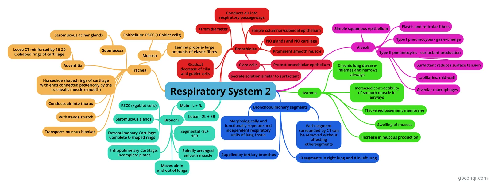
Mind map: Respiratory system
Map content
Respiratory System 2
Trachea
- Horseshoe shaped rings of cartilage with ends connected posteriorly by the trachealis muscle (smooth)
- Conducts air into thorax
- Withstands stretch
- Transports mucous blanket
- Mucosa
- Epithelium: PSCC (+Goblet cells)
- Lamina propria- large amounts of elastic fibres
- Submucosa
- Seromucous acinar glands
- Adventitia
- Loose CT reinforced by 16-20 C-shaped rings of cartiliage
Bronchi
- PSCC (+goblet cells)
- Seromucous glands
- Extrapulmonary Cartilage: Complete C-shaped rings
- Intrapulmonary Cartilage: incomplete plates
- Spirally arranged smooth muscle
- Moves air in and out of lungs
- Main - L + R,
- Lobar - 2L + 3R
- Segmental -8L+ 10R
Bronchopulmonary segments
- Morphologically and functionally seperate and independent respiratory units of lung tissue
- Supplied by tertiary bronchus
- Each segment surrounded by CT can be removed without affecting othersegments
- 10 segments in right lung and 8 in left lung
Bronchioles
- <1mm diameter
- Conducts air into respiratory passageways
- Simple columnar/cuboidal epithelium
- NO glands and NO cartilage
- Prominent smooth muscle
- Gradual decrease of cilia and goblet cells
- Clara cells
- Protect bronchiolar epithelium
- Secrete solution similar to surfactant
Alveoli
- Simple squamous epithelium
- Type I pneumocytes - gas exchange
- Type II pneumocytes - surfactant production
- Surfactant reduces surface tension
- Elastic and reticular fibres
- capillaries: mid-wall
- Alveolar macrophages
Asthma
- Chronic lung disease- inflames and narrows airways
- Increased contractibility of smooth muscle in airways
- Thickened basement membrane
- Swelling of mucosa
- Increase in mucous production
Information
- Description
- Mind Map on Respiratory System 2, created by Colleen Curley on 19/04/2016.
- Created by
- Colleen Curley
- Views
- 65
- Tags
- Rating