
Notes
The Heart
Content of the notes (1)
Page 1
Role of the cardiovascular system:
Primary transport system in the body
Delivers nutrients oxygen
Removes waste
Role of the heart:
Pump blood to the lungs
Pump blood around the body
Where is it located?
Thorax
Mediastinum - structure between the lungs
Left side, with the apex near the sternum
Shape and structure:
Conical shape
Within the pericardial sac:
- OUTER LAYER: Parietal pericardium - tough and dense tissue
-INNER LAYER: Serous pericardium - secretes fluid to lubricate
Three layers:
- OUTER LAYER: Epicardium (secretes mucus)
- MIDDLE LAYER: Myocardium (heart muscle- does all the hard work)
- INNER LAYER: Endocardium (lining of the heart)
Heart chambers:
Four chambers - left and right side separated by the cardiac septum
TOP CHAMBERS (cranial) - L&R atrium
BOTTOM CHAMBERS (caudal) - L&R ventricles pump blood out of the heart
How does the heart beat?
Myocardial cells contract without nerves supplying them
Cells are linked by intercolated discs that all the contractions to continue between them
Areas of myocardial tissue control the flow of the contraction:
- SINO ATRIAL NODE IN RA - Pacemaker starts the contraction which spreads across atria and prevented from entering ventricles by insulating barrier
- ATRIOVENTRICULAR NODE (AV NODE) - Receives the contraction inbetween ventricles and passes to Bundle of His
- BUNDLE OF HIS
- PURKINJE FIBRES - Spreads through ventricles by Purkinje Fibres, contracting from the apex up towards the base- stimulates myocardial cells - pushes blood through the heart
What happens inside the heart?
Four valves ensures that the blood moves in the right direction
ATRIOVENTRICULAR VALVES -area between the atria and ventricles
- RIGHT AV VALVE (AKA Tricuspid valve) - three fibrous tissue cusps
- LEFT AV VALVE (AKA Bicuspid valve/Mitral valve) - two fibrous cusps
Valves are prevented from inverting by the subvalvular apparatus so it doesn't open the wrong way
At the exit of the ventricles are the semilunar valves:
- PULMONARY VALVE - at exit of the right ventricle (lungs)
- AORTIC VALVE - at exit of the left ventricle (aorta)
How does blood flow through the heart?
Blood is first pumped to the lungs and then around the body
Pumped simultaneously by two sides of the heart
The left side is more muscled than the right as it pumps blood to the whole body
BLOOD VESSELS INVOLVED:
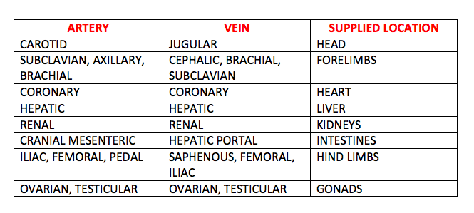
VENA CAVA: brings blood to the RA (caudal VC from abdomen, cranial VC from the head)
PULMONARY ARTERY: takes blood from the RV to the lungs (the only artery to carry deoxygenated blood)
PULMONARY VEIN: brings blood from the lungs to the LA (the only vein to carry oxygenated blood)
AORTA: Takes blood from LV to the body
VEINS = DEOXYGENATED TOWARDS TH HEART, BLOOD CARRIED AT LOW PRESSURE, VALVES PREVENT BLOOD POOLING
ARTERY = OXYGENATED AWAY FROM THE HEART, HIGH PRESSURE, THICK MUSCULAR WALLS, SMOOTH MUSCLE
CAPILLARIES= MIX AS BLOOD SWAPS, BRANCHING NETWORK IN PERFUSED BODY TISSUE, EASILY DAMAGED, ALLOWS GASES, NUTRIENTS AND FLUID TO LEAVE THE BLOOD AND ENTER TISSUE, ONE CELL THICK
The Hepatic Portal System
Key role of liver is to detoxify the blood and use key nutrients from digestion
Blood from small intestine is taken directly to the liver by the hepatic portal vein
Liver also supplied by hepatic artery which provides its own nutrients
Blood from both vessels leave the liver by the hepatic vein
Blood sampling
Needs to be arterial blood for blood gasses
Easy sampling - venous blood as closer to surface of skin, less risk of haemorrhaging, veins are thinner, low pressure
DOG: Jugular, Cephalic, Lateral Saphenous
CAT: Jugular, Cephalic, Medial Saphenous
Fetal Circulation
Oxygen comes from placenta via the umbilical vein, not the lungs
Some nutrients from umbilical vein is first dropped off at liver
Bypasses by ductus venosus to quickly get oxygen rich blood quickly to the heart
At the heart oxygenated blood bypass the lungs via the foramen ovale or ductus arteriosus
This oxygen rich blood is sent to brain and rest of body
Blood returns to the placenta via the umbilical artery
What is blood pressure?
The force exerted on the walls of the blood vessels (recorded in mmHg/milimetres of mercury)
Blood pressure drives blood through capillary beds allowing nutrients and waste exchange
Controlled long term through renal mechanisms/ short term via CVS
Monitored around the body by Baroreceptors which is sent to Medulla (brain)
Medulla sends messages via the sympathetic nervous system and parasympathetic nervous system
Makes changes to systemic vascular resistance and cardiac output (factors that can quickly affect BP)
Medulla receives messages also from chemoreceptors located in cartoid artery and aorta which monitors O2, CO2 and PH
Systemic Vascular Resistance (SVR)
Size of the blood vessels
When blood vessels constrict (smaller), BP goes UP (vasoconstriction)
When blood vessels dilate (larger), BP goes DOWN (vasodilation)
How much blood comes out of the heart?
Measured in mL/min
Influenced by: how fast the heart is pumping (HR) and how much blood is has to pump (SV)
CARDIAC OUTPUT = STROKE VOLUME X HEART RATE
Taking a pulse rate
Count for 15 seconds x 4 / 30 seconds x 2 / 1 minute
Assess character: weak/strong/thready/bounding
Compare with heart sound
Missing pulses are called pulse deficits
Auscultation
Lubb - S1 (L & R atrioventricular valves) dubb - S2 (L&R semi lunar valves)
Sound made by closure of heart valves
Information
- Description
- Veterinary Nursing Note on The Heart, created by Lexi Davey on 21/04/2019.
- Created by
- Lexi Davey
- Views
- 3
- Rating