
Notes
Chapter 15: Homeostasis
Content of the notes (8)
Page 1
15.1 The principles of homeostasis
Homeostasis is the process of keeping a constant internal environment. It is important for the body to have a stable environment so that cells can function properly. In order to do this, there are several things that need to be regulated in the body:
•The amount of water in the body (osmoregulation)
•The amount of glucose in the body (glucoregulation)
•The body’s temperature (thermoregulation)
•The amount of nitrogenous waste in the body
Negative Feedback is where deviation away from the norm produces a response which returns the system back to the norm. Positive Feedback is where deviation away from the norm produces a response which further increases the level away from the norm. Homeostasis is the maintenance of a constant internal environment.
Positive and negative feedback
Homeostasis depends on sensory receptors detecting small changes in the body, and effectors working to restore the status quo. These precise control mechanisms in the body are based on feedback systems that enable the maintenance of a relatively steady state around a narrow range of conditions.
Negative feedback: Most of the feedback systems in the body involve negative feedback. A small change in one direction is detected by sensory receptors. As a result, effectors work to reverse the change and restore conditions to their base level. Negative feedback systems work to reverse the initial stimulus. You have seen negative feedback in action in the control of blood sugar levels by insulin and glucagon. Negative feedback systems are also important in many other aspects of homeostasis including temperature control and the water balance of the body. Thermoregulation works by having thermoreceptors detect changes in the body temperature. and will send information to the hypothalamus which controls a variety of effectors that respond to a decrease in body temperature. Blood vessels near the skin constrict, reducing blood flow (and consequent heat loss) to the environment. Skeletal muscles will contract rapidly in response to a decrease in body temperature as the shivering helps to generate heat, which increases body temperature.
Positive feedback: These systems are not as common in the body. In a positive feedback system, a change in the internal environment of the body is detected by sensory receptors, and effectors are stimulated to reinforce that change and increase the response. One example occurs in the blood clotting cascade. When a blood vessel is damaged, platelets stick to the damaged region and they release factors that initiate clotting and attract more platelets. These platelets also add to the positive feedback cycle and it continues until a clot is formed. Another example of a positive feedback mechanism is seen during childbirth. The head of the baby presses against the cervix, stimulating the production of the hormone oxytocin. Oxytocin stimulates the uterus to contract. pushing the head of the baby even harder against the cervix and triggering the release of more oxytocin. This cycle continues until the baby is born.
Endotherms, such as birds and mammals, use metabolic heat to maintain a stable internal temperature, often one different from the environment.
Ectotherms, like lizards and snakes, do not use metabolic heat to maintain their body temperature but take on the temperature of the environment.
Page 2
15.2 Thermoregulation in ectotherms
Organisms are constantly heating up and cooling down as a result of their surroundings. These changes depend on a number of physical processes. These include exothermic chemical reactions, latent heat of evaporation, radiation, convection, and conduction. Thermoregulation in both ectotherms and endotherms is controlled mainly by the preoptic area of the anterior hypothalamus. Such homeostatic control is separate from the sensation of temperature.
Most animals are ectotherms and use their surroundings to warm their bodies (ectotherm means 'outside heat'). Their core body temperature is heavily dependent on their environment. Ectotherms include all the invertebrate animals, along with fish, amphibians, and reptiles. Many ectotherms living in water do not need to thermoregulate . The high heat capacity of water means that the temperature of their environment docs not change much. Ectotherms that live on land have a much bigger problem with temperature regulation. The temperature of the air can vary dramatically both between seasons and even over the period of a day from the middle of the day to the end of the night. As a result ectotherms have evolved a range of strategies that enable them to cool down or warm up.
Behavioural responses
Ectotherms display a number of behaviours which increase or reduce the radiation they absorb from the Sun. Sometimes they need to warm up to reach a temperature at which their metabolic reactions happen fast enough for them to be active. They may bask in the Sun, orientate their bodies so that the maximum surface area is exposed to the Sun, and even extend areas of their body to increase the surface area exposed to the Sun. For example, lizards often bask for long periods of time to get warm enough to move fast and hunt their prey, and insects such as locusts and butterflies orientate themselves for maximum exposure to the sun and spread their wings to increase the available surface area to get warm enough to fly. Similar behaviour can also be seen in limas when, to absorb more heat from the sun, they will close their eyes and open out their arms facing the sun.
Ectotherms can increase their body temperature through conduction by pressing their bodies against the warm ground. They also get warmer as a result of exothemic metabolic reactions. Galapagos iguanas will contract their muscles and vibrate increasing cellular metabolism to raise their body temperature. Similarly, moths and butterflies may vibrate their wings to warm their muscles before they take night. Ectotherms some times need to cool down to prevent their core temperature reaching a point where enzymes begin to denature. To cool down, many of the warming processes arc reversed. Ectotherms shelter from the sun by seeking shade, hiding in cracks in rocks. or even digging burrows. They will press their bodies against cool, shady earth or Stones. or move into available water or mud. They turn their bodies so that the minimum surface area is exposed to the sun, and minimise their movements to reduce the metabolic heat generated.
Physiological responses
Much of the thermoregulation by ectotherms is the result of behavioural responses but some of them have physiological responses as well. Dark colours absorb more radiation than light colours. Lizards living in colder climates tend to be darker coloured than lizards living in hotter countries so that they get warmer. Some ectotherms also alter their heart rate 10 increase o r decrease the metabolic rate and sometimes to affect the warming or cooling across the body surfaces. Ectotherms a re always more vulnerable fluctuations in the environment than endotherms. However, by using a variety of behavioural and physiological strategies many of them can maintain relatively stable core temperatures. They need less food than endotherms as they use less energy regulating their temperatures. and so they ca n survive in some very difficult habitats where food is in short supply. During periods of cold, some ectotherms enter a state of torpor, in which their metabolism slows or, in some cases, such as the wood frog, effectively stops. The torpor might last overnight or last for a season, or even for years, depending on the species and circumstances.
There is a difference between exothermic and endothermic despite the two terms seeming interchangeable. Whereas the thermodynamic terms "exothermic" and "endothermic" respectively refer to processes that give out heat energy and processes that absorb heat energy, in biology this is effectively inverted. The metabolic term "ectotherm" is given to organisms that rely largely on external heat to achieve a full working temperature, and "endotherm" refers to organisms that produce heat from within as a major factor in controlling their bodily temperature.
Page 3
15.3 Thermoregulation in endotherms
Receptors are used in homeostasis to detect changes in the internal environment. The peripheral temperature receptors are in the skin and detect changes in the surface temperature. Temperature receptors in the hypothalamus detect the temperature of the blood deep in the body. The temperature of the skin is much more likely to be affected by external conditions than the temperature of the hypothalamus.
The combination of the two gives the body great sensitivity and allows it to respond not only to actual changes in the temperature of the blood but to pre-empt possible problems that might result from changes in the external environment. The temperature receptors in the hypothalamus act as the thermostat of the body, controlling the responses that maintain the core temperature in a dynamic equilibrium of 37°C give or take 1.
Methods of thermoregulation
Endotherms use their internal exothermic metabolic activities to keep them warm, and energy-requiring physiological responses to help them cool down. They also have passive ways of heating up and cooling down, to reduce the energy demands on their bodies. Like ectotherms, endotherms have a range of behavioural responses to temperature changes that include basking in the sun, pressing themselves to warm surfaces, wallowing in water and mud to cool down. and digging burrows to keep warm or cool Some animals even become dormant through the coldest weather (hibernation) or through the hottest weather (aestivation is a period of prolonged or deep sleep similar to hibernation but occurs in summer or during dry seasons to avoid heat stress rather than cold).
Humans have additional behavioural adaptations to help control body temperature- clothes are worn to stay warm, houses are built, and then heated up or cooled down to maintain the ideal temperature. In spite of these behavioural responses, endotherms mainly rely on physiological adaptations to maintain a stable core body temperature, regardless of the environmental conditions or the amount of exercise being done. These adaptations include the peripheral temperature receptors, the thennorcgularory cenrres of the hypothalamus, the skin, and muscles.
Centres for thermoregulation
The physiological responses of endotherms to changes in the core temperature are the result of complex homeostatic mechanisms involving negative feedback control from the hypothalamus, there are two control centres:
The heat loss centre: is activated when the temperature of the blood flowing through the hypothalamus increases. It sends impulses through autonomic motor neurones to effectors in the skin and muscles, triggering responses that act to lower the core temperature.
The heat gain centre: This is activated when the temperature of the blood flowing through the hypothalamus decreases. lr sends impulses through the autonomic nervous system to effectors in the skin and the muscles, triggering responses that act to raise the core temperature. The interaction of the sensory receptors, the autonomic nervous system, and the effectors in a sophisticated feedback system enables endotherms to maintain a very stable core body temperature regardless of environmental conditions or activity levels.
Decreasing temperature
When endotherms get too warm, measures need to be taken to decrease the temperature back to the ideal, this can be achieved through physical and behavioural methods.
vasodilation: The arterioles near the surface of the skin dilate when the temperature rises. The vessels that provide a direct connection between the arterioles and the venules (the arteriovenous shunt vessels) constrict. This forces blood through the capillary networks close to the surface of the skin. The skin flushes and cools as a result of increased radiation. If the skin is pressed against cool surfaces. then the cooling results from conduction.
increased sweating: As the core temperature starts to increase, rates of sweating also increase. Sweat sp reads out across the surface of the skin. Some mammals, like humans and horses, have sweat glands all over their body. As the sweat evaporates from the surface of the skin, heat is lost which cools the blood below the surface. In some animals, the sweat glands are restricted to the less hairy areas of the body such as the paws. These animals often open their mouths and pant when they get hot, again losing heat as the water evaporates. In human beings. around l dm3 of sweat is lost by evaporation on a normal day. If the conditions are very hot and dry or the person is exercising very hard. up to 12dm3 of sweat a day can be lost. Kangaroos and cats often lick their front legs to keep cool in high temperatures.
Flattening of hair or feathers: As the body temperature begins to increase. the erector pili muscles (the hair erector muscles) in the skin relax, as a result, the hair or feathers of the animal lie flat to the skin. This avoids trapping an insulating layer of air. It has little effect in humans. Endotherms that live in hot climates often have anatomical adaptations as well as the behavioural and physiological adaptations already described. These minimise the effect of high temperatures and maximise the ability of the animal to cool down through the surface area of the body. They include a relatively large surface area: volume ratio to maximise cooling (e.g. include large ears and wrinkly skin). and pale fur or feathers to reflect radiation. Birds counteract overheating by gular fluttering, or rapid vibrations of the gular (throat) skin. Down feathers trap warm air acting as excellent insulators just as hair in mammals acts as a good insulator.
Increasing temperature
shivering: A response to cold in humans and other warm-blooded animals. When the core body temperature drops, the shivering reflex is triggered to maintain homeostasis. Skeletal muscles begin to shake in small movements, creating warmth by expending energy. Shivering can also be a response to a fever, as a person may feel cold. During fever the hypothalamic set point for temperature is raised. The increased set point causes the body temperature to rise (pyrexia), but also makes the patient feel cold until the new set point is reached. Severe chills with violent shivering are called rigors. Rigors occur because the patient's body is shivering in a physiological attempt to increase body temperature to the new set point.
vasoconstriction: The arterioles ncar the surface of the skin constrict. The arteriovenous shunt vessels dilate, so very little blood flows through the capillary networks close to the surface of the skin. The skin looks pale, and very little radiation takes place. The warm blood is kept well below the surface.
Decreased sweating: As the core temperature falls, rates of sweating decrease and sweat production will stop entirely. This greatly reduces cooling by the evaporation of water from the surface of the skin, although some evaporation from the lungs still continues.
Raising the body hair or feathers: As the body temperature falls, the erector pili muscles in the skin con tract, pulling the hair or feathers of the animal straight up which traps an insulating layer of air and reduces cooling through the skin. The effect can be quite dramatic and it is a very effective way to reduce heat loss to the environment in many animals. In humans this has little effect although you can observe the hairs being pulled upright. In cold weather, birds fluff their feathers and animals raise their fur to thicken the insulating layer. The same response in people (goosebumps) is not as effective because of our limited body hair.
Page 4
15.4 Excretion, homeostasis, and the liver
Excretion in mammals
All mammals produce metabolic wastes, but the main ones are listed below.
Carbon dioxide: one or the waste products of cellular respiration which is excreted from the lungs.
Bile pigments: formed from the breakdown of haemoglobin from old red blood cells in the liver. They are excreted in the bile from the liver into the small intestine via the gall bladder and bile duct. They colour the faeces.
Nitrogenous waste products (urea): formed from the breakdown of excess amino acids by the liver. All mammals produce urea as their nitrogenous waste. Fish produce ammonia while birds and insects produce uric acid. Urea is excreted by the kidneys in the urine.
The liver
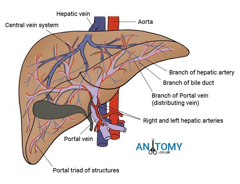
The liver is one of the major body organs involved in homeostasis. It is a reddish-brown organ which makes up about 5% of the total body mass (the largest internal organ of the body).It lies just below the diaphragm and is made up of several lobes. The liver is very fast growing and damaged areas generally regenerate very quickly. The liver has a very 1ich blood supply - about I dm3 of blood nows through it every minute. Oxygenated blood is supplied to the liver by the hepatic artery and removed from the liver and returned to the heart in the hepatic vein.
The liver is also supplied with blood by a second vessel, the hepatic portal vein. This carries blood loaded with the products of digestion straight from the intestines to the liver and this is the starting point for many metabolic activities of the liver. Up to 75% of the blood flowing through the liver comes via the hepatic portal vein.
The liver carries out many different complex functions but the cells are surprisingly simple and uniform in appearance. Liver cells or hepatocytes have large nuclei. prominent Golgi apparatus, and lots of mitochondria. indicating that they are metabolically active cells. They divide and replicate, even if around 65% of the liver is lost, it will regenerate in a matter of months.
The blood from the hepatic artery and the hepatic portal vein is mixed in spaces called sinusoids which are surrounded by hepatocytes. This mixing increases the oxygen content of the blood from the hepatic portal vein, supplying the hepatocytes with enough oxygen for their needs. The sinusoids contain Kupffer cells, which act as the resident macrophages of the liver, ingesting foreign particles and helping to protect against disease. The hepatocytes secrete bile from the breakdown of the blood into spaces called canaliculi, and from these the bile drains into the bile ductules which take it to the gall bladder.
Histology of the liver
The liver is connected to the rest of the body through four transporting vessels:
Hepatic artery: supplies the liver with oxygenated blood from the heart
Hepatic vein: carries deoxygenated blood away from the liver
Hepatic portal vein: brings blood rich in products of digestion from the dodenum (first part of the small instestine) and ingested harmful substances can be removed or broken down straight away.
Bile duct: takes bile that is produced by the liver to the gall bladder to be stored.
Branches of the bile duct, hepatic artery and hepatic portal vein form the portal track/triad.
Functions of the liver
Detoxification: The level of toxins in the body always tends to increase. Apart from urea, many other metabolic pathways produce potentially harmful substances. We also take in a wide variety of toxins by choice such as alcohol and other drugs. The liver is the site where most of these substances are detoxified and made harmless.
Carbohydrate metabolism: Hepatocytes are closely involved in the homeostatic control of glucose levels in the blood by their interaction with insulin and glucagon. When blood glucose levels rise, insulin levels rise and stimulate hepatocytes to convert glucose to the storage carbohydrate glycogen. About 100g of glycogen is stored in the liver. Similarly, when blood sugar levels start to falL the hepatocytes convert the glycogen back to glucose under the influence of the hormone glucagon.
Deamination of excess amino acids: The liver plays a vital role in protein metabolism where hepatocytes synthesise most of the plasma proteins. Hepatocytes also carry out transamination (the conversion of one amino acid into another). This is important because the diet does not always contain the required balance of amino acids but transamination can overcome the problems this might cause.
The most important role of the liver in protein metabolism is in deamination - the removal of an amine group from a molecule. The body cannot store either proteins or amino acids. Any excess ingested protein would be excreted and therefore wasted if it were not for the action of the hepatocytes. They deaminate the amino acids, removing the amino group, and converting it first into ammonia which is very toxic and the n to urea. Urea is toxic in high concentrations but not in the concentrations normally found in the blood. Urea is excreted by the kidneys (you will learn about the role of the kidneys in excretion and water balance in Topics 15.5 and 15.6). The remainder of the amino acid can then be fed into cellular respiration or converted into lipids for storage.
https://www.abpischools.org.uk/topic/homeostasis-kidneys/6
Page 5
5.5 The structure and function of the mammalian kidney
Structure of the kidneys
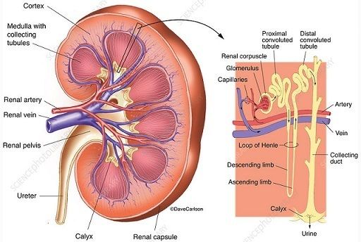
The kidneys are supplied with blood at arterial pressure by the renal arteries that branch off from the abdominal aorta. Blood that has circulated through the kidneys is removed by the renal vein that drains into the inferior vena cava. About 90-120cm3 of blood passes through the kidneys every minute. All of the blood in the body passes through the kidneys about once an hour. The kidneys filter J 80dm3 of blood a day, producing l- 2 dm3 of urine. The final volume depends on many different factors.
The kidneys are made up of millions or small structures called nephrons that act as filtering units. The sterile liquid produced by the kidney tubules is called urine. The urine passes out of the kidneys down tubes called ureters. lt is collected in the bladder, a muscular sac that can store around 40Q-600 cm3 of urine. When the bladder is getting full, the sphincter at the exit to the bladder opens and the urine passes out of the body down the urethra.
The kidney consists of three main areas, the cortex, the medulla, and the pelvis. The cortex is the dark outer layer, This is where the filtering of the blood takes place and it has a very dense capillary network carrying the blood from the renal artery to the nephrons. The medulla is lighter in colour- it contains the tubules of the nephrons that form the pyramids of the kidney and the collecting duas. The pelvis (which is Latin for basin) of the kidney is the central chamber where the urine collects before passing out down the ureter.
Nephrons
ln the nephrons, the blood is filtered and then the majority of the filtered material is returned to the blood, removing nitrogenous wastes and balancing the mineral ions and water. Each nephron is around 3 cm long and there are around 1.5 million nephrons in each kidney. This provides the body with several kilometres of tubules for the reabsorption of water, glucose, salts, and other substances back into the blood. The structure of a nephron, in order, is listed.
Bowman's capsule: cup-shaped structure that contains the glomerulus, a tangle of capillaries. More blood goes into the glomerulus than leaves it due to the ultra filtration processes that take place.
Proximal convoluted tubule: the first, coiled region of the tubule after the Bowman's capsule, found in the cortex of the kidney. This is where many of the substances needed by the body are reabsorbed into the blood.
Loop of Henle: a long loop of tubule that creates a region with a very high solute concentration in the tissue nuid deep in the kidney medulla . The descending loop runs down from the cortex through the medulla to a hairpin bend at the bottom of the loop. The ascending limb travels back up through the medulla to the cortex.
Distal convoluted tubule: a second twisted tubule where the fine-tuning of the water balance of the body takes place. The kidney and osmoregulation). Further regulation of the ion balance and pH of the blood also takes place in this tubule.
Collecting duct: the urine passes down the collecting duct through the medulla to the pelvis. More fine-Luning of 1hc water balance takes place- the walls of this part of the tubule arc also sensitive to ADH.
The nephron has a network of capillaries around it which finally lead into a venule and then to the renal vein. The blood that leaves the kidney has greatly reduced levels of urea, but the levels of glucose and other substances such as amino acids needed by the body a rc almost the same as when the blood entered the kidneys (may be slightly less as some glucose will have been used for selective reabsorption). The mineral ion concentration in the blood has also been restored to ideal levels.
The four mechanisms used to create and process the filtrate are filtration, reabsorption, secretion and excretion. Filtration occurs in the glomerulus and is largely passive: it is dependent on the intracapillary blood pressure. About one-fifth of the plasma is filtered as the blood passes through the glomerular capillaries; four-fifths continues into the peritubular capillaries. Normally the only components of the blood that are not filtered into Bowman's capsule are blood proteins, red blood cells, white blood cells and platelets.
Ultrafiltration
The first stage in the removal of nitrogenous waste and osmoregulation of the blood is ultrafiltration. Ultrafiltration in the kidney tubules is a specialised form of the process that results in the formation of tissue fluid in the capillary beds of the body and it is the resuIt of the structure of the glomerulus and the cells lining the Bowman's capsule.
The glomerulus is supplied with blood by a re la tively wide afferent (incoming) arteriole from the renal artery. The blood leaves through a narrower efferent (outward) arteriole and as a result there is considerable pressure in the capillaries of the glomerulus. This forces the blood out through capillary wall- it acts rather like a sieve . Then the fluid passes through the basement membrane - scientists are increasingly recognising the basement membrane as an important factor in the filtration process. The basement membrane is made up of a ne two rk of collagen fibres and o th er proteins that make up a second 'sieve'. Most of the plasma contents can pass through the basement membrane but the blood cells and many proteins are retained in the capillary because of their size.
The wall ol the Bowman's capsule also involves special cells called podocytes that act as an additional filter. They have extensions called pedice/s that wrap around the capillaries, forming slits that make sure any cells, platelets, or large plasma proteins that have managed to get through the epithelial cells and the basement membrane do not get through into the tubule itself. The filtrate which enters the capsule contains glucose, salt. urea, and many other substances in the same concentrations as they arc in the blood plasma. The process is so efficient that up to 20% of the water and solutes arc removed from the plasma as it passes through the glomerulus. The volume of blood that is filtered through the kidneys in a given time is known as the glomerular filtration rate.
Reabsorption
Ultrafiltration removes urea, the waste product of protein breakdown, from the blood but it also removes a lot of water along with the glucose, salt, and other substances which are present in the plasma. Many of these substances are needed by the body- for example, glucose is used for cellular respiration and is never normally excreted. The ultrafiltrate is also hypotonic to (less concentrated than) the blood plasma. The main function of the nephron after the Bowman's capsule is to return most of the filtered substances back to the blood.
Once the substances have been removed from the nephron, they diffuse into the extensive capillary network which surrounds the tubules down steep concentration gradients. These are maintained by the constant flow of blood through the capillaries. The filtrate reaching the loop of Henle at the end of the proximal convoluted tubule is isotonic (at same concentration) with the tissue fluid surrounding the tbule and isotonic with the blood. At this stage over 80% of the glomerular filtrate has been reabsorbed back into the blood. This remains the same regardless of the conditions in the body.
The loop of henle
The loop of Henle is a U-shaped tube that extends from the proximal tubule. It consists of a descending limb and an ascending limb. It begins in the cortex, receiving filtrate from the proximal convoluted tubule, extends into the medulla as the descending limb, and then returns to the cortex as the ascending limb to empty into the distal convoluted tubule. The primary role of the loop of Henle is to concentrate the salt in the interstitium, the tissue surrounding the loop. The changes that take place in the descending limb of the loop of Henle depend on the high concentrations of sodium and chloride ions in the tissue fluid of the medulla that arc the result of events in the asceidning limb of the loop.
Considerable differences aid in distinguishing the descending and ascending limbs of the loop of Henle. The descending limb is permeable to water and noticeably less permeable to salt, and thus only indirectly contributes to the concentration of the interstitium. As the filtrate descends deeper into the hypertonic interstitium of the renal medulla, water flows freely out of the descending limb by osmosis until the tonicity of the filtrate and interstitium equilibrate. The hypertonicity of the medulla (and therefore concentration of urine) is determined in part by the size of the loop of Henle.
Unlike the descending limb, the thin ascending limb is impermeable to water, a critical feature of the countercurrent exchange mechanism employed by the loop. The ascending limb actively pumps sodium out of the filtrate, generating the hypertonic interstitium that drives countercurrent exchange. In passing through the ascending limb, the filtrate grows hypotonic since it has lost much of its sodium content. This hypotonic filtrate is passed to the distal convoluted tubule in the renal cortex
Distal convoluted tubule
The distal convoluted tubule has a different structure and function to that of the proximal convoluted tubule. Cells lining the tubule have numerous mitochondria to produce enough energy (ATP) for active transport to take place. Much of the ion transport taking place in the distal convoluted tubule is regulated by the endocrine system. In the presence of parathyroid hormone, the distal convoluted tubule reabsorbs more calcium and secretes more phosphate. When aldosterone is present, more sodium is reabsorbed and more potassium secreted. Atrial natriuretic peptide causes the distal convoluted tubule to secrete more sodium. If the body lacks salt, sodium ions will be actively pumped out of the distal convoluted tubule with chloride ions following down an electrochemical gradient. Water can also leave the distal tubule, concentrating the urine, if the walls of the tubule are permeable in response to ADH. The distal convoluted tubule also plays a role in balancing the pH of the blood.
The collecting duct
The collecting duct passes down through the concentrated tissue fluid of the renal medulla. This is the main site where the concentration and volume of the urine produced is determined. Water moves out of the collecting duct by diffusion down a concenrrarion gradient as it passes through the renal medulla. As a result the urine becomes more concentrated. The level of sodium ions in the surrounding fluid increases through the medulla from the cortex to the pelvis. This means water can be removed from the collecting duct all the way along its length, producing very hypertonic urine when the body needs to conserve water. The permeability of the collecting duct to water is controlled by the level of ADH, which determines how much or little water is reabsorbed. You will learn more about the role of the collecting duct in maintaining the water potential of the blood in Topic 15.6, The kidney and osmoregulation.
Page 6
15.6 The kidney and osmoregulation
Osmoregulation
Osmoregulators tightly regulate their body osmolarity, maintaining constant internal conditions. They are more common in the animal kingdom. Osmoregulators actively control salt concentrations despite the salt concentrations in the environment. An example is freshwater fish. The gills actively uptake salt from the environment by the use of mitochondria-rich cells. Water will diffuse into the fish, so it excretes a very hypotonic (dilute) urine to expel all the excess water. A marine fish has an internal osmotic concentration lower than that of the surrounding seawater, so it tends to lose water and gain salt. It actively excretes salt out from the gills. Most fish are stenohaline, which means they are restricted to either salt or fresh water and cannot survive in water with a different salt concentration than they are adapted to.
Page 7
15.7 Urine and diagnosis
Urine and pregnancy testing
The human embryo implants in the uterus, around six days after conception. The site of the developing placenta then begins to produce a chemical called human chorionic gonadotrophin (hCG). Some of this hormone is found in the blood and the urine of the mother. Until the 1960s, the most reliable available pregnancy test was to inject the urine from a pregnant woman into an African clawed toad (Xenopus laevis). If she was pregnant, the hCG triggered egg production in the wad within 8-12 hours of the injection. It could not be used until the woman was several weeks pregnant.
Modern pregnancy tests still test for hCG in the urine, but they rely on monoclonal antibodies. Some are so sensitive that pregnancy can be detected within hours of implantation.
Making monoclonal antibodies
Monoclonal antibodies are antibodies from a single clone of cells that are produced to target particular cells or chemicals in the body.
A mouse is injected with hCG so it makes the appropriate antibody. The B-cells that make the required antibody are then removed from the spleen of the mouse and fused with a myeloma, a type of cancer cell which divides very rapidly. This new fused cell is known as a hybridoma. Each hybridoma reproduces rapidly, resulting in a done of millions
of 'living factories' making the desired antibody. These monoclonal antibodies are collected, purified and used in a variety of ways.
These are the main stages in a pregnancy test:
the wick is soaked in the first urine passed in the morning- this will have the highest levels of hCG.
The test contains mobile monoclonal antibodies that have very small coloured beads attached to them. They will only bind to hCG. If the woman is pregnant the hCG in her urine binds to the mobile monoclonal antibodies and forms a hCG/antibody complex (complete with coloured bead).
The urine carries on along the test structure until it reaches a window.
Here there are immobilised monoclonal antibodies arranged in a line or a pattern such as a positive (+) sign that only bind to the hCG/antibody complex. if the woman is pregnant, a coloured line or pattern appears in the first window.
The urine continues up through the test to a second window.
Here there is usually a line of immobilised monoclonal antibodies that bind only to the mobile antibodies, regardless of whether they are bound to hCG or not. This coloured line forms regardless of whether the woman is pregnant - it simply indicates thai 1he Lest is working
Urine and anabolic steroids
Some athletes have tried to cheat in competitions by using anabolic steroids. Anabolic steroids are drugs that mimic the action of the male sex hormone testosterone and they stimulate the growth of muscles. They are,sz however, excreted in the urine. By testing the urine using gas chromatography and mass spectrometry, scientists can show that an individual has been using these drugs. which are banned in all sports. The urine sample is vaporised with a known solvent and passed along a wbe. The lining of the tube absorbs the gases and is analysed to give a chromatogram that can be read to show the presence of the drugs.
Urine and drug testing
Urine is tested for tlhc presence of many different drugs, inducting alcohol. Because drugs or metabolites - the breakdown products of drugs- are filtered through the kidneys and stored in the bladder, iL is possible to find drug traces in the mine some time after a drug has been used.
If someone is suspected of having taken an illegal drug, they may be asked to provide a urine sample and this will be divided into two. The first sample may be tested by an immunoassay, using monoclonal antibodies to bind to the drug or its breakdown product. If this shows positive, the second sample may be run through a gas chromatograph/ mass spectrometer to confirm the presence of the drug.
Page 8
15.8 Kidney failure
There are a number of reasons why the kidneys may fail. They include kidney infections, where the structure of the podocytes and the tubules themselves may be damaged or destroyed; raised blood pressure that can damage the structure of the epithelial cells and basement membrane of the Bowman's capsule; and genetic conditions such as polycystic kidney disease where the healthy kidney tissue is replaced by fluid-filled cysts or damaged by pressure from cysts. lf the kidneys are infected or affected by high blood pressure this may cause:
Protein in the urine- if the basement membrane or podocytes of the Bowman's capsule are damaged, they no longer aa as filters and large plasma proteins can pass into the filtrate and are passed out in the urine.
Blood in the urine- another symptom that the filtering process is no longer working.
If the kidneys fail completely, the concentrations of urea and mineral ions build up in the body. The effects include:
Loss of electrolyte balance- if the kidneys fail, the body cannot excrete excess sodium, potassium, and chloride ions. This causes osmotic imbalances in the tissues and eventual death.
Build-up of toxic urea in the blood - if the kidneys fail the body cannot get rid of urea and it can poison the cells.
High blood pressure - the kidneys play an important role in controlling the blood pressure by maintaining the water balance of the blood. If the kidneys fail, the blood pressure increases
and this can cause a range of health problems including heart problems and strokes.
Weakened bones as the calcium/phosphorus balance in the blood is lost.
Pain and stiffness in joints as abnormal proteins build up in the blood.
Anaemia -the kidneys are involved in the production of a hormone called erythropoietin that stimulates the formation of red blood cells. When the kidneys fail it can reduce the production of red blood cells causing tiredness and lethargy.
Measuring glomerular filtration rate
Kidney problems almost always affect the rate at which blood is filtered in the Bowman's car sules of the nephrons. The glomerular filtration rate (GFR) is widely used as a measure to indicate kidney disease. The rate of filtration is not measured directly- a simple
blood test measures the level of creatinine in the blood. Creatinine is a breakdown product of muscles and it is used to give an estimated glomerular filtration rate (eGFR). The units arc cm 3/ min. If the levels of creatinine in the blood go up. it is a signal that the kidneys arc not working properly. Certain factors need to be taken into account in the calculations to work out GFR. For example, GFR decreases steadily with age even if you arc healthy, and men usually have more muscle mass and therefore more creatinine than women.
Hemodialysis and peritoneal dialysis are not perfect. They do some of the work of the kidney, but it is not as good as a real kidney. So people who need dialysis are not as healthy. They must take medicines. For example, in kidney failure, the kidneys do not make any erythropoetin. Doctors have to give people erythropoetin so they make enough red blood cells.
Haemodialysis
This involves the use of a dialysis machine. It is usually carried out in hospital although sometimes patients will have a machine in their own home. Blood leaves the patient's body from an artery and flows into the dialysis machine, where it flows between partially permeable dialysis membranes. These membranes mimic the basement membrane of the Bowman's capsule. On the other side of the membranes is the dialysis fluid. During dialysis it is vital that patients lose the excess urea and mineral ions that have built up in the blood. It is equally important that they do not lose useful substances such as glucose and some mineral ions.
The loss of these is prevented by careful control of the dialysis fluid. It contains normal plasma levels of glucose to ensure there is no net movement of glucose out of the blood. The dialysis fluid also contains normal plasma levels of mineral ions, so any excess mineral ions in the blood move out by diffusion down a concentration gradient into the dialysis fluid, thus restoring the correct electrolyte balance of the blood. The dialysis fluid contains no urea meaning there is a very steep concentration gradient from the blood to the fluid, and as a result, much of the urea leaves the blood. The blood and the dialysis fluid flow in opposite directions to maintain a countercurrent exchange system and maximise the exchange that takes place.
The whole process of dialysis depends on diffusion clown concentration gradients- there is no active transport. Dialysis takes about eight hours and has to be repeated regularly. Patients with kidney failure who rely on haemodialysis have to remain attached to a dialysis machine several times a week for many hours. They also need to manage their diets carefully, eating relatively lillie protein and salt and monitoring their fluid intake to keep their blood chemistry as stable as possible. The only time they can cat and drink what they like is at the beginning of the dialysis process.
Peritoneal dialysis
Peritoneal dialysis is done inside the body- it makes use of the natural dialysis membranes formed by the lining of the abdomen, that is. the peritoneum. It is usually done at home and the patient can carry on with their normal life while it takes place. Before treatment starts, a cut (incision) is made near your belly button and a thin tube called a catheter is inserted through the incision and into the space inside your abdomen (the peritoneal cavity). This is left in place permanently. The dialysis fluid is introduced into the abdomen using a catheter. It is left for several hours for dialysis to take place across the peritoneal membranes, so that urea and excess mineral ions pass out of the blood capillaries, into the tissue nuid, and out across the peritoneal membrane into the dialysis fluid. The fluid is then drained off and discarded, leaving the blood balanced aga in and the urea and excess minerals removed.
Complications may include infections within the abdomen, hernias, high blood sugar, bleeding in the abdomen, and blockage of the catheter. Use is not possible in those with significant prior abdominal surgery or inflammatory bowel disease. It requires some degree of technical skill to be done properly.
Kidney transplants
Long-term dialysis has some serious side effects. The best solution for the patient is a kidney transplant, where a single healthy kidney from a donor is placed within the body. The blood vessels arc joined and the ureter of the new kidney is inserted imo the bladder. If the transplant is successful the kidney will function normally for many years.
The main problem with transplanted organs is the risk of rejection. The antigens on the donor organ differ
from the antigens on the cells of the recipient and the immune system is likely to recognise this. This can result in rejection and the destruction of the new kidney. There are a number of ways of reducing the risk of rejection. The match between the antigens of the donor and the recipient is made as close as possible. For example, a donor kidney can be used with a 'tissue type' very similar to the recipient (from people with the same blood group).
The recipient is given drugs to suppress their immune response (immunosuppressant drugs) for the rest of their lives. This helps to prevent the rejection of their new organ. immunosuppressant drugs are improving all the time and the need for a really close tissue match is becoming less important. The disadvantage of taking immunosuppressant drugs is that they prevent the patients from responding effectively to infectious diseases. They have to take great care if they become ill in any way. However. most people feel this is a small price to pay for a new, functioning kidney.
Transplanted organs don't last forever with the average transplanted kidney functioning for around 9- 10 years, although some have continued working for around 50 years. Once the organ starts to fail the patient has to rerum to dialysis and wait until another suitable kidney is found.
Transplants Vs dialysis
Dialysis is much more readily available than donor organs, so it is there whenever kidneys fail. It enables the paticm to lead a relatively normal life. However, patients have to monitor their diet carefully and need regular sessions on the machine. Long-term dialysis is much more expensive than a transplant and can eventually cause damage to the body.
If a patient receives a kidney transplant they are free from the restrictions which come with regular dialysis sessions and dietary monitoring. This is generally the ideal scenario for patients waiting for a transplant. The main source of donor kidneys is from people who die suddenly, often from road accidents, strokes, and heart attacks. In the UK, organs can be taken from people if they carry an organ donor card or arc on the online donor register. Alternatively, a relative of someone who has died suddenly can give their consent.
Unfortunately for people needing a transplant, there is a shortage of donor kidneys. Many people do not register as donors. In addition, as cars become safer, fewer people die suddenly in traffic accidents. Whilst good. this means there arc fewer potential donors. At any one time there arc thousands of people undergoing kidney dialysis. Most will not have the opportunity to have a kidney transplant. In 2013-14, 3257 people in the UK had kidney transplants. However, there were still around 6000 people on dialysis waiting for a kidney. There arc increasing numbers of live donor transplants. where a family member of someone with a tissue match donates a kidney. In 2013-14. 2143 people received kidneys from donors who had died, and 1114 were from living donors. In 2011, scientists grew functioning embryonic kidney tissue from stem cells. Going forward, the hope is that whole new kidneys can be grown perhaps even without the antigens which trigger the immune reaction so that patients don't need to take immunosuppressant drugs.
Information
- Description
- A-level Biology (Module 5: Communication, homeostasis, and energy ) Note on Chapter 15: Homeostasis, created by Emily Rowland-Rawson on 21/01/2020.
- Created by
- Emily Rowland-Rawson
- Views
- 3
- Rating