
Notes
Biology: Cell Structure & Organisation
Content of the notes (4)
Page 1
Cell Structure & Organisation
Cells are the fundamental units of life, in which metabolic reactions occur. The detailed structure of a cell, as revealed by the electron microscope, is called its ultrastructure. Simple organisms comprise only one cell, that is, they are unicellular. More complex organisms consist of many cells and are multicellular. They have specialised cells which carry out particular functions. Although cells have features in common, they differ in their internal structure, related to their different functions. There are two types of cells: prokaryotic cells and eukaryotic cells. Eukaryotic cells have a distinct nucleus and membrane-bound organelles. Prokaryotic cells include those of bacteria and have a simpler structure, such as the absence of a nucleus.
Page 2
Cells & Their Organisation
Cell Structure
All cells are surrounded by a membrane made of phospholipids and proteins. Biological membranes are so thin that their structure cannot be distinguished in the light microscope, and in the electron microscope, they appear as a single line.
Eukaryotic cells contain membrane-bound organelles, which are enclosed in the cytoplasm. The advantage is that potentially harmful chemicals, such as enzymes, are isolated and molecules with particular functions, such as chlorophyll, can be concentrated in one area. Membranes provide a large surface area for the attachment of enzymes involved in metabolic processes, and the provide a transport system inside the cell.
When looking at images of cells, it is important to understand the units of measurement. The standard unit of measurement is the metre (m). Biology works in the range of nanometres (nm), when considering molecules, to kilometres (km), when considering ecosystems. But for cells and organelles, micrometres (um), are the most convenient unit.
An organelle is a specialised structure with a specific function inside a cell.
The three principles of cell theory are:
1. All living organisms are composed of one or more cells.
2. The cell is the basic unit of life.
3. Cells can only arise from preexisting cells.
Units
SI stands for Systeme Internationale, the system that defines which units are used for scientific communication. These are the SI units of length:
1000nm = 1um
1000um = 1mm
1000mm = 1m
1000m = 1km
Nucleus
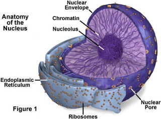
This is the most prominent feature in the cell. It is usually spherical and 10-20um in diameter. It contains DNA which, with protein, comprises the chromosomes. The chromosomes direct protein synthesis because they are the site of transcription. The DNA also provides a template for DNA replication.
The nucleus has a number of components:
1. It is bounded by two membranes, called the nuclear envelope, with pores which allow the passage of large molecules, such as mRNA, and ribosomes. The outer membrane is continuous with the endoplasmic reticulum. Refer to the "nuclear envelope" rather than the "nuclear membrane" because the nucleus is surrounded by two membranes.
2. The granular material in the nucleus is nucleoplasm. It contains chromatin, which is made of coils of DNA bound to protein. During cell division, chromatin condenses in chromosomes.
3. Within the nucleus are one or more small spherical bodies, each called a nucleolus. They are the sites of formation of rRNA, a constituent of ribosomes.
Mitochondria
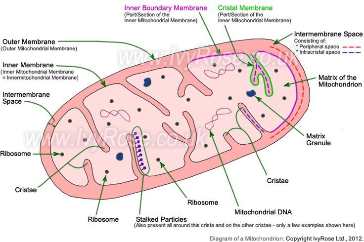
Mitochondria are often cylindrical and 1-10um in length. They comprise:
1. Two membranes, separated by a narrow, fluid-filled inter-membrane space. The inner membrane is folded inwards to form cristae.
2. An organic matrix, which is a solution containing many compounds, including lipids and proteins.
3. Small (70S) ribosomes and a small circle of DNA which enable mitochondria to make some of their own proteins and self-replicate.
The function of mitochondria is to produce ATP in aerobic respiration. Some of the reactions occur in the matrix and other on the inner membrane. The cristae provide a large surface area for the attachment of enzymes involved in respiration.
Metabolically active cells, such as muscle cells, need a plentiful supply of ATP. They contain many mitochondria, reflecting the high metabolic activity taking place.
Being cylindrical, mitochondria have a larger surface area than a sphere of the same volume, in other words, their surface area to volume ratio is bigger. Compared with a sphere, being a cylinder reduces the diffusion distance between the edge and the centre, making aerobic respiration more efficient.
Chloroplasts
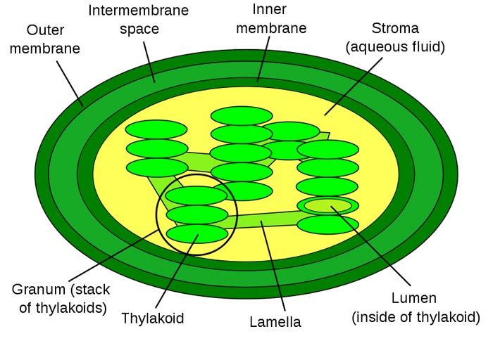
Chloroplasts occur in the cells of photosynthesising tissue. In many plants the highest concentration is in the palisade mesophyll cells, just below the upper surface of the leaf. Each chloroplast is surrounded by two membranes, comprising the chloroplast envelope. Not all cells contain chloroplasts. Root cells, for example, being underground, would have no use for them.
1. The stroma is fluid-filled and contains some of the products of photosynthesis, including lipid droplets and starch grains, which can take up a large part of the stroma.
2. Like mitochondria, they contains 70S ribosomes and circular DNA which enable them to make some of their own proteins and self-replicate.
3. Within the stroma are many closed, flattened sacs called thylakoids. A stack of thylakoids is a granum. Each granum comprises between two and a hundred parallel sacs. The photosynthetic pigments, such as chlorophyll, are found in the thylakoids. This arrangement produces a large surface area, efficient for trapping light energy.
Endosymbiotic Theory
Endosymbiosis is a theory that describes the origin of chloroplasts and mitochondria. As far back as 1883, the division of chloroplasts was seen to closely resemble that of free-living cyanobacteria. In the 1920s, the idea that mitochondria were once independent bacteria was also suggested.
Mitochondria and chloroplasts have 70S ribosomes and circular DNA. It is suggested that at least 1.5x10^9 years ago, some ancient bacteria with very fluid membranes engulfed others and maintained a symbiotic relationship with them. Some of those engulfed were very good at turning glucose and oxygen into ATP, evolving eventually into mitochondria. Some could turn carbon dioxide and water into glucose, evolving eventually into chloroplasts. In 1967, Lynn Margulis published a paper titled "The origin of mitosing cells" which substantiated the idea of endosymbiosis. Now, biologists have plenty of evidence that both chloroplasts and mitochondria have their origins in free-living cells.
Endoplasmic Reticulum (ER)
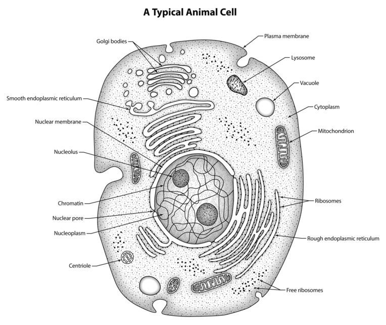
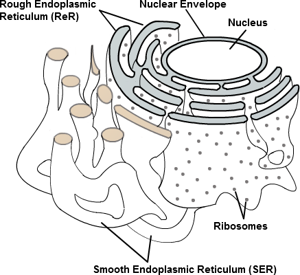
The endoplasmic reticulum is an elaborate system of parallel double membranes forming flattened sacs with interconnected, fluid-filled spaces between them, called cisternae. These are pink in the electron micrograph. The ER is connected with the nuclear envelope. This system allows the transport of materials through the cell. There are two types of ER:
1. Rough ER (RER) has ribosomes on the outer surface and transports the proteins made there. RER is present in large amounts in cells that make a lot of protein, such as cells making amylase in the salivary glands.
2. Smooth ER comprises membranes that lack ribosomes. It is associated with the synthesis and transport of lipids.
Cells that store large quantities of carbohydrates, proteins and lipids, including liver and secretory cells, have extensive ER,
Ribosomes
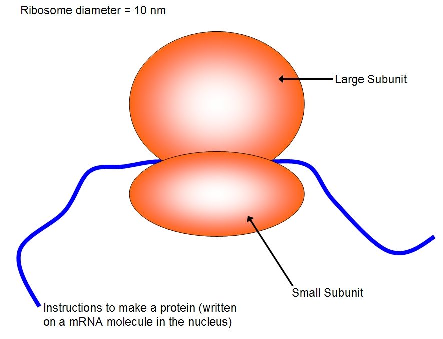
Ribosomes are smaller in prokaryotic that eukaryotic cells. In prokaryotic cells they are 70S in size, whereas those in the cytoplasm of eukaryotic cells are 80S, where they occur singly or attached to membranes on the RER. Ribosomes have one large and one small subunit. They are assembled in the nucleolus from ribosomal RNA (rRNA) and protein. They are important in protein synthesis, as they are the site of translation, where mRNA is used to assemble the polypeptide chain.
To measure the size of ribosomes, scientists see how fast they sink through a solution spun very fast in an ultracentrifuge. Larger and denser structures ink faste. Sedimentation rate is measured in S units. (S stands for Svedburgh, the Swedish scientists who invented the ultracentrifuge.)
Ribosomes are much smaller than the nucleus and mitochondria. They often appear as black dots in electron micrographs. In diagrams, they are often shown like this:
Golgi Body/ Apparatus/ Complex
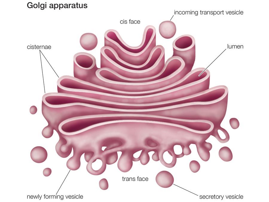
The structure of the Golgi body resembles the ER, but it is more compact. Vesicles containing polypeptides pinch off from the RER and fuse with the stack of membranes which constitute the Golgi body. Proteins are modified and packaged in the Golgi body. At the other end of the Golgi body, vesicles containing the modified proteins are pinched off. These may carry proteins elsewhere in the cell or move to and fuse with the cell membrane, secreting the modified proteins by exocytosis.
The functions of the Golgi body include:
1. Producing secretory enzymes, packaged into secretory vesicles.
2. Secretory carbohydrates, e.g. for the formation of plant cell walls.
3. Producing glycoprotein.
4. Transporting and storing lipids.
5. Forming lysosomes, containing digestive enzymes.
Lysosomes
Lysosomes are small, temporary vacuoles surrounded by a single membrane, formed by being pinched off from the Golgi body. They contain and isolate potentially harmful digestive enzymes from the remainder of the cell. They release these enzymes when the cell needs to recycle worn out organelles. The enzymes in lysosomes can also digest material that has been taken into the cell, e.g. lysosomes fuse with the vesicle made with a white blood cell engulfs bacteria by phagocytosis and their enzymes digest the bacteria.
Some cells are programmed to die in a process called apoptosis, such as in spaces between the fingers and toes in the embryo. Lysosomes and their enzymes have an essential role in this.
Centrioles
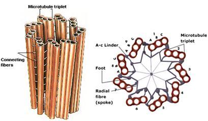
Centrioles occur in all animal cells and most protoctistans but not in the cells of higher plants. They are located just outside the nucleus. Centrioles are two rings of microtubules, making hollow cylinders positioned at right angles to one another. Together they are sometimes called the centrosome. During cell division, centrioles organise the microtubules that make the spindle.
Vacuole
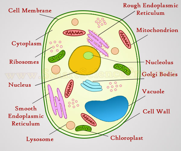
Most plant cells have a large permanent vacuole which consists of a fluid-filled sac bounded by a single membrane, the tonoplast. Vacuoles contain cell sap, a solution with stores chemicals such as glucose, amino acids and minerals, and may store vitamins and pigments, as in oranges. Vacuoles have a major role in supporting soft plant tissues.
Animal cells contain vacuoles but, unlike in plant cells, these are small, temporary vesicles and may occur in large numbers.
Cell Wall
The cell wall of a plant cell consists largely of cellulose. Cellulose molecules are held together in microfibrils, which are aggregated into fibres, embedded in a polysaccharide matrix called pectin. The cell wall has the following functions:
1. Transport - the gaps between the cellulose fibres make the cell wall fully permeable to water and dissolved molecules and ions. The space outside the cells through which solution moves, is called the apoplast. The apoplast pathway is the main way the waste crosses the plant root.
2. Mechanical strength - the structure of cellulose microfibrils and their laminated arrangement make the cell wall very strong. When the vacuole is full of solution, the cell contents push against the cell wall, which resists expansion and the cell becomes turgid, supporting the plant.
3. Communication between cells - cell walls have pores, called pits, through which strands of cytoplasm, called plasmodesmata (singular = plasmodesma), pass. The plasmodesma occurs where there is no cellulose thickening between two cells. The strand of cytoplasm runs from one cell to the next. The network of cytoplasm in connected cells is called the symplast. The symplast pathway is important in water transport through a plant.
Plant cells each have up to about 100000 plasmodesmata connecting them to other cells, with about 10perum^2. Plants do not have a nervous system, and it may be that this high connectivity through the cytoplasm can serve a similar function.
Differences between Plant and Animal Cells
Animal cells
Cell wall: Absent
Chloroplasts: Absent
Plasmodesmata: Absent
Vacuole: present - small, temporary; scattered throughout cell
Centrioles: Present
Energy store: Glycogen
Plant cells
Cell wall: Present - surrounds cell membrane
Chloroplasts: Present - in cells above ground
Plasmodesmata: Present
Vacuole: Present - large, permanent, central; filled with cell sap
Centrioles: Absent
Energy store: Starch
Organelles are Interrelated
Organelles have been described here separately but their functions within the cell are often related:
1. The nucleus contains chromosomes in which the DNA encodes proteins.
2. Nuclear pores in the nuclear envelope allow mRNA molecules, transcribed off the DNA, to leave the nucleus and attach to ribosomes in the cytoplasm or on the rough ER.
3. Ribosomes contains rRNA, transcribed from DNA located on the nucleolus.
4. Protein synthesis occurs on ribosomes, producing proteins in their primary structure.
5. Polypeptides made on the ribosomes are moved through the RER and are packaged into vesicles. The vesicles bud off the RER and carry the polypeptides to the Golgi body, where they are chemically modified and folded.
6. The Golgi body produces vesicles containing newly synthesised proteins. They may be lysosomes, containing digestive enzymes, used within the cell. They may be secretory vesicles, which carry the proteins to the cell membranes for exocytosis.
7. Phospholipids and triglycerides move through the smooth endoplasmic reticulum to various destinations in the cell.
Page 3
Prokaryotic Cells, Eukaryotic Cells and Viruses
The cells of prokaryotes may resemble the first living cells. The oldest fossil prokaryotes are from rocks formed 3.5 billion years ago, so they must have evolved before then, within the first billion years of Earth's history. The cells of eukaryotes probably evolved from prokaryotic cells and the oldest fossilised examples are from rocks about 1.5 billion years old. Fungi, Protoctista, plants and animals all have eukaryotic cells.
Viruses are not made of cells and are not classified with living organisms. They seem to exist at the interface between living and non-living organisms.
Prokaryote: A single-celled organism lacking membrane-bound organelles, such as a nucleus, with its DNA free in the cytoplasm.
Eukaryote: An organism containing cells that have membrane-bound organelles, with DNA in chromosomes within the nucleus.
Prokaryotic Cells
An example of a prokaryotic cell is a bacterium. The major distinguishing feature of prokaryotic cells is that they have no nucleus, or any internal membranes, so, unlike eukaryotic cells, they have no membrane-bound organelles. In some prokaryotes, infolding of the cell membrane in a mesosome or photosynthetic lamellae increases the membrane's surface area. They are unicellular and rarely form multicellular structures.
All prokaryotes have DNA molecules loose in the cytoplasm, a peptidoglycan (murein) cell wall, 70S ribosomes, cytoplasm and cell membrane. Some prokaryotes also have a slime coat, flagella (one, some or many), photosynthetic lamellae holding photosynthetic pigments, mesosome - site of aerobic respiration and plasmids.
Comparison of Eukaryotic and Prokaryotic Cells
Prokaryotes
Length: Small, 1-10um
Organelles: None
DNA: free in cytoplasm
Nuclear envelope: none
Plasmids: may be present
Cell wall: Peptidoglycan (murein)
Chloroplasts: none, but may use photosynthetic lamellae for photosynthesis
Mitochondria: none, but may use mesosome for aerobic respiration
Mesosome: present in some
Ribosomes: 70S; free in cytoplasm
Eukaryotes
Length: Larger, 10-100um
Organelles: membrane-bound
DNA: combined with protein in chromosomes
Nuclear envelope: double membrane
Plasmids: absent
Cell wall: Cellulose in plants; chitin in fungi
Chloroplasts: in plants and some Protoctista
Mitochondria: present
Mesosome: absent
Ribosomes: 80S; free in cytoplasm or attached to ER
Viruses
Viruses are so small that they cannot be seen in the light microscope. They pass through filters that can trap bacteria and although experiments in the late nineteenth century suggested their existence, they could not be seen until the electron microscope was invented.
They are not made of cells and so they are described as "acellular". There are no organelles, no chromosomes and no cytoplasm. Outside a living cell, a virus exists as an inert "virion". However, when they invade a cell, they are able to take over the cell's metabolism and multiply inside the host cell. Each virus particle is made up of a core of nucleic acid, either DNA or RNA, surrounded by a protein coat, the capsid. In some viruses, a membrane derived from the host cell surrounds the capsid.
Cells of all groups of organisms can be infected with viruses. The viruses that attack the bacteria are called bacteriophages. A well-known bacteriophage is T2, which attack Escherichia Coli (E. Coli).
Viruses can be crystallised, not a property associated with living organisms. The only characteristic of life that viruses show is their ability to reproduce, which contributes to the long debate as to whether they can be regarded as being alive.
The new field of paleovirology examines the evolution of viruses. The probably emerged several different times very early in the history of life.
Viruses cause a variety of diseases including:
1. In humans, there are viruses flu, chickenpox, cold, HIV, mumps, rubella, Ebola.
2. In plants, there are viruses tobacco mosaic virus, cauliflower mosaic virus.
3. In birds, there is avian flu.
4. In other mammals, viruses include swine flu, cowpox, feline leukaemia virus (FLV).
Page 4
Levels of Organisation
Differentiation and Specialisation
Single-celled organisms carry out all life functions within a single cell. Multicellular organisms have specialised cells, forming tissues and organs, which have various structures and roles. Stem cells have the potential to become any cell type in the body. The development of a cell into a specific type is called differentiation. As they differentiate, cells become specialised in structure and in the chemical reactions that they perform.
Tissues
Cells near each other in the embryo often differentiate in the same way and group together as a tissue.
A tissue is a group of cells with the same structure and function working together.
Mammalian tissues
Mammals have several tissue types including epithelial, muscular and connective tissue.
1. Epithelial tissue
Epithelial tissue forms a continuous layer, covering or lining the internal and external surfaces of the body. Epithelia have no blood vessels but may have nerve endings. These cells sit on a basement membrane, made of collagen and protein and they vary in shape and complexity. They often have a protective or secretory function.
The simplest form is simple cuboidal epithelium, in which cells have a cube shape (cuboidal) and the tissue is just one cell thick (simple). It occurs in the proximal convoluted tubule of the kidney nephron and the ducts of salivary glands.
Information
- Description
- A level Biology (Core Concepts) Note on Biology: Cell Structure & Organisation, created by Phoebe Johnson on 27/01/2017.
- Created by
- Phoebe Johnson
- Views
- 15
- Rating