
Mapa mental
Protein interactions (and analysis)
Conteúdo do mapa
Protein interactions (and analysis)
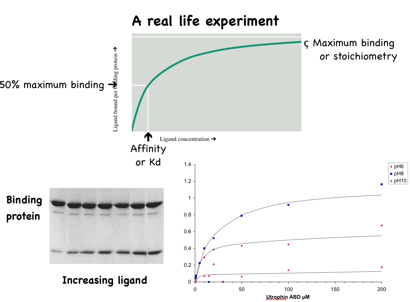
Measuring the strength of protein interactions
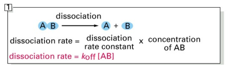
- Theory
- Typical applications
- Can be used to measure any protein-protein interaction in solution or on a surface providing you have a suitable detection system
- PAGE (Poly-acrylamide Gel Electrophoresis)
- Western Blot
- Far western or blot overlay
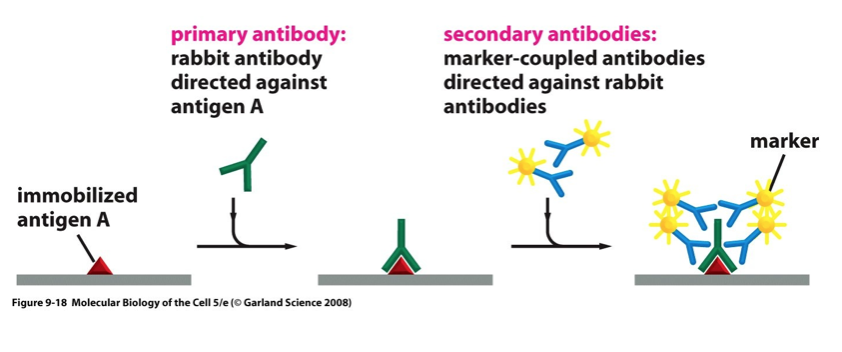
- ELISA (Enzyme-Linked Immunosorbent Assay
- Can be used to measure any protein-protein interaction in solution or on a surface providing you have a suitable detection system
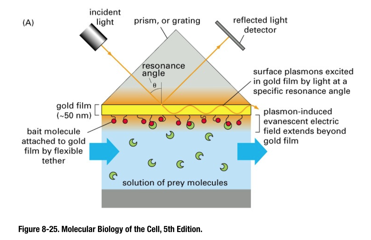
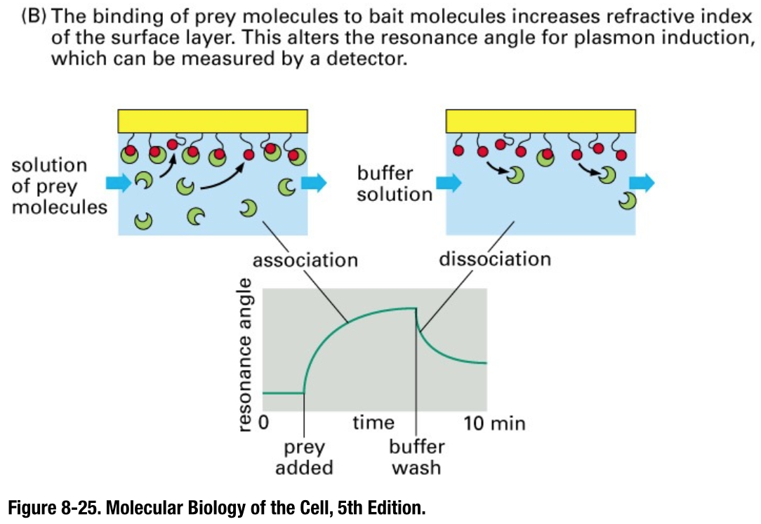
Surface plasmon resonance
- aka ‘Biacore’
- The Kd of a given protein-protein interaction can be read directly from traces like these
- however one limitation is the attachment of proteins to a solid support which may cause conformational change and affect the result
Genetic methods of identifying protein interactions
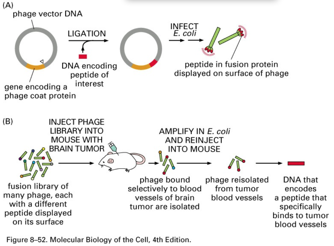
- Phage display libraries
- Use of random peptide libraries is a powerful technique with many applications
- Identification of specific epitopes as drug or antibody targets, mapping of protein-protein binding interfaces, generation of artificial antibodies, epitope mapping of antibodies to identify important binding sites etc.
- Use of random peptide libraries is a powerful technique with many applications
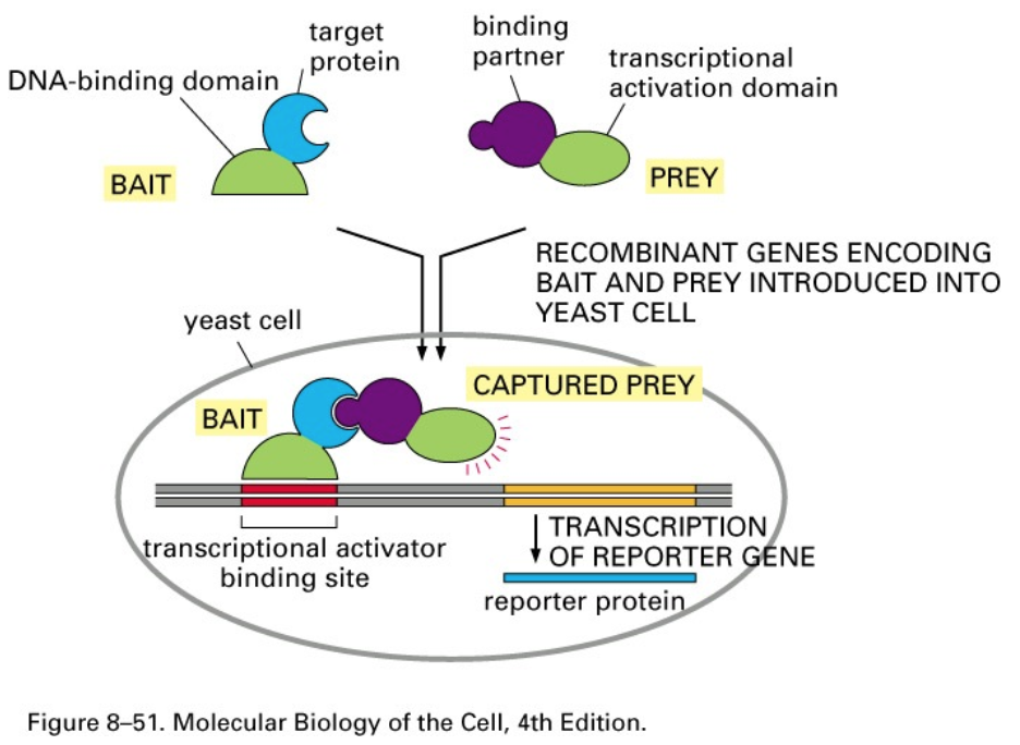
- Yeast two-hybrid screening
- Bait protein cDNA cloned into bait plasmid in the host yeast strain.
- Prey usually a whole library of cDNA’s transfected with the bait plasmid.
- Interaction between bait and prey allows DNA binding and activation domains to activate a reporter gene.
- Yeast colony survives on a restrictive growth media.
- Prey plasmid recovered and sequenced to find interacting protein.
- Yeast colony survives on a restrictive growth media.
- Interaction between bait and prey allows DNA binding and activation domains to activate a reporter gene.
- Prey usually a whole library of cDNA’s transfected with the bait plasmid.
- Bait protein cDNA cloned into bait plasmid in the host yeast strain.
- Phage display libraries
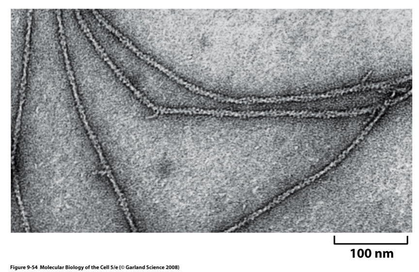
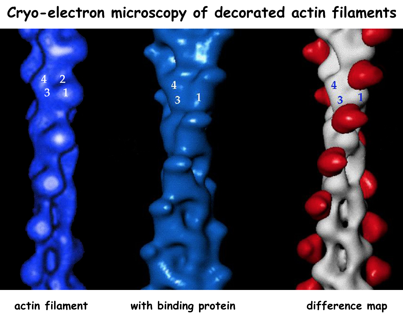
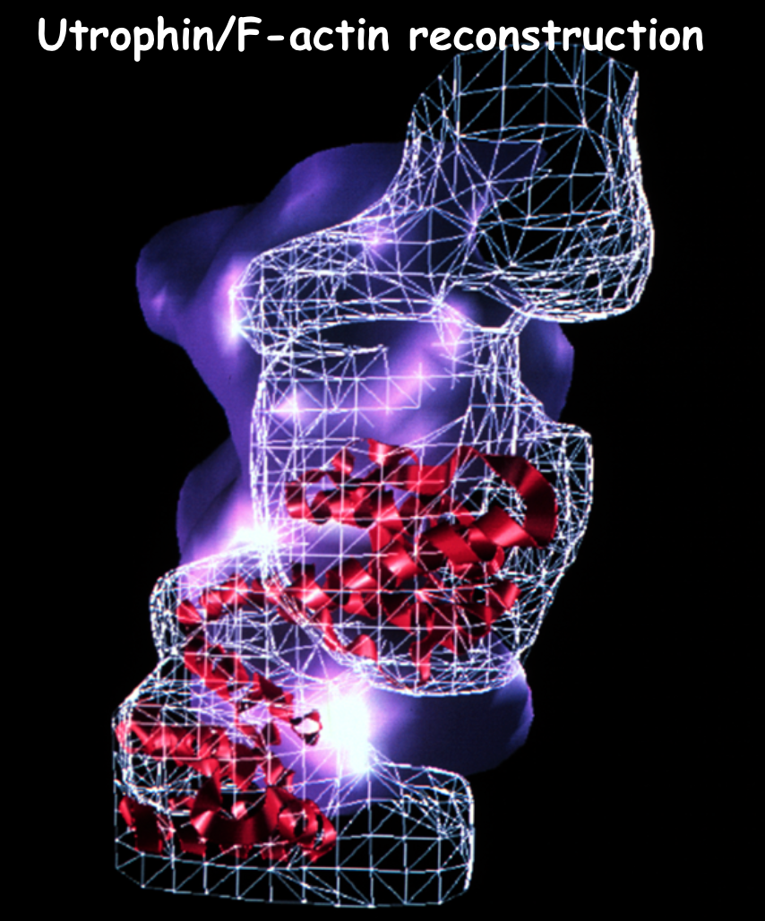
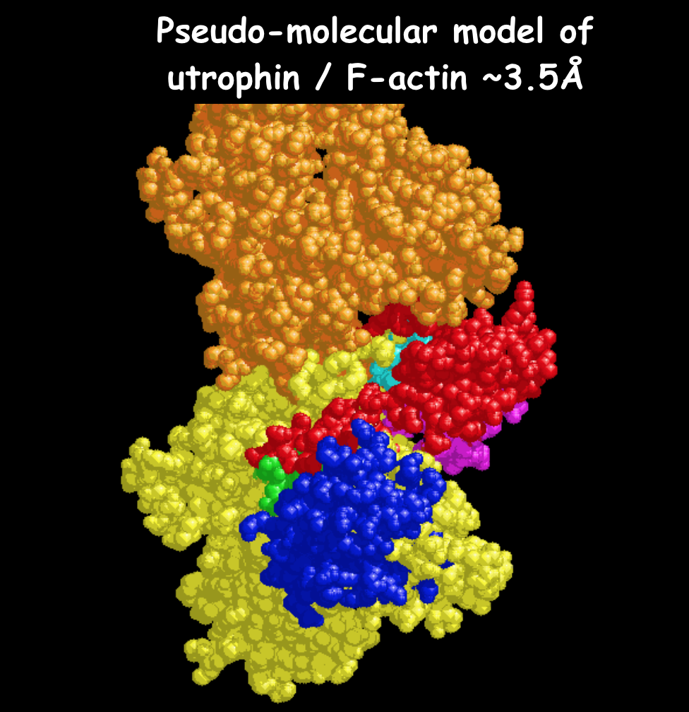
Electron Microscopy II - actin
- EM of actin filaments with a binding protein attached.
- EM uses negative stain (shown) or vitreous ice (cryo-EM) to preserve the specimen.
- Image analysis is then employed to build up an average structure.
- EM uses negative stain (shown) or vitreous ice (cryo-EM) to preserve the specimen.
- EM of actin filaments with a binding protein attached.
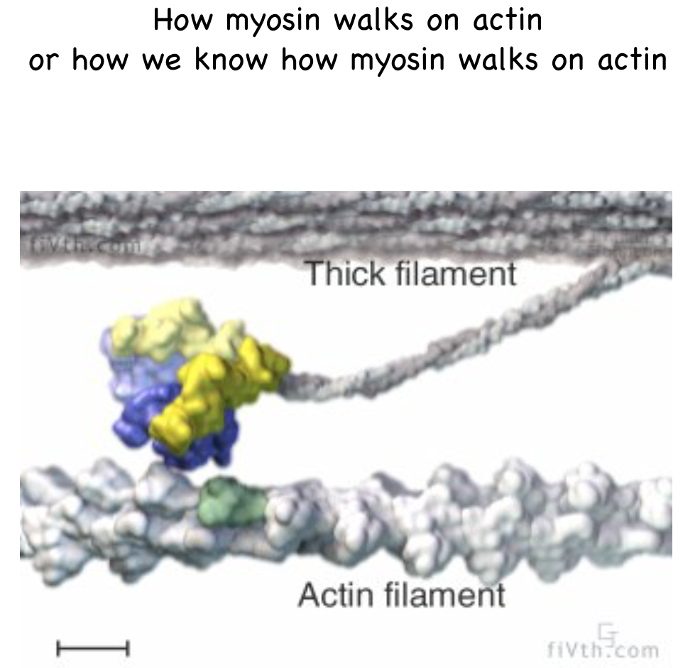
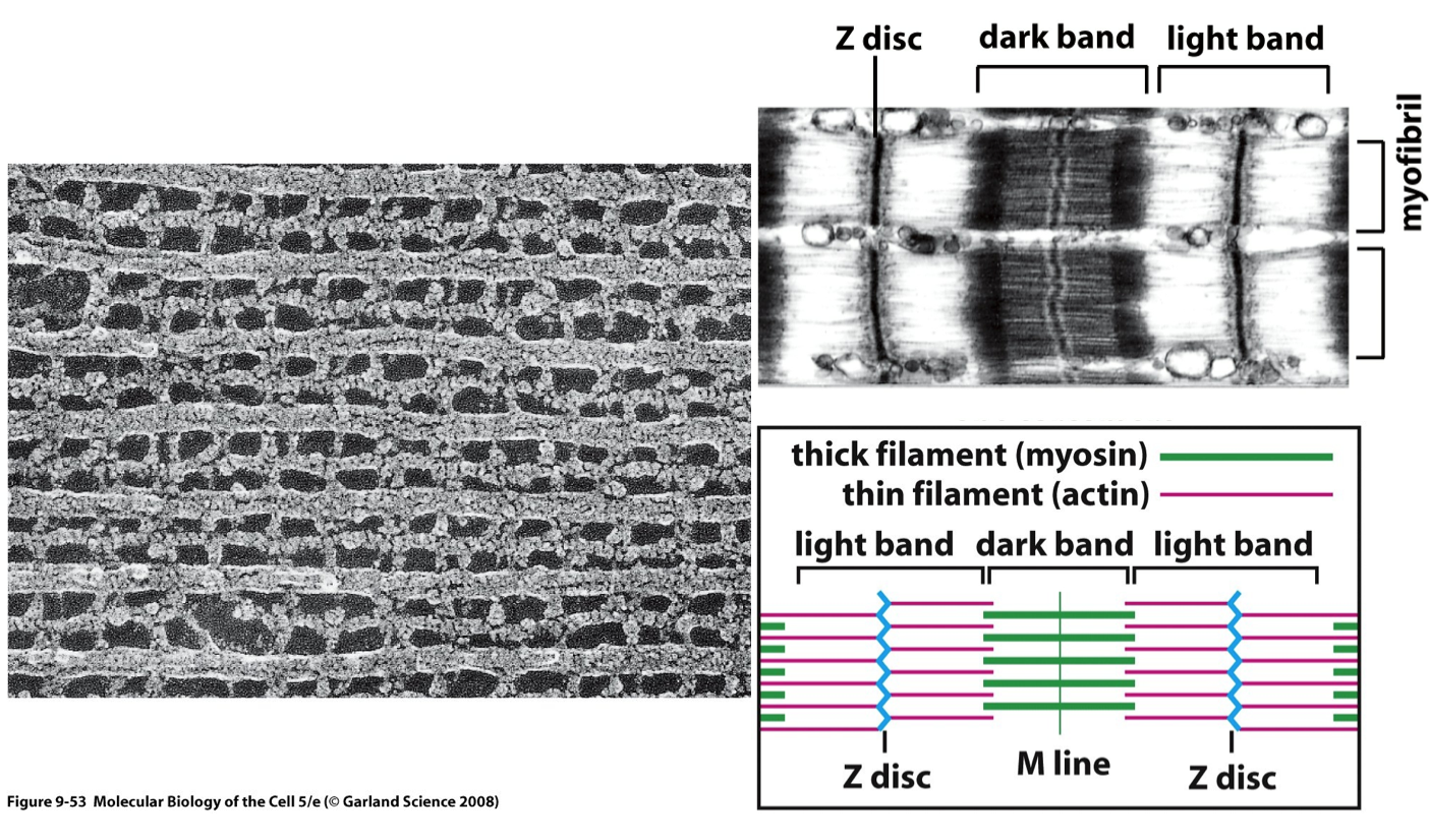
A protein machine of the cell - actomyosin
- Muscle ultrastructure
Imagens e arquivos do mapa
Informações
- Descrição
- Undergraduate BMS238 Cell and molecular biology (Proteins) Mapa Mental sobre Protein interactions (and analysis), criado por Kristi Brogden em 16-08-2014.
- Criado por
- Kristi Brogden
- Visualizações
- 9
- Avaliação