17247619
Descrição
Quiz por Imam Shaik, atualizado more than 1 year ago
|
|
Criado por Imam Shaik
aproximadamente 5 anos atrás
|
|
Questão 1
Questão
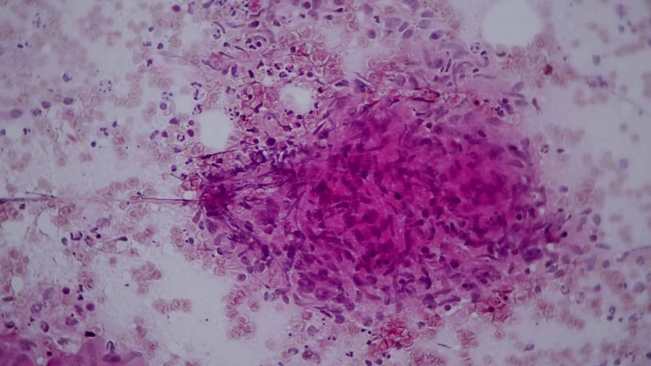
Photomicrograph A shows FNAC findings of the patient’s lymph node.
Image:
4 (binary/octet-stream)
Responda
-
Caseating granuloma
Questão 2
Questão
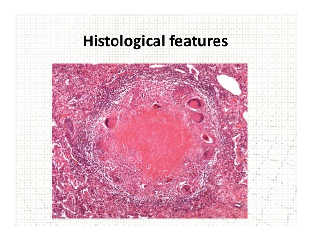
Photomicrograph B is a corresponding histopathological findings of the patient’s lymph node.
1. Name the lesion observed in photomicrograph B.
2. Describe at least THREE microscopic features of the lesion in photomicrograph B .
Image:
5 (binary/octet-stream)
Responda
-
Caseous necrosis
-
epitheloid cells/granuloma
-
Langhans gaint cells
-
mononuclear cells/lymphocytes
-
fibrosis
Questão 3
Questão
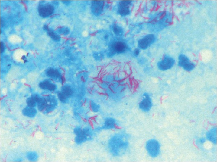
Photomicrograph C shows the findings from a Ziehl-Neelsen stain of the FNAC.
OSPE 3
1. Give your observation from the stained FNAC above.
2. Describe the morphology of this organism.
Image:
6 (binary/octet-stream)
Responda
-
AFB positive
-
mycobacterium
Quer criar seus próprios Quizzes gratuitos com a GoConqr? Saiba mais.