
Flashcards
Digestion
Cards in this deck (38)
- 1What is the difference between the peritoneum and mesentery?Peritoneum- serous lined membrane that covers the digestive viscera
Mesentery- folding of the peritoneum that attaches the stomach, small intestine, pancreas, spleen, and other organs to the posterior wall of the abdomen - 2What are peritoneal and retroperitoneal organs?Peritoneal- organ is completely covered by visceral layer of peritoneum- small intestine, large intestine, liver, gallbladder, pancreas, spleen
Retroperitoneal- organ is outside or behind the peritoneal cavity and partially covered by the visceral peritoneum- kidneys, adrenal glands, ureters, bladder, rectum, ovaries, uterus - 3Alimentary canalAlso the gastrointestinal (GI) tract
Includes: mouth, pharynx, esophagus, stomach, small and large intestine - 4Accessory organsIncludes: teeth, salivary glands, gallbladder, liver, pancreas
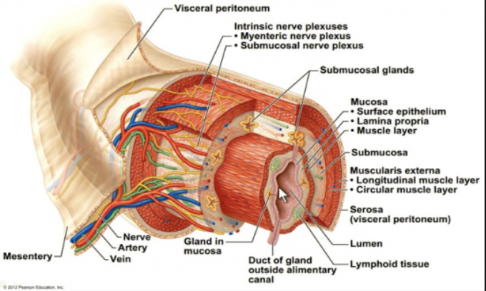
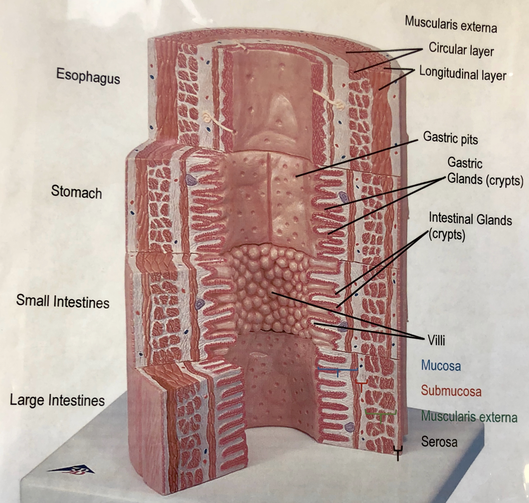
- 5Four tunics of alimentary canal wall layersMucosa, submucosa, muscularis externa, serosa (adventitia)
- 6MucosaEpithelium- stratified squamous (nonkeratinized) in the mouth, esophagus, and anus - simple columnar in stomach, small intestine, and large intestine
Lamina Propria- areolar connective tissue with blood vessels
Muscularis mucosae- thin layer of smooth muscle - 7SubmucosaAreolar connective tissue and dense irregular connective tissue
Relatively thick, highly vascular, serve the mucosa
Has glands and nerve plexuses - 8Muscularis ExternaCircular layer - inner layer of smooth muscle - in stomach for segmentation
Longitudinal layer - outer layer of smooth muscle - in stomach for peristalsis
*Stomach also has an oblique later for mechanical/grinding of food
Segmentation and peristalsis of digested food along the tract - 9Serosa (Adventitia)Dense irregular connective tissue
Provides lubrication and reduces friction - 10Alimentary canal wall
- 11—Gastric Pits of the Stomach!
Three types of cells...
1. Chief- release pepsinogen (cut by HCl to create pepsin and pepsin breaks down protein)
2. Parietal- HCl and intrinsic factor (important for absorption of vitamin B12)
3. Enteroendocrine- release hormones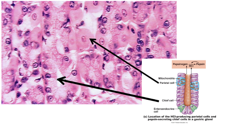
- 12—Figure of the stomach's histology
How to identify: simple columnar epithelium, gastric pits are forked and straight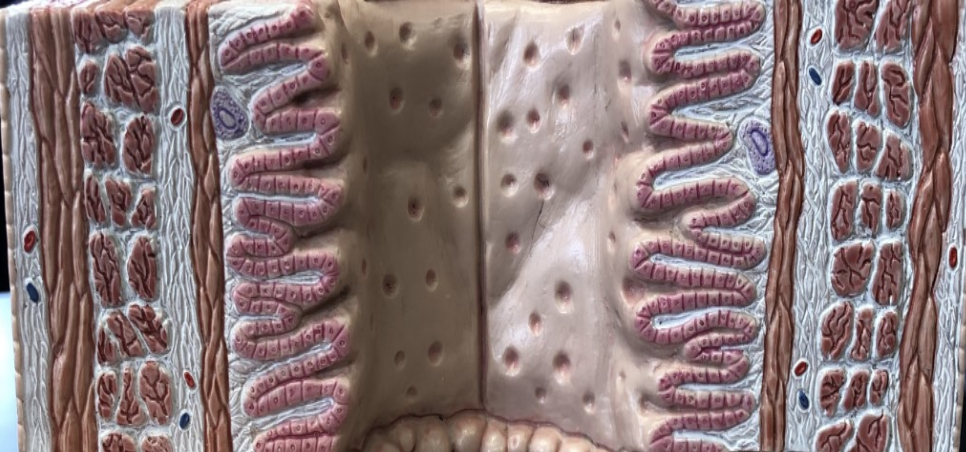
- 13—Mucosa-top
Submucosa- middle light
Muscularis externa- bottom
Serosa- can't see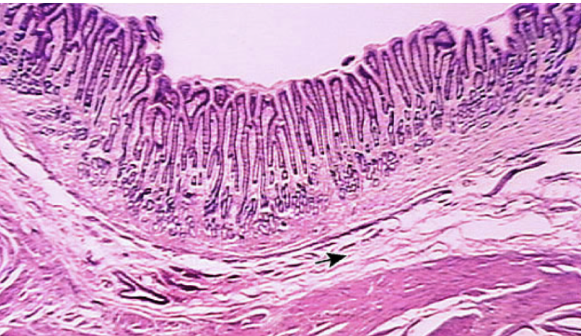
- 14—Mucosa of the gastroesophageal junction
Why can a hiatal hernia occur here? - The esophagus sphincter muscle normally closes tightly. With a hiatal hernia, the sphincter new position may keep it from completely closing. The backflow of digestive juices may damage the esophagus - LACK OF TRUE EXTERNAL / INTERNAL SPHINCTER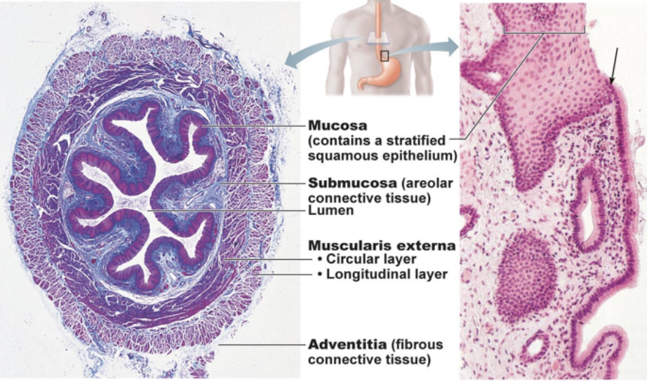
- 15—Side one is the esophagus- many layers of stratified squamous
Side 2 is the stomach- ridges- simple columnar folded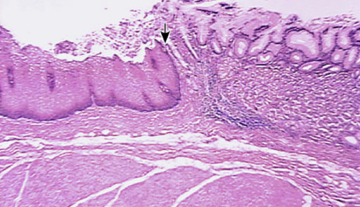
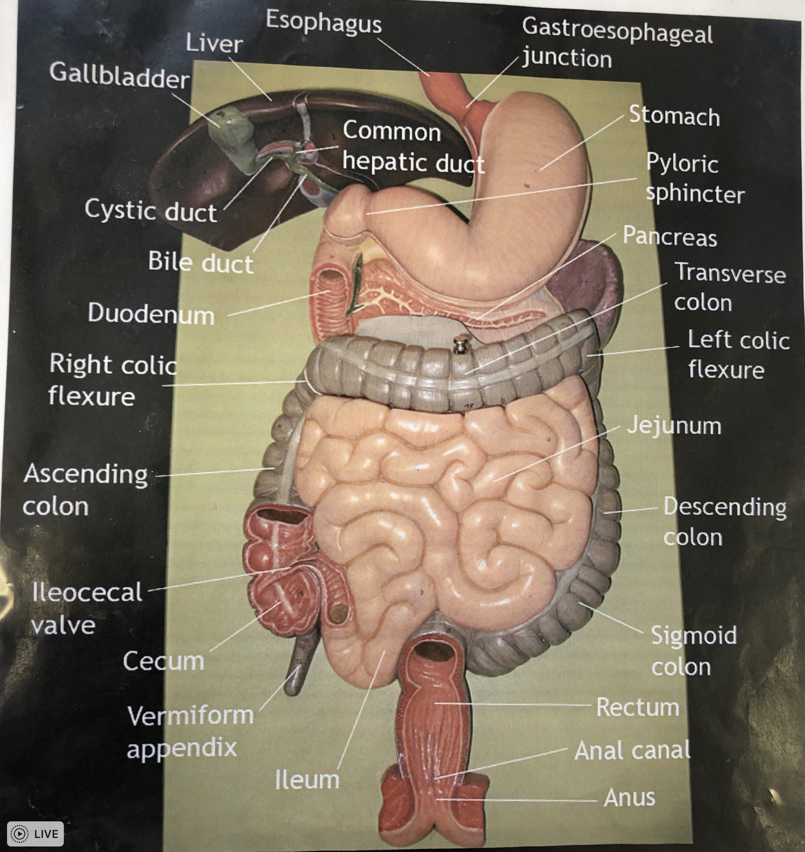
- 16—Main regions of the small intestine! (DJ Ileum)
*All regions contain paneth cells
Duodenum- 10cm - extends from pyloric sphincter and curves around the head of the pancreas - most important area for digestion - highest percentage of digestive enzymes - *Brunner's glands - release basic mucus to to raise pH
Jejunum- 8 feet - umbilical region
Ileum- 12 feet - terminal portion and joins the large intestine at the ileocecal valve - *Peyer's patches = MALTs -help kill off pathogens on food we eat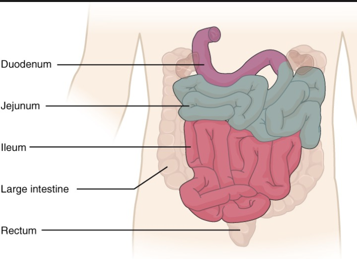
- 17What are the pyloric sphincter and ileocecal valve?Pyloric sphincter- band of smooth muscle at the junction between the pylorus of the stomach and the duodenum of the small intestine
Ileocecal valve- valve between the small intestine and the large intestine that prevents material from flowing back into the small intestine - 18In which region would you find the pancreatic duct and bile duct?Pancreatic duct is outside of the parietal peritoneum and the bile duct is inside the parietal peritoneum
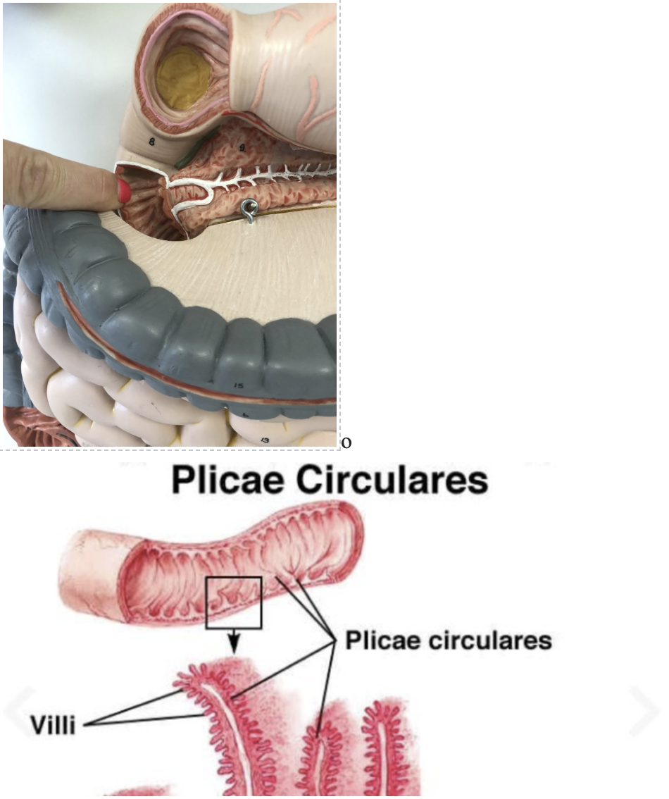
- 19The small intestine is marked by some distinctive histological features, including villi, microvilli, and intestinal crypts. Distinguish between these three anatomical features.Villi are finger like projections out of the epithelia of the small intestine
Microvilli are projections out of the villi
Intestinal crypts are pits in the epithelia of the small intestine - how we absorb fats
These increase surface area of the small intestine which increases absorption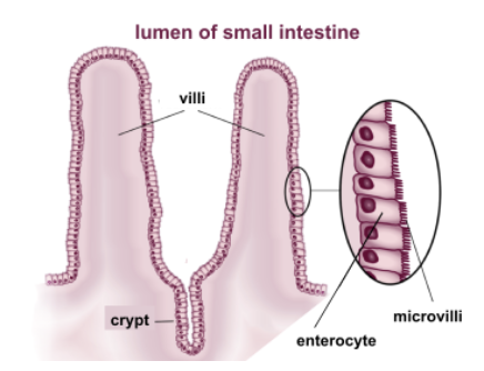
- 20—Plicae circulares - folds in the small intestine - especially in the lower parts of the duodenum and jejnum
- 21Important histological features of the small intestineBrush boarder cells, paneth cells, brunner's glands, peyer's patches
- 22—Brush Boarder cells!
Simple columnar epithelium that is carpeted with microvilli
The microvilli project from the epithelium like bristles on a brush
Absorption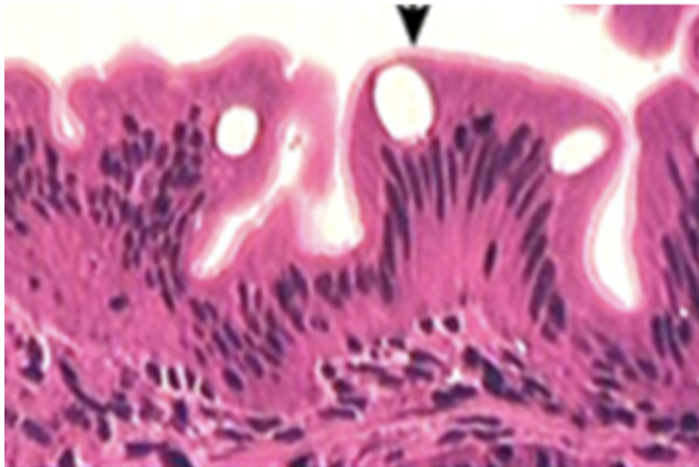
- 23—Paneth cells!
Everywhere in the small intestine
Have enzymes - acid hydrolases - kill off bacteria in food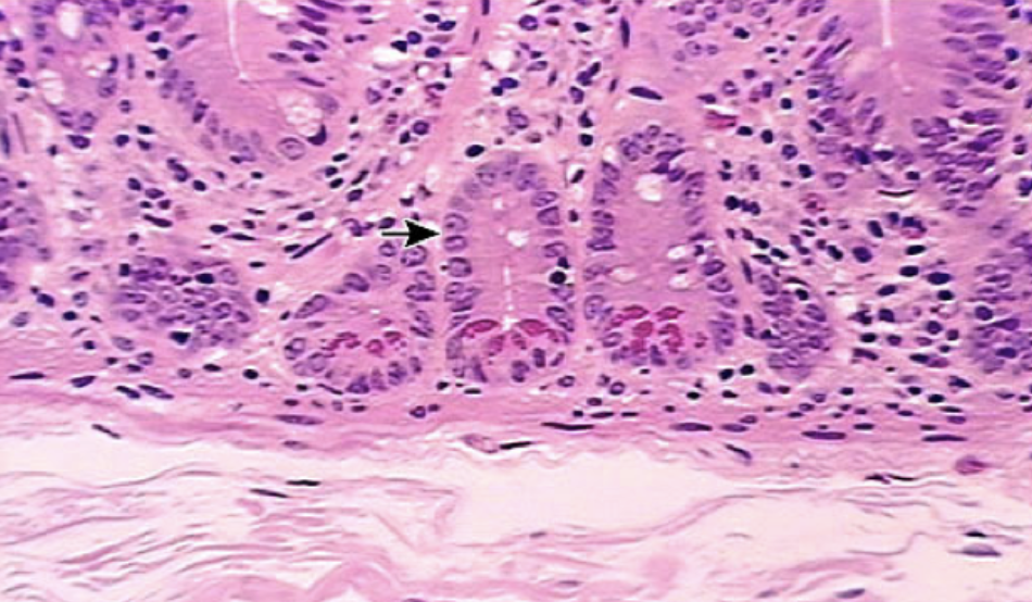
- 24—Brunner's glands!
Duodenum
Arrow pointing to villus
Mucous glands, mostly in duodenum, produce alkaline mucus to raise pH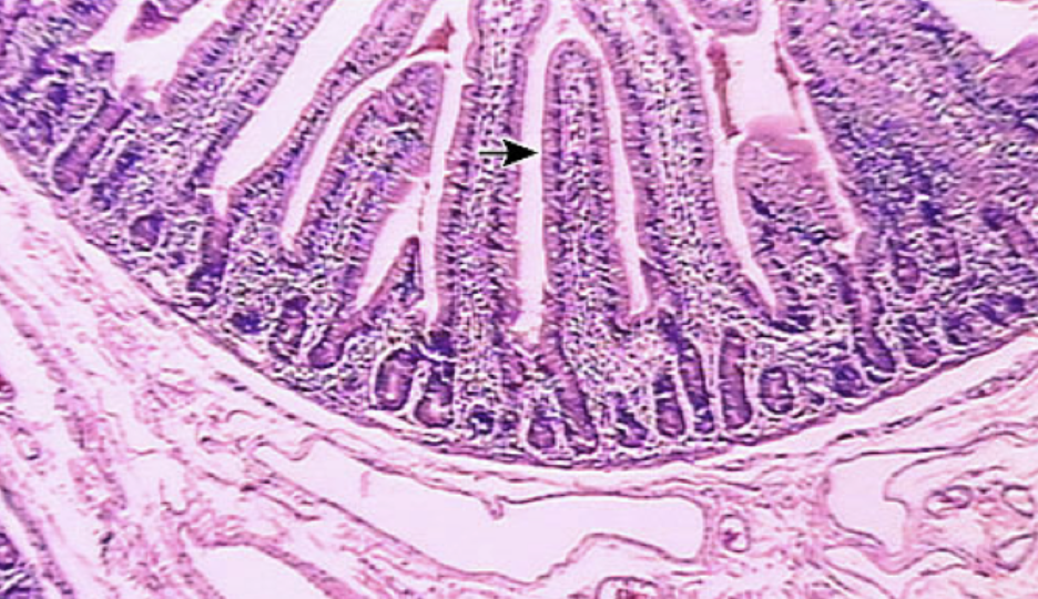
- 25—Peyer's Patches!
Ileum
MALTS = mucosa associated lymphatic tissue
Kill off pathogens in food that we eat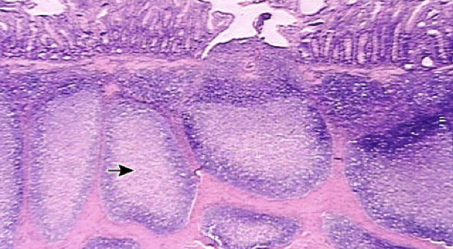
- 26—Figure of small intestine histology
How to identify: layer of simple columnar epithelium, villi (round balls in the middle) irregular intestinal crypt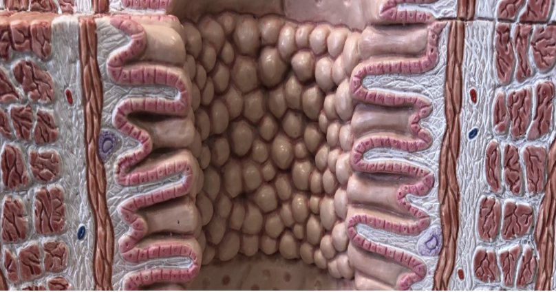
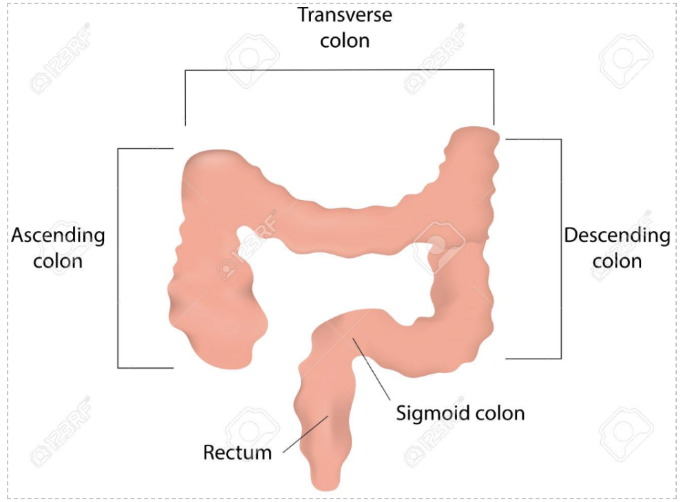
- 27Divisions of the large intestineCecum, colon, rectum, anus
- 28—Ascending colon, transverse colon, descending colon, sigmoid colon
- 29Unlike the small intestine, the large intestine does not have villi or microvilli. Why not?Because the chemical digestion is completed in the small intestine before the chyme reaches the small intestine
You do not need these for absorption of water, you only need them for the absorption of nutrients - 30The large intestine is marked with lots of goblet cells. What is the importance of these cells in the large intestine?Goblet cells release mucus
So much mucus is needed in the large intestine in order to defectate - 31What role does the lymphoid tissue surrounding the appendix play?Allows the appendix to reset microbiome, can survive without it but it helps having an appendix due to the ability to reset the microbiome
- 32—Figure of histology of the large intestine
How to identify: simple columnar, regular intestinal crypts, most goblet cells for secreting mucus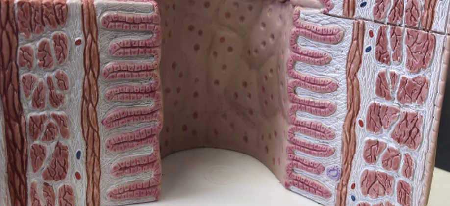
- 33—Goblet cells
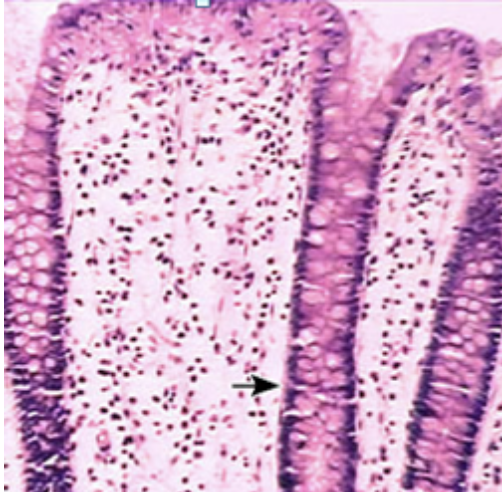
- 34—Figure of histology of the esophagus
How to identify: small layer of stratified squamous epithelial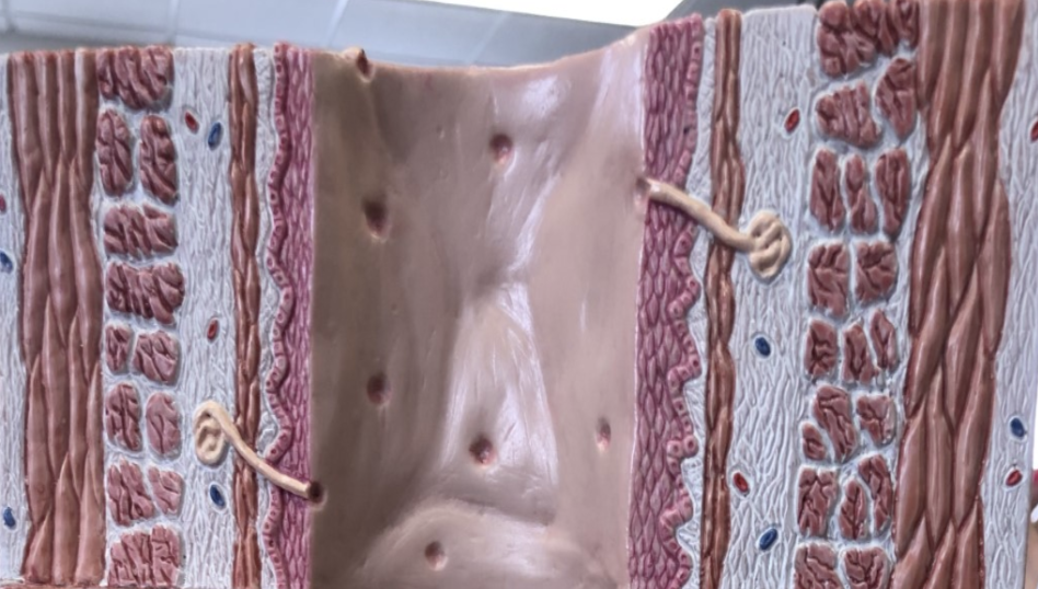
- 35—Small intestine!
Villus & simple columnar epithelium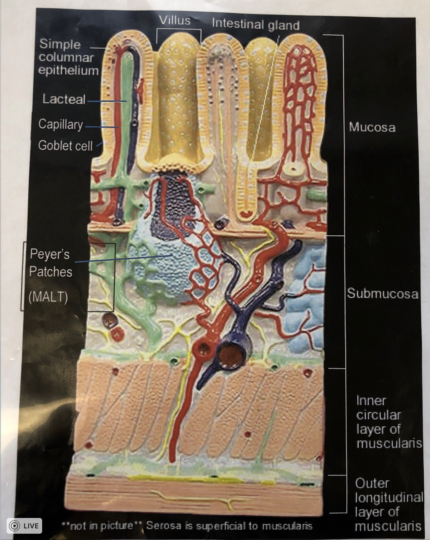
- 36—Small intestine
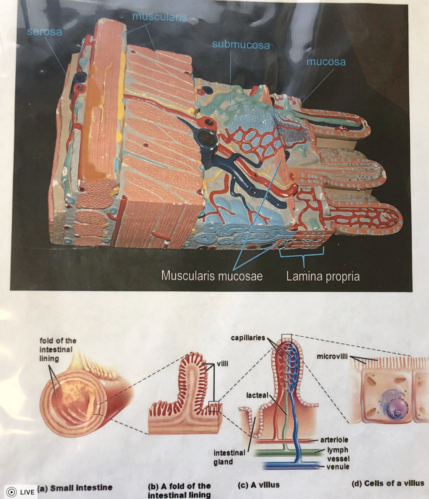
- 37—Four tunics of alimentary canal wall layers
- 38—Digestive system
Information
- Description
- Nursing Flashcards on Digestion, created by Megan Dowd on 01/04/2019.
- Created by
- Megan Dowd
- Views
- 4
- Rating