
Mind map
Microscopy
Map content
Microscopy
Light Microscopes
- A micrscope is an instrument that allows you to magnify an object. They were first discovered in the 16/17th century.
- There are different ways in which samples and spiciest can be prepared.
- DRY MOUNT: solid specimens are viewed whole or cut into think slices (sectioning). Its placed on the slide and a cover slip
- WET MOUNT: Specimens are supposed in a locked. A cover slip is placed from an angle.
- SQUASH SLIDES: A wet mount is prepared, then the lens tissue is used to press the cover slide.
- SMEAR SLIDES: The edge of a side is used to smear the sample, creating a thin even coast on another slide, and a cover slide is placed top.
- The cytosol of cells and other structures are transparent. Stains increase contrast and different components take to stains differently.
- To prepare a sample for staining, it is placed on a slide and allowed to air dry. This is then heat fixed by passing through a flame. The speciment will adhere to the microscope slide and will then take up stains
- Crystal violet is positively charged and is attracted to negatively charged materials in the cytoplasm. and congo red is negatively charged and is replied by cytosol.
- There are different ways in which samples and spiciest can be prepared.
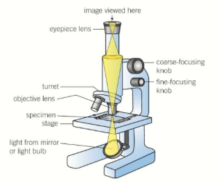
- A compound light microscope has two lenses - objective which is near the specimen and an eyepiece lens.
- The objective lens produces a magnified image, which is magnified again by the eyepiece lends.
- This allows for higher magnification and reduced chromatic aberration.
- Illumination is usually provided by a light underneath the sample. Opaque specimens can be illuminated from above with some.
- The objective lens produces a magnified image, which is magnified again by the eyepiece lends.
- A micrscope is an instrument that allows you to magnify an object. They were first discovered in the 16/17th century.
Laser scanning confocal
- Light microscopes have developed. Conventional optical microscopes use visible light to illuminate specimens and lens to produce a magnified image.
- In fluorescent microscopes, a higher light intensity is used to illuminate the specimen thats been treated with a dye. Fluorescence is the absorption and re radiation of light. Light of a longer wavelength and lower energy is emitted and used to produce an image.
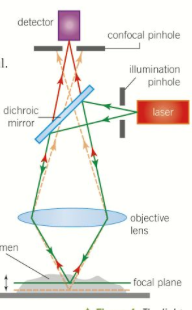
- A laser scanning confocal microscope moves a single spot of focused light across a specimen, causing fluorescence from he components with a dye. The light from the specimen is filtered through a pinhole aperture. One light from close is detected.
- Light other parts of the specimens would reduce resolution and cause blurring. This unwanted radiation doesn't pass the pinhole and isn't detected. A laser is used instead of light to get higher intensities.
- Thin sections are examined and the spot is moved across the specimen. A 2D image is produced, 3D can be made if images are taken from different focal planes.
- It is non invasive and is used in diagnosis of the eye. It can see the distribution of molecules within cells meaning it is used in drug development.
- The beam splitter is dichroic mirror, which reflects one wavelength but allows other wavelengths to pass through. The position of the two pinholes means the light waves from he laser follow the same path as light waves. This means they have the same focal plane.
- It is non invasive and is used in diagnosis of the eye. It can see the distribution of molecules within cells meaning it is used in drug development.
- Thin sections are examined and the spot is moved across the specimen. A 2D image is produced, 3D can be made if images are taken from different focal planes.
- Light other parts of the specimens would reduce resolution and cause blurring. This unwanted radiation doesn't pass the pinhole and isn't detected. A laser is used instead of light to get higher intensities.
- Light microscopes have developed. Conventional optical microscopes use visible light to illuminate specimens and lens to produce a magnified image.
Electron Microscope
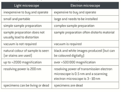
- In light microscopy, resolution is the limiting factor. In electron microscopy, a beam of electrons with a wavelength less than 1nm is used to illuminate the specimen.
- This means more detail can be seen.
- Very expensive piece of equipment and can only be used inside a carefully controlled environment in a dedicated space.
- Specimens can be damaged by the electron beam.
- As the preparation process is very complex, there is a problem with artefacts.
- In a transmission electron microscope, a beam of electrons is transmitted through a specimen and focused to produce an image. The best resolution with he resolving power of 0.5nm
- In scanning electron microscope a beam of electrons is sent across the surface of the specimen and the reflected electrons are collected. The resolving power is from 3-10nm so the resolution is not as good as with TEM. It creates stunning 3D images of surfaces are produced.
- The inside of an electron microscope is a vacuum to ensure the electron beams travel in straight lines. Specimen preparation involves freezing, staining with heavy metals and dehydration. Samples for TEM will be set in resin and retained. Samples for SEM may be fractured to expose the inside and then coated with heavy metals.
- An artefact is a strucutre detail caused by processing the specimen. The bubbles get trapped under the cover slip (light), or changes in the cells happen for electron (distortion of organelles, empty spaces in cytoplasm, continuity in membranes).
- In light microscopy, resolution is the limiting factor. In electron microscopy, a beam of electrons with a wavelength less than 1nm is used to illuminate the specimen.
Calculations
- Magnifying an object is how many times larger the image is than the actual size, not the detail. The resolution also needs to be increased. Resolution is limited by the diffraction of light as it passes through samples
- Diffraction is the tendency of light waves to spread as they pass close to physical structures.
- Resolution can be increased by using beams of electrons which have a shorter wavelength. The beams are still diffracted but the shorter wavelength mean that beams can be much closer before they overlap. This means objects that much smaller and closer together can be seen without diffraction blurring the image.
- To measure the size of a sample under a microscope, you use an eyepiece graticule.
- You align the micrometer scale with the eyepiece and take a reading. 1 graticule division = number of micrometers/graticule divisions
- 1000 nm= 1 um, 1000um = 1mm, 1000 mm = 1m
- Diffraction is the tendency of light waves to spread as they pass close to physical structures.
- Magnification = size of image/actual size of object
- Magnifying an object is how many times larger the image is than the actual size, not the detail. The resolution also needs to be increased. Resolution is limited by the diffraction of light as it passes through samples
Images and files in the map
Information
- Description
- A level Biology (2 - FOUNDATIONS IN BIOLOGY ) Mind Map on Microscopy, created by Molly Butcher on 27/03/2017.
- Created by
- Molly Butcher
- Views
- 0
- Rating