
Notes
Processes within cells
Content of the notes (8)
Page 1
How HIV Infects The Body
Soft moist mucous membrane is present inside mouth, nose, digestive tract.
HIV can reach a Lymphnode in 3 ways:
1. Infectious synapse
2. Dendritic cell endocytoses it and exocytoses it into Lymphnode.
Dendritic cell endocytoses a group of HIV particles and brings that endocytosed group to the lymphnode where it releases the particles that then infect other cells.
3. HIV can directly infect the dendritic cell at some point.
It will start pumping out copy after copy of HIV particles.
How HIV Infects A T Helper Cell
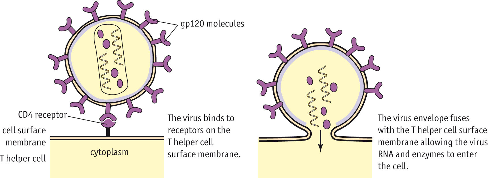
Once in the lymphnode, the HIV is able to infect T helper cells (CD4 lymphocytes). GP120 molecule on the surface of an HIV particle binds to the CD4 Receptor on the T helper cell as it is a complimentary. This pulls the two cells closer and the HIV cell penetrates the membrane of the CD4 Lymphocyte, inserting its genetic material inside (viral RNA, Reverse Transcriptase, Integrase) in order to hijack the cell and be able to take over its mechanisms in order to produce new HIV particles.
> Macrophages also have CD4 Receptors so HIV can infect them.
Page 2
HIV Hijacks Protein Synthesis
One capsid with RNA and viral enzymes enters the cell. Reverse transcriptase takes viral RNA and our nucleotides floating in the cytoplasm to revert viral RNA into single-stranded DNA. Synthesizes complementary strand of DNA for the single strand, double-stranded viral DNA now made.
The viral enzyme, integrase, brings viral DNA to T cell nucleus through a nuclear pore. Integrase makes a cut in human T cell DNA and integrates viral DNA into the genetic code. This step confirms lifelong infection of HIV. Now...
1. Gene will not be transcribed into mRNA and therefore is a latent HIV infection.
2. DNA transcription enzyme, RNA polymerase, transcribes viral DNA into mRNA strands which leave the nucleus and are transcribed on a ribosome on rER. They make way through ER, reach cell membrane and cluster. Are then released after protease cleaves up the polyprotein to create a mature HIV particle. Exocytosed and able to infect other cells.
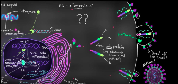
NOTE: Reverse Transcriptase can make a lot of errors in this reverse transcription, resulting in mutations of HIV particles.
the virus can develop resistance to certain antiviral medications
it's very hard to make a vaccine because small changed in viral genes can make the vaccine ineffective
Page 3
Protein Synthesis
TRANSCRIPTION
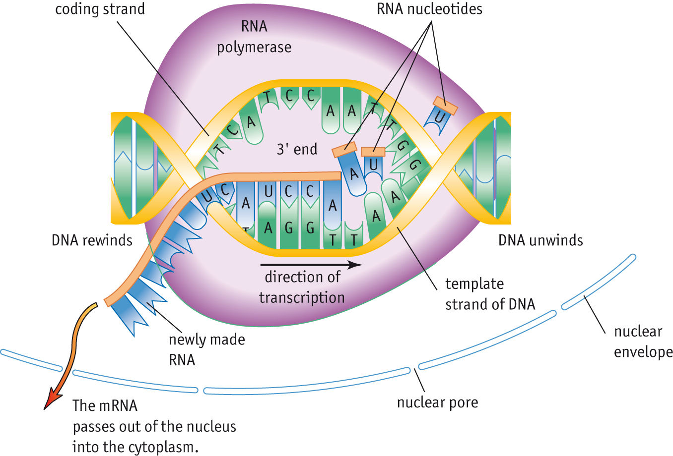
RNA Polymerase attaches to DNA and hydrogen bonds between base pairs break, DNA molecule unwinds. The sequence on the template strand (antisense strand because once transcribes, makes a strand with the same base sequence as the original sense strand of DNA) is transcribed and makes a mRNA molecule with the same base sequence as the DNA coding strand (sense strand).
Complementary RNA nucleotides align and form phosphodiester bonds to produce a mRNA molecule.
TRANSLATION
Turning a sequence of bases in the genetic code into a sequence of amino acids. In the genetic code, several triplets can code for more than one amino acid and so it is degenerate. Each triplet code is adjacent therefore the code is non-overlapping.
In the cytoplasm, mRNA attaches to a ribosome. Ribosomes are composed of two subunits. The larger subunit contains two tRNA binding sites. mRNA attaches to the smaller subunit so that two mRNA codons face the two binding sites of the larger subunit.
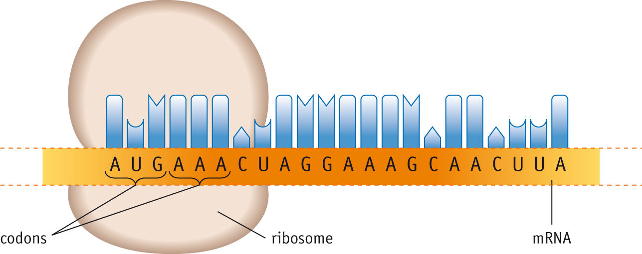
The first codon exposed on the ribosome is always the start codon AUG, the tRNA molecule carrying methionine has the complementary code UAC and it hydrogen bonds to the start codon AUG. The next codon is facing the next binding site.
Ribosome holds the mRNA, tRNAs, amino acids and associated enzyme in place whilst a peptide bond forms between the two amino acids.
PEPTIDE BOND:
Forms in a condensation reaction between the amine group of one amino acid and the carboxylic acid group of the next. This forms a dipeptide.
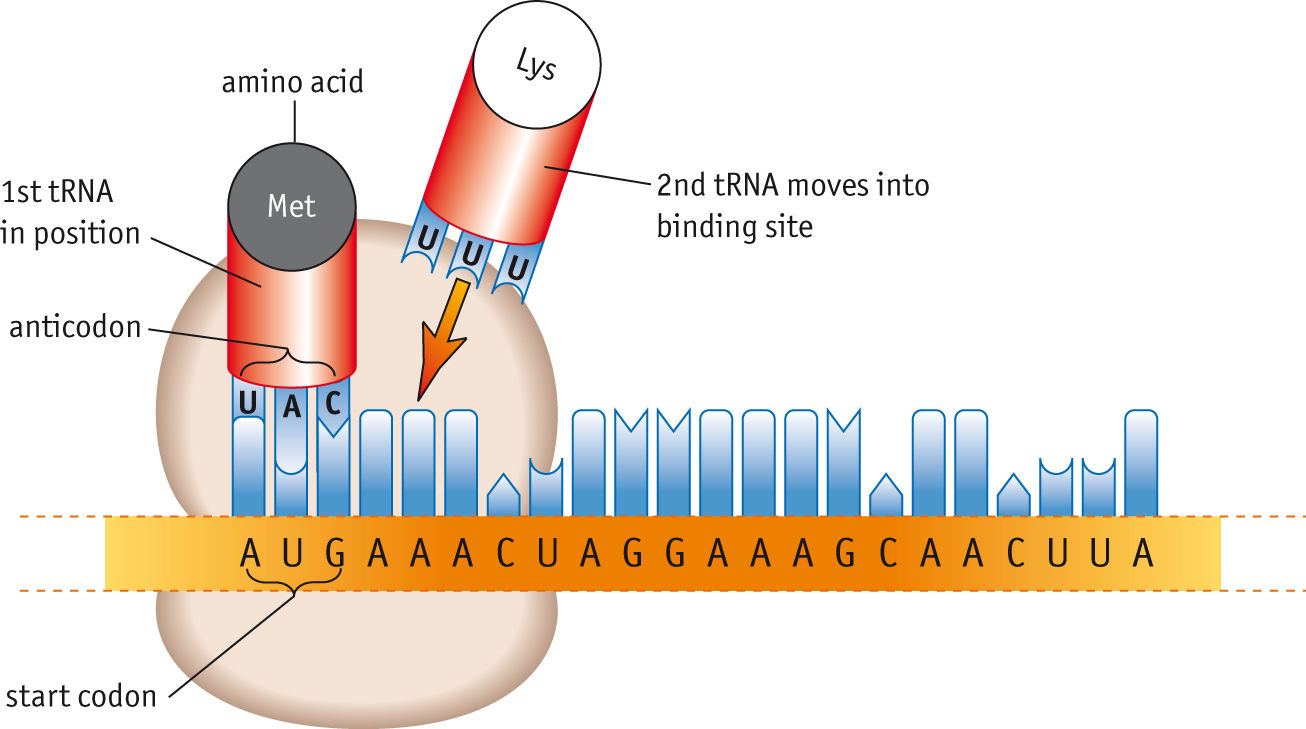
> NOTE: tRNA molecules do not get used up - they continuously return to the cytoplasm to pick up new amino acids until a stop codon is reached.
Page 4
Lines of Body Defence
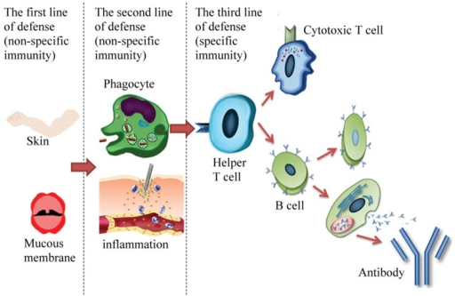
There are three lines of defense in the body:
The first line of defense:
Non-specific Immunity
Mucous membranes such as in the nose and digestive tract
Skin barriers such as skin flora and lysozyme (enzyme that breaks down bacterial cell walls)
The second line of defense:
Non-specific Immunity
Phagocytosis
Inflammatory Response - Damaged white blood cells and mast cells in connective tissue below the skin and around blood vessels release chemicals (such as histamine) that cause arterioles in the area to dilate, increasing blood flow in the capillaries. Vessels then leak and plasma fluid is released. WBC's and antibodies leak from the blood into the tissue causing oedema (swelling).
The third line of defense
Specific Immunity
Involves Helper T Cells stimulating a range of other cells such as B effector cells and T killer cells.
Results in antibodies
Page 5
B Cell activation
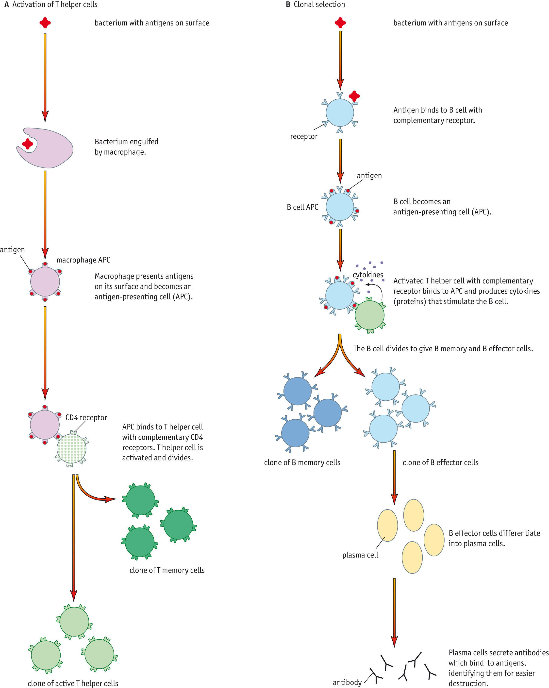
A bacterium with antigens on its surface binds to the complementary B cell receptor and the B cell becomes an antigen presenting cell (APC). A T cell with complementary receptor binds to the B cell and produces cytokines (these are proteins) which stimulate the B cell.
What does the B cell do?
The B cell divides to give B memory and B effector cells. The effector cells differentiate into plasma cells which then produce antibodies. These bind to antigens, marking them as identification for easier destruction by the specific immune response.
Page 6
T Helper Cell Activation - Specific Immune Response
When a piece of biological material is engulfed by a macrophage, it displays fragments on its cell surface membrane as they are displayed as 'non-self' antigens. These alert the immune system of the presence of foreign antigens in the body.
T helper cell with complementary CD4 receptors binds to the antigen presented on the surface of the APCs. This activates the T helper cell and it divides producing a clone of memory T cells and a clone of active T cells.
Page 7
Activation Of T Killer Cells
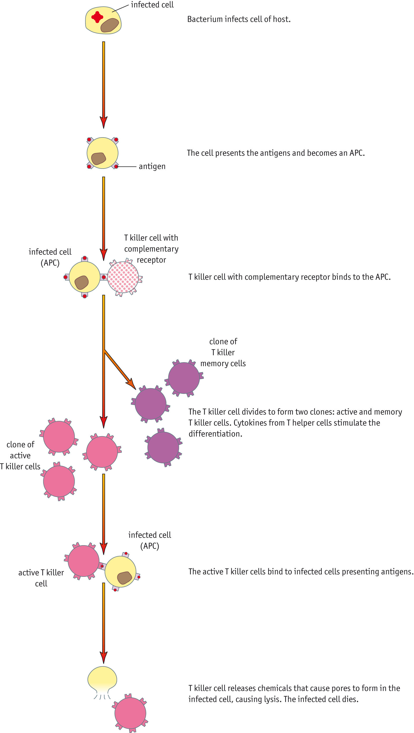
If a bacterium or virus infects a body cell, a foreign antigen is presented on its surface and a receptor on a T killer cell binds to this as it is complimentary. The T killer cell divides to form active clones and memory clones and this is stimulated by cytokines produced from T helper cells. Active T Killer cells then bind to cells presenting foreign antigens. T Killer cell releases chemicals that cause pores to form in the infected cell, causing cell lysis. Infected cells died and pathogens released can be marked by antibodies for destruction by macrophages.
LYSIS: Ions and water flow into an infected cell causing it to swell and burst.
NOTE: Without cytokines, there would not be enough T killer cells produced to fight a viral infection.
Page 8
Sarcomere Shortening: The Sliding Filament Theory
Information
- Description
- A level Biology (SNAB: Processes) Note on Processes within cells, created by Cecily Russell on 07/01/2018.
- Created by
- Cecily Russell
- Views
- 0
- Rating