
Microscopy
Slides in this set (4)
-
- Cells are studied using microscopes
- Microscopes let us see things that we cant see with the naked eye. The microscopy techniques we can use have developed over the years as technology and knowledge have improved.
- Light microscopes use light and lenses to form an image of a specimen and magnify it. They let us see individual cells and large subcellular structures, like nuclei.
- Electron microscopes use electrons instead of light to form an image. They have a much higher magnification than light microscopes.
- They also have higher resolution. Resolution is the ability to distinguish between two points, so a higher resolution gives a sharper image.
- Electron microscopes let us see much smaller things in more detail, like the internal structure of mitochondria and chloroplasts. They even let us see tinier things like ribosomes and plasmids.
-
- Preparing a slide:
- Place the specimen onto the slide
- Add drops of stain (iodine for a plant and methylblue for animal) to the specimen. Stains like iodine make features such as the nucleus easier to see.
- Place a cover slip over the specimen and make sure air bubbles are removed
- Place the slide on the stage and adjust the objective lens and focus
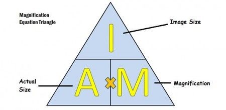
- Calculating magnification:
- I = image size I = A x M
- A = actual size A = ! / M
- M = magnification M = I / A
-
GCSE Style Questions (calc needed)
- 1. A red blood cell is 7.5 micrometres in diameter. It is magnified 2000 times. Calculate the diameter of the image seen through the microscope in mm.
- Answer:
- I = A x M
- I = 7.5 x 2000
- I = 15,000 mm
- 2. A sperm cell has a tail 40 micrometres long and a student draws it 40 mm long. Calculate the magnification.
- Answer:
- M = I / A M = 40 mm / 40 micrometres
- M = 4000 / 4
- M = 1000
-
The Light Microscope and Observing Cells
- A light microscope uses a series of lenses to produce a magnified image of an object:
- The object is placed on a rectangular glass slide
- The slide is placed on a stage with a light source below
- Light shines through the object and into the objective lens
- The light passes through the eyepiece lens and from there into your eye.
- When you observe cells, it is usual to make a drawing of what you see. Very often there is so much to see that you can only aim to draw part of it.
- Use a pencil rather than pen or colours
- Outline the features as accurately as you can
- Use as little shading as possible
- Label your drawing with the name of the sample and the total magnification you used
Information
- Description
- Using pages 12 and 13 or your CGP GCSE Biology for AQA (Grade 1-9) Revision guide.
- Created by
- Sas D · Jan 2018
- Updated
- Jan 7, 2018
- Views
- 90
- Rating